. 2007 Oct 1;21(19):2410-21.
doi: 10.1101/gad.439307.

Accelerated aging and failure to segregate damaged proteins in Sir2 mutants can be suppressed by overproducing the protein aggregation-remodeling factor Hsp104p

Affiliations

Accelerated aging and failure to segregate damaged proteins in Sir2 mutants can be suppressed by overproducing the protein aggregation-remodeling factor Hsp104p

Nika Erjavec et al. Genes Dev. .

Abstract

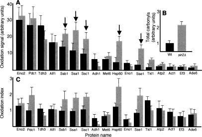
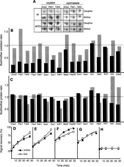
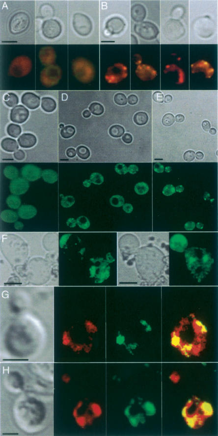
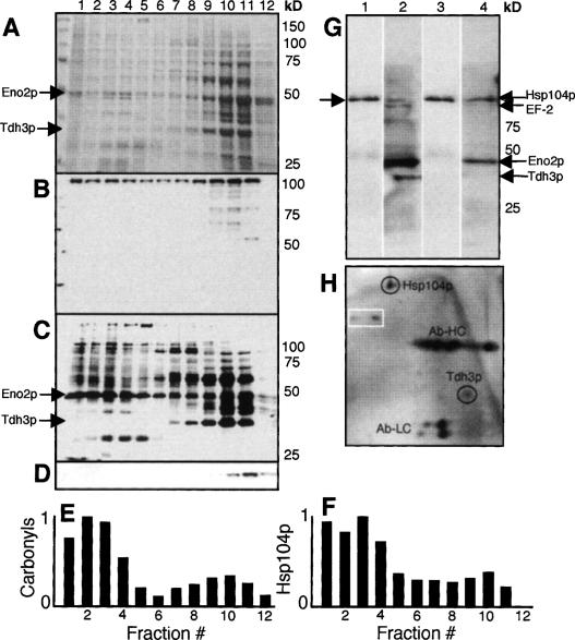
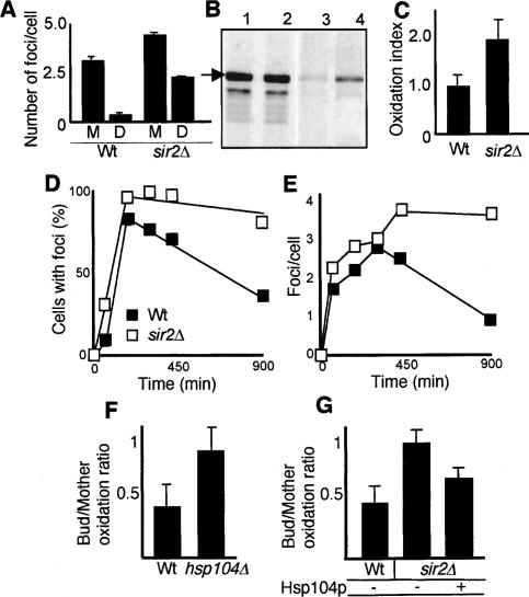
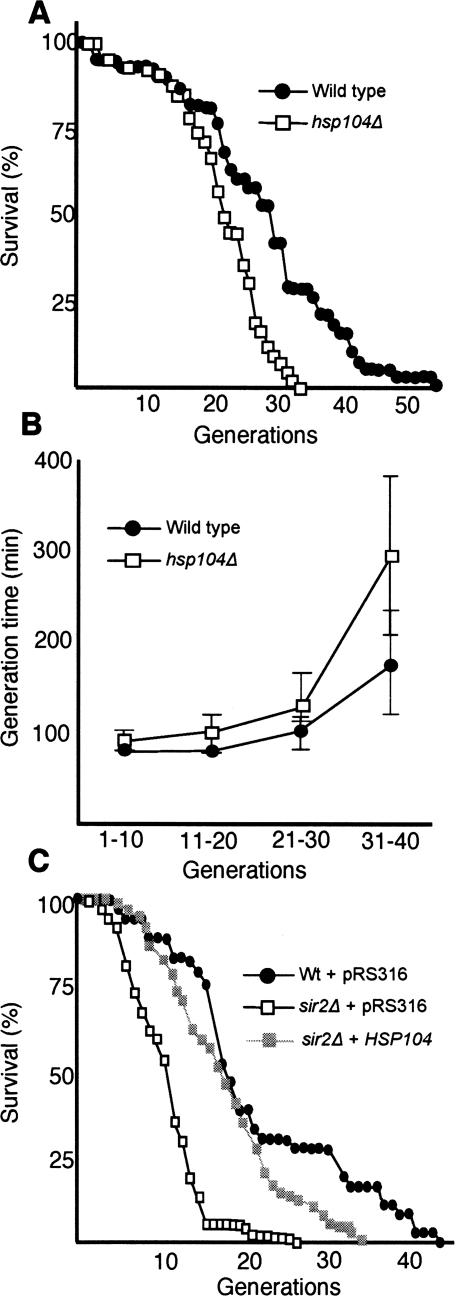
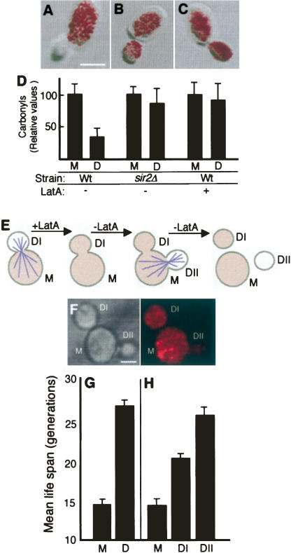
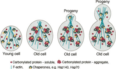
The levels of oxidatively damaged, carbonylated, proteins increase with the replicative age of yeast mother cells. We show here that such carbonylated proteins are associated with Hsp104p-containing protein aggregates and that these aggregates, like oxidized proteins, are retained in the progenitor cell during cytokinesis by a Sir2p-dependent process. Deletion of HSP104 resulted in a breakdown of damage asymmetry, and overproduction of Hsp104p partially restored damage retention in sir2Delta cells, suggesting that functional chaperones associated with protein aggregates are required for the establishment of damage asymmetry and that these functions are limited in sir2Delta cells. In line with this, Hsp104p and several Hsp70s displayed elevated damaged in sir2Delta cells, and protein aggregates were rescued at a slower rate in this mutant. Moreover, overproduction of Hsp104p suppressed the accelerated aging of cells lacking Sir2p, and drugs inhibiting damage segregation further demonstrated that spatial quality control is required to rejuvenate the progeny.


Figures

Figure 1.
Figure 1.
Cellular chaperones are preferential targets of age-dependent protein oxidation in the short-lived sir2Δ mutant. (A) Oxidation signal of single proteins from 10- to 12-generations-old wild-type (black bars) and sir2Δ (gray bars) cells. The specific oxidation signal was derived by comparing the carbonylation intensity of single proteins with the total carbonylation signal from all proteins. Protein identities are found in Table 1; arrows point to chaperones. (B) Comparison of total carbonylation (normalized to protein concentration) of the whole proteome from aged wild-type and sir2Δ cells. (C) Oxidation index of single proteins from aged wild-type (black bars) and sir2Δ (gray bars) cells. The specific oxidation index was calculated by normalizing the carbonylation intensity (carbonylation signal in the specific protein per total carbonylation signal) of single proteins to their abundance (Coomassie-derived signal in specific protein per total Coomassie signal on the gel). An index >1 indicates a protein whose oxidation level exceeds that of the average carbonylated protein. Values shown are the average of at least four different 2D gels and two independent sorting events; error bars in A–C represent the standard deviation of the mean.
Figure 2.
Figure 2.
Damage asymmetry is the result of a specific Sir2p-mediated retention of oxidized proteins in the mother cell during cytokinesis. (A) 2D PAGE carbonylation (oxyblot) and abundance (Coomassie) of the three major carbonylated proteins—Eno2p, Pdc1p, and Thd3p—in wild-type (top rows) and sir2Δ (bottom rows) mother and daughter cells. The mother cells are 10–12 generations old. (B) Bud/mother oxidation ratio in aged wild type (black bars) and sir2Δ (gray bars). The ratio was calculated for each of the identified proteins, by comparing the specific oxidation signals (see Fig. 1) of daughter cells and their 10- to 12-generations-old mothers. (C) Bud/mother protein ratio in aged wild type (black bars) and sir2Δ (gray bars). The ratio was determined by comparing the specific abundance of daughter and mother proteins as seen by Coomassie staining. In both, B and C, the values shown are the average of at least four different 2D gels and two independent sorting events. Budding wild-type (D,F) and sir2Δ (E,G) cells subjected to FRAP analysis. Signal recovery of Eno2p-GFP (D,E) and Tdh3p-GFP (F,G), expressed as the percentage of the signal prior to bleaching, gives a measure of the rate of de novo protein synthesis between mothers (filled squares) and their daughters (empty squares). (H) Controls were carried out by blocking protein synthesis with cyclohexamide, prior to bleaching. Representative curves are shown in each case.
Figure 3.
Figure 3.
Carbonyl-damaged proteins form aggregates associated with Hsp104p in vivo in aged yeast cells. Protein carbonylation signal in young wild-type cells (A) and in aged (10–12 generations old) mother cells (B). Bright-field pictures (top) and carbonyl immunodetection (bottom) are shown. (C–F) Hsp104-GFP distribution in young cells subjected to no stress (C), mild H2O2 stress (D), and severe H2O2 stress (E), and in unstressed 10- to 12-generations-old mother cells (F). The corresponding bright-field pictures are also shown; 95% of cells in the population displayed the hereby shown phenotype. Intense cytosolic Hsp104-GFP foci correspond to sites of electron-dense aggregates (Lum et al. 2004). Foci of carbonylated proteins and Hsp104p in H2O2 stressed cells (G) and in unstressed 12-generations-old cells (H). From left to right are shown a bright-field image, a carbonyl image, a Hsp104 foci, and a merge between carbonyls and Hsp104 foci. Bars, 5 μm.
Figure 4.
Figure 4.
Carbonylated proteins are enriched in high-molecular aggregates and copurify with Hsp104p. Proteins were size-fractionated by sucrose gradient centrifugation, and total protein patterns (A), abundance of Hsp104p (B), carbonylated proteins (C), and actin (D) were analyzed in each fraction. Total protein was determined by Coomassie brilliant blue staining, while Hsp104p, actin, and carbonylated proteins were determined by immunodetection as described in Materials and Methods. The abundance of carbonylated proteins (E) and Hsp104p (F) in each size fraction normalized to total protein in the fractions. (G) Hsp104p (lanes 1,3) and carbonylated proteins (lanes 2,4) from oxidatively stressed (lanes 1,2) and unstressed (lanes 3,4) cells copurified by pull-down of Hsp104-TapTag as described in Materials and Methods. (H) 2D gel showing copurification of one of the major carbonylated proteins, Tdh3p, upon immunoprecipitation of Hsp104p. The identity of Tdh3p was confirmed by mass spectrometry. The heavy (Ab-HC) and light (Ab-LC) chains of the antibody are depicted on the 2D gel. The spots in the white box mark nonspecific proteins coming down also in the control (bead only).
Figure 5.
Figure 5.
Hsp104p-dependent PQC is required for damage segregation and is diminished in cells lacking Sir2p. (A) Distribution of Hsp104-GFP foci in mothers (M) and daughters (D) during cytokinesis of 10–12-generations-old wild-type (n = 55) and sir2Δ mutant (n = 74) cells. (B) Hsp104p levels (lanes 1,2) and Hsp104 carbonylation (lanes 3,4) in wild-type (lanes 1,3) and sir2Δ (lanes 2,4) cells. The arrow indicates Hsp104p. (C) Carbonyl levels normalized to total levels of Hsp104p in wild-type and sir2Δ mutant cells. (D) Fraction of cells in the population displaying Hsp104p foci before and during exposure to 1 mM H2O2 for 120 min, and after removal of the oxidant. Between 200 and 400 cells were scored at each time point and were defined as having either a diffuse cytosolic, nonaggregated distribution of Hsp104-GFP or, conversely, a discrete, aggregated one (see also Lum et al. 2004). Results are shown for both wild type (black squares) and sir2Δ (open squares). (E) The average number of Hsp104-GFP foci per cell before, during, and after oxidative stress (as in C); between 58 and 198 cells were analyzed at each time point. (F) Retention of protein damage in 12-generations-old wild-type and hsp104Δ mother cells, as determined by in situ carbonylation. (G) Effects of overproducing Hsp104p on damage asymmetry in sir2Δ cells. Shown are values for the wild type carrying the vector control (n = 44), sir2Δ carrying the vector control (n = 53), and sir2Δ overexpressing HSP104 (n = 43). Error bars correspond to the standard error (A–C) or the standard deviation of the mean (F–G).
Figure 6.
Figure 6.
Hsp104 is required for yeast longevity, and its overproduction suppresses the reduced life span of Sir2p-deficient cells. (A) Replicative life span of wild-type (black circles) and the hsp104Δ mutant (open squares) on YPD. The standard error for the wild type was ±0.4 (n = 78) and for the hsp104Δ mutant was ±0.1 (n = 81). (B) Generation times of wild type (black circles) and the hsp104Δ mutant (open squares) as a function of their replicative age. Error bars correspond to the standard deviation of the mean. (C) Replicative life span of control wild-type (black circles) and sir2Δ cells harboring the empty vector (open squares), and sir2Δ cells overproducing Hsp104p (gray squares). The standard error for wild type was ±0.2 (n = 72), for sir2Δ cells was ±0.7 (n = 74), and for sir2Δ cells overproducing Hsp104p was ±0.2 (n = 74). Cells were grown on selective YNB-Uracil medium. All life-span measurements were performed in duplicate or triplicate.
Figure 7.
Figure 7.
Transient depolymerization of the actin cytoskeleton eliminates damage asymmetry and limits the replicative potential of the daughter cell. Overlays of transmission images and in situ detection of carbonyls in untreated wild-type cells (A) and sir2Δ (B) and wild-type (C) cells treated with 200 μM Lat-A. (D) Quantification of carbonyls in mothers (M) and daughters (D) of cells treated as in A–C. (E) Schematic representation of the experiment in which a transient depolymerization of the actin cytoskeleton is achieved by a short-term Lat-A treatment. After removal of the drug, the mother (M) gives rise to two daughters nearly simultaneously: a first one affected by the treatment (DI), and a second one (DII) generated at a new bud site once the cytoskeleton had oriented itself again. Damaged proteins (shaded color) are distributed equally between mother and daughter DI, and this damage symmetry persists after cell separation. As the actin cytoskeleton is reoriented, damaged proteins are largely prevented from being inherited by daughter DII. (F) In situ carbonylation of cells exposed to a transient Lat-A treatment, as described above. Bar, 5 μm. (G) Mean replicative life span of 10–12-generations-old mothers and their daughters treated with DMSO only. The standard error (error bars in the figure) for mother cells (M) was ±0.4 and for daughter cells (D) was ±0.8. (D) Mean replicative life span of age-matched mothers (M) and their “twin” daughters, DI and DII, after treatment with Lat-A. Error bars correspond to the standard error of three independent measurements. The standard error for mother cells was ±1.0, for DI daughter cells was ±0.6, and for DII daughter cells was ±1.2.
Figure 8.
Figure 8.
Model for the proposed segregation of damaged aggregated proteins during yeast cytokinesis. Actin cables align themselves along the bud–mother axis during bud emergence and persist as such throughout cytokinesis. This allows for a vectorial transport of cargo between mother and bud that is mediated by adaptor proteins. In addition, it has been shown that the actin cytoskeleton provides a scaffold for large aggregates—e.g., prion Sup35-derived aggregates—and that this resembles the mammalian aggresomes (Ganusova et al. 2006). Hsp104p is implicated in the interaction between prion protein aggregates and the cytoskeletal components, thus helping the cell to localize misfolded prion proteins and prevent them from causing damage (Ganusova et al. 2006). Likewise, we suggest that Hsp104p may provide a bridge between carbonylated aggregates and the cytoskeleton that not only prevents proteotoxicity but reduces the inheritance of these aggregates to the progeny.

Similar articles

Cited by

References

    1. Aguilaniu H., Gustafsson L., Rigoulet M., Nyström T., Gustafsson L., Rigoulet M., Nyström T., Rigoulet M., Nyström T., Nyström T. Protein oxidation in G0 cells of Saccharomyces cerevisiae depends on the state rather than rate of respiration and is enhanced in pos9 but not yap1 mutants. J. Biol. Chem. 2001;276:35396–35404. - PubMed
    1. Aguilaniu H., Gustafsson L., Rigoulet M., Nyström T., Gustafsson L., Rigoulet M., Nyström T., Rigoulet M., Nyström T., Nyström T. Asymmetric inheritance of oxidatively damaged proteins during cytokinesis. Science. 2003;299:1751–1753. - PubMed
    1. Bota D.A., Davies K.J., Davies K.J. Lon protease preferentially degrades oxidized mitochondrial aconitase by an ATP-stimulated mechanism. Nat. Cell Biol. 2002;4:674–680. - PubMed
    1. Davies K.J. Protein damage and degradation by oxygen radicals. I. General aspects. J. Biol. Chem. 1987;262:9895–9901. - PubMed
    1. Desnues B., Cuny C., Gregori G., Dukan S., Aguilaniu H., Nyström T., Cuny C., Gregori G., Dukan S., Aguilaniu H., Nyström T., Gregori G., Dukan S., Aguilaniu H., Nyström T., Dukan S., Aguilaniu H., Nyström T., Aguilaniu H., Nyström T., Nyström T. Differential oxidative damage and expression of stress defence regulons in culturable and non-culturable cells of Escherichia coli cells. EMBO Rep. 2003;4:400–405. - PMC - PubMed

Publication types

MeSH terms

Substances