. 2007 Oct 1;21(19):2433-47.
doi: 10.1101/gad.437807.

Activation of the UNC5B receptor by Netrin-1 inhibits sprouting angiogenesis

Affiliations

Activation of the UNC5B receptor by Netrin-1 inhibits sprouting angiogenesis

Bruno Larrivée et al. Genes Dev. .

Abstract

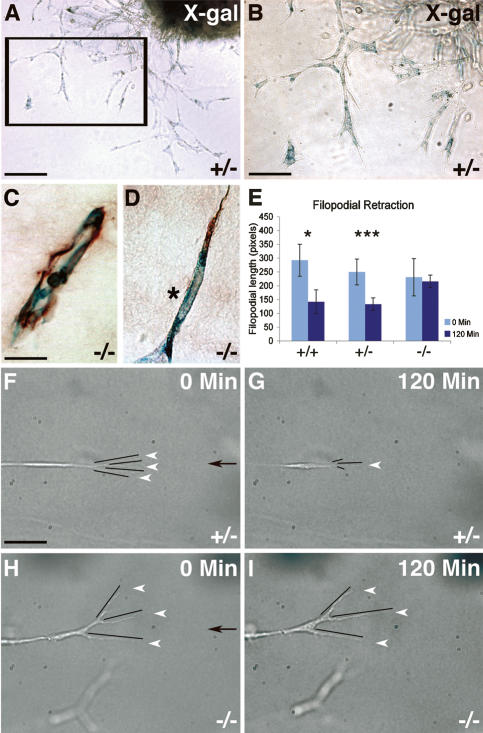
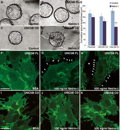
Netrins are secreted molecules with roles in axonal growth and angiogenesis. The Netrin receptor UNC5B is required during embryonic development for vascular patterning, suggesting that it may also contribute to postnatal and pathological angiogenesis. Here we show that unc5b is down-regulated in quiescent adult vasculature, but re-expressed during sprouting angiogenesis in matrigel and tumor implants. Stimulation of UNC5B-expressing neovessels with an agonist (Netrin-1) inhibits sprouting angiogenesis. Genetic loss of function of unc5b reduces Netrin-1-mediated angiogenesis inhibition. Expression of UNC5B full-length receptor also triggers endothelial cell repulsion in response to Netrin-1 in vitro, whereas a truncated UNC5B lacking the intracellular signaling domain fails to induce repulsion. These data show that UNC5B activation inhibits sprouting angiogenesis, thus identifying UNC5B as a potential anti-angiogenic target.


Figures

Figure 1.
Figure 1.
Unc5b expression during postnatal and pathological neovascularization. (A–D) Unc5b plap (A,C) reporter expression in the retina of P12 heterozygous mice double-stained with IsolectinB4 (B,D). C and D are higher magnifications of boxed areas in A and B, respectively. (C,D) Unc5b is expressed in arteries (a), but most IsolectinB4-labeled capillaries are negative. (E–H) Unc5b plap (E,G) reporter expression in the OIR retina of P17 mice double-stained with IsolectinB4 (F,H). G and H are higher magnifications of boxed areas in E and F, respectively. (G,H) Unc5b is up-regulated in arteries and sprouting vessels. (*) Arterio–venous shunt; (v) vein. (I) Whole-mount X-gal staining of skin of an adult heterozygous unc5b-LacZ-plap mouse shows weak unc5b expression in arteries (a) but not in veins (v), and expression in hair follicles (arrowheads). (J,K) bFGF-containing matrigel plug injected into a heterozygous unc5b-LacZ-plap mouse. Whole-mount X-gal staining (J) of the freshly dissected plug (K) shows up-regulation of unc5b expression in vessel sprouts (arrowheads) and arteries (a) but not veins (v). (L) Section of a matrigel plug stained with X-gal and PECAM-1. Note unc5b expression in endothelial cells (arrowheads) and a smooth muscle cell (*). (M–P) PC3 tumor cells implanted for 7 d into a heterozygous unc5b-LacZ-plap mouse. Whole-mount AP (M) collagen-IV (N) double staining of a thick section of a tumor nodule. Note unc5b expression in tumor arteries (a), but not veins (v), and in sprouting capillaries (arrowheads). (O,P) Higher magnification of a sprout (yellow arrowhead in M and N) expressing the unc5b lac-Z reporter (O) and double-stained with Collagen-IV (P). Bars: A,B,E,F, 225 μm; C,D,L–N, 110 μm; G,H, 45 μm; I, 1500 μm; J,K, 2000 μm; O,P, 25 μm.
Figure 2.
Figure 2.
Inhibition of matrigel neovascularization by Netrin-1. (A–D) Matrigel plugs in C57/Bl6 wild-type mice (+/+). (A,B) Quantification of hemoglobin content (A) and VEGFR-2 protein levels (B). bFGF is at 300 ng/mL, and Netrin-1 (Net) is at 300 or 1000 ng/mL. n = 10 mice per growth factor treatment; two independent groups of mice for A and B. (C,D) Vessel morphology in matrigel plugs containing bFGF (C) or bFGF and Netrin-1 (both at 300 ng/mL) (D) after IsolectinB4 perfusion. (D) Note reduction of vessel branching in the presence of Netrin-1. (E–G) Whole-mount X-gal staining of matrigel plugs in unc5b+/− mice. Note little invasion of unc5b-expressing vessels (dashed areas) in plugs containing Netrin-1 alone (E), robust stimulation of neovascularization by bFGF (F), and inhibition of neovascularization in plugs containing both factors at 300 ng/mL (G). (H) Quantification of serial paraffin sections prepared from matrigel plugs. The number of sections invaded by at least one blood vessel was scored on 100–200 sections per plug. n = 1 control, 3 unc5b+/− mice treated with 1000 ng/mL Netrin-1, 4 unc5b+/− and 5 unc5b−/− mice treated with bFGF and with bFGF and Netrin-1. (I,J) Higher magnification of vascular front (corresponding to boxed areas in F and G, respectively). Note numerous sprouts extending into the gel in the presence of bFGF alone (I, arrowheads), while less sprouts are observed in the presence of both bFGF and Netrin-1 (300 ng/mL) (J, arrowhead). (K,L) Overview of X-gal-stained plugs in unc5b−/− mice. Note similar vascularization (dashed areas) in absence (K) and presence (L) of Netrin-1. (M,N) Vascular front (corresponding to boxed areas in K and L, respectively) in matrigel plugs injected into unc5b−/− mice. Note similar numbers of sprouts in the absence (M) and presence (N) of Netrin-1. Bars: C,D, 75 μm; E–G,K,L, 233 μm; I,J,M,N, 26 μm.
Figure 3.
Figure 3.
Unc5b-expressing endothelial tip cells retract filopodia in response to Netrin-1 aortic ring assays. (A,B) Sprouts growing from heterozygous unc5b lacZ-plap aortic rings into collagen gels stain with X-gal. B is a higher magnification of boxed area in A. (C,D) X-gal/PECAM-1 double staining of sections prepared from explants show lumenized vessels close to the explant (C) and double-staining of tip cells (D). (*) Tip cell nucleus. (E–I) Tip cell response to gradients of recombinant Netrin-1 recorded by time-lapse videomicroscopy. (E) Quantification of filopodial length at the beginning (t = 0) and the end of the time-lapse movies (t = 120 min). (F–I) Responses of individual tip cells from unc5b+/− (F,G) and unc5b−/− (H,I) explants to gradients of recombinant Netrin-1 (gradient source indicated by black arrows in F and H). Still images from time-lapse movies at the indicated time points. White arrowheads point to filopodia. Filopodial length is indicated by black lines. Note filopodial retraction in G, but not in I. Total number of tip cells analyzed: six +/+, 17 +/−, and 16 −/−. Error bars, SEM; (*) P < 0.05; (***) P < 0.001, Mann-Whitney U-test. Bars: A, 170 μm; B, 85 μm; C,D, 20 μm; F–I, 45 μm.
Figure 4.
Figure 4.
Repulsion of PAECs expressing FL UNC5B in the presence of Netrin-1. (A–E) Gelatin-coated microcarrier beads were seeded with parental, UNC5B FL, or UNC5B ΔCD (UNC5B CD) PAECs and embedded in fibrin gels supplemented with 10% FBS or 10% FBS and 300 ng/mL Netrin-1. (E) Endothelial sprout formation was quantified after 24 h (cumulative sprout length per bead). n = 4 (UNC5B FL) or 3 (parental and UNC5B CD) experiments performed in triplicate. Error bars: SEM; (**) P < 0.025, Mann-Whitney U-test. (F–H) Confocal images of UNC5B FL PAECs treated with BSA (F) or with recombinant mouse Netrin-1 at 100 ng/mL (G) or 500 ng/mL (H) for 30 min. Note dose-dependent receptor aggregation at filopodial extensions (arrowheads) and cell retraction (*). (I–K) UNC5B CD PAECs treated with BSA (I) or with recombinant mouse Netrin-1 (J,K). Note absence of retraction. Bars: A–D, 115 μm; F–K, 30 μm.
Figure 5.
Figure 5.
Expression of netrins and their receptors in human tumor cell lines. (A) Three-hundred nanograms of total RNA extracted from the indicated tumor cell lines were reverse-transcribed with oligo-dT and amplified with primer pairs specific for the indicated genes and tubulin. Two independent RT and PCR reactions (32 and 40 cycles, respectively) were performed, with similar results. (−) No amplification; (+−) weak or no amplification at 32 cycles, but band detectable after 40 cycles; (+) band present in both amplifications; (++) strong band; (+++) very strong band. (B) Gels representing 40 cycle amplifications of netrin-1, netrin-3 and netrin-4. Note absence of netrin expression in Miapaca pancreatic cancer cells and Mel2a melanoma cells (outlined in red) and presence of netrin-4 in PC3 prostate cancer cells (outlined in blue).
Figure 6.
Figure 6.
Repulsion of UNC5B FL PAECs by Netrin-1-overexpressing tumor cells. (A–C) Confocal images from cocultures of PAECs expressing UNC5B FL (green) with the indicated control-infected tumor cell lines labeled with the lipophilic fluorescent dye PKH26 (red). Note mixing of the two populations after 24 h of culture. (D–F) PAEC UNC5B FL cells cocultured with the indicated Netrin-1-transduced tumor cell lines. Note formation of UNC5B GFP aggregations (arrowheads) next to Netrin-1-producing tumor cells and segregation of PAECs from tumor cells. (G–I) UNC5B ΔCD PAECs do not segregate from Netrin-1-secreting tumor cells. (J) Quantification of the overlap of tumor cells by endothelial cells. The area of PAECs covered by tumor cells was measured on six images per well in three independent experiments. Error bars: SEM; (*) P < 0.05, Mann-Whitney U-test. Bars, 80 μm.
Figure 7.
Figure 7.
Netrin-1 overexpression reduces sprouting of unc5b-expressing vessels and delays tumor angiogenesis. (A,B) Mel2a control tumor 4 wk post-implantation. (B) Representative section hybridized with an antisense riboprobe against unc5b showing expression on sprouting vessels invading the tumor (blue, arrowheads). (A) Netrin-1 immunostaining (red) of the same section. Note absence of immunoreactivity in control tumor tissue. Red staining is due to background immunofluorescence of erythrocytes contained in blood vessels. (C,D) Mel2a Netrin-1-overexpressing tumor 3 wk post-implantation. Note that unc5b-positive vessels (blue, arrowheads) avoid Netrin-1-positive tumor areas. (E) Quantification of PECAM-1-positive and unc5b-positive blood vessels per surface area of the tumor in control and Netrin-1-overexpressing Mel2a tumors. (F) Growth curve of Mel2a tumor xenografts implanted in the dorsa of NMRI nu/nu mice. n = 10 mice per group. (G,H) Miapaca control tumor 6 wk post-implantation. Note unc5b-positive sprouts (blue, arrowheads) in the Netrin-1-negative tumor tissue. (I,J) Miapaca Netrin-1-overexpressing tumor 6 wk post-implantation. Note absence of unc5b-positive sprouts in Netrin-1-overexpressing tumor tissue. (K) Quantification of PECAM-1-positive and unc5b-positive blood vessels per surface area in control and Netrin-1-overexpressing Miapaca tumors. (L) Growth curves of Miapaca tumor xenografts implanted in the dorsa of NMRI nu/nu mice. n = 10 mice per group. Data in E and K represent the quantification of two or three tumors per group and three to five sections per tumor. Error bars: SEM; (*) P < 0.05, Mann-Whitney U-test. Bars: A–D, 70 μm; G–J, 125 μm.
Figure 8.
Figure 8.
Netrin-1-induced inhibition of tumor vessel sprouting requires UNC5B. (A–C) Whole-mount Collagen-IV staining of thick sections from PC3 control (A) and Netrin-1-overexpressing (B,C) tumors implanted into unc5b heterozygous (A,B) and homozygous (C) mutant mice. The tumor nodule forming beneath the skin is outlined (dashed, red). Vessels invading the tumor are outlined (dashed, white). Vascular area within the tumors is reduced by Netrin-1 in unc5b heterozygous (B) compared with control (A), but not homozygous mutant (C), mice. (D) Computer-assisted quantification of vascular area in tumor nodules. n = 3 tumors per group; four thick sections per tumor (corresponding to the entire tumor) were photographed and quantified. (E–J) Higher magnifications of vessels sprouting from the skin into the tumor area. (E,G,I) Whole mounts were double-stained with Collagen-IV and X-gal. In the overlays shown in F, H, and J, Collagen-IV staining appears as purple color on the dark-blue X-gal staining. Note double-positive vessels (arrowheads) sprouting into control PC3 tumors (F), blunted appearance of vessel sprouts in Netrin-1-overexpressing tumors implanted into unc5b heterozygous mice (H), and restored sprouting when Netrin-1-overexpressing tumors are implanted into unc5b homozygous mutant mice (J). Error bars: SEM; (*) P < 0.05, Mann-Whitney U-test. Bars: A–C, 800 μm; E–J, 130 μm.

Similar articles

Cited by

References

    1. Ackerman S.L., Kozak L.P., Przyborski S.A., Rund L.A., Boyer B.B., Knowles B.B., Kozak L.P., Przyborski S.A., Rund L.A., Boyer B.B., Knowles B.B., Przyborski S.A., Rund L.A., Boyer B.B., Knowles B.B., Rund L.A., Boyer B.B., Knowles B.B., Boyer B.B., Knowles B.B., Knowles B.B. The mouse rostral cerebellar malformation gene encodes an UNC-5-like protein. Nature. 1997;386:838–842. - PubMed
    1. Augustin H.G. Tubes, branches, and pillars: The many ways of forming a new vasculature. Circ. Res. 2001;89:645–647. - PubMed
    1. Baluk P., Morikawa S., Haskell A., Mancuso M., McDonald D.M., Morikawa S., Haskell A., Mancuso M., McDonald D.M., Haskell A., Mancuso M., McDonald D.M., Mancuso M., McDonald D.M., McDonald D.M. Abnormalities of basement membrane on blood vessels and endothelial sprouts in tumors. Am. J. Pathol. 2003;163:1801–1815. - PMC - PubMed
    1. Benimetskaya L., Wittenberger T., Stein C.A., Hofmann H.P., Weller C., Lai J.C., Miller P., Gekeler V., Wittenberger T., Stein C.A., Hofmann H.P., Weller C., Lai J.C., Miller P., Gekeler V., Stein C.A., Hofmann H.P., Weller C., Lai J.C., Miller P., Gekeler V., Hofmann H.P., Weller C., Lai J.C., Miller P., Gekeler V., Weller C., Lai J.C., Miller P., Gekeler V., Lai J.C., Miller P., Gekeler V., Miller P., Gekeler V., Gekeler V. Changes in gene expression induced by phosphorothioate oligodeoxynucleotides (including G3139) in PC3 prostate carcinoma cells are recapitulated at least in part by treatment with interferon-β and -γ. Clin. Cancer Res. 2004;10:3678–3688. - PubMed
    1. Carmeliet P., Tessier-Lavigne M., Tessier-Lavigne M. Common mechanisms of nerve and blood vessel wiring. Nature. 2005;436:193–200. - PubMed

Publication types

MeSH terms

LinkOut - more resources