. 2007 Oct 1;21(19):2495-508.
doi: 10.1101/gad.439107.

The nature of telomere fusion and a definition of the critical telomere length in human cells

Affiliations

The nature of telomere fusion and a definition of the critical telomere length in human cells

Rebecca Capper et al. Genes Dev. .

Abstract

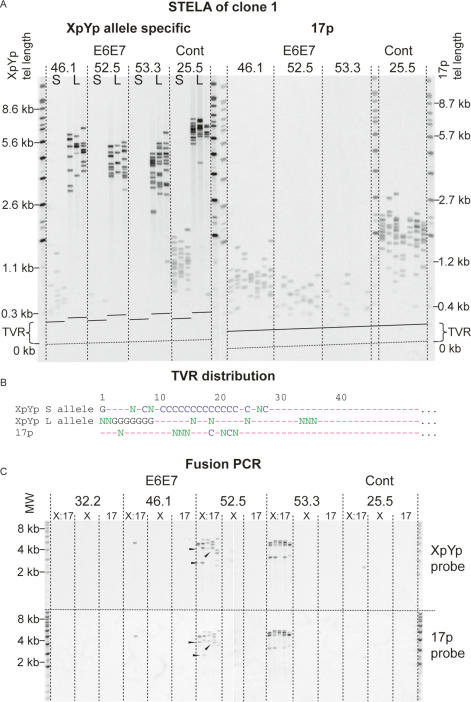
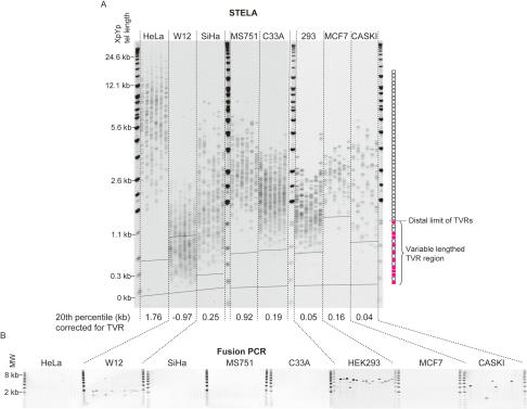
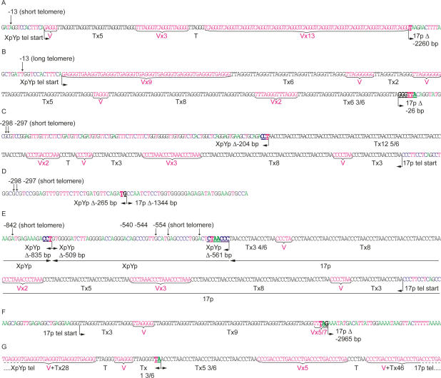
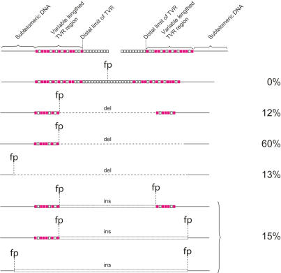
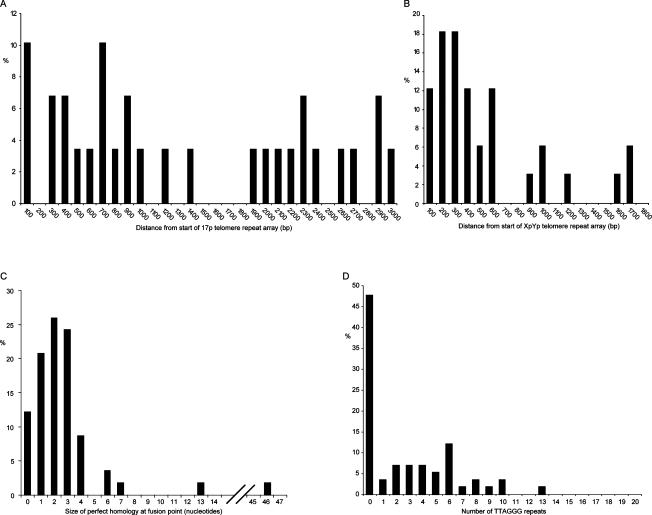
The loss of telomere function can result in telomeric fusion events that lead to the types of genomic rearrangements, such as nonreciprocal translocations, that typify early-stage carcinogenesis. By using single-molecule approaches to characterize fusion events, we provide a functional definition of fusogenic telomeres in human cells. We show that approximately half of the fusion events contained no canonical telomere repeats at the fusion point; of those that did, the longest was 12.8 repeats. Furthermore, in addition to end-replication losses, human telomeres are subjected to large-scale deletion events that occur in the presence or absence of telomerase. Here we show that these telomeres are fusogenic, and thus despite the majority of telomeres being maintained at a stable length in normal human cells, a subset of stochastically shortened telomeres can potentially cause chromosomal instability. Telomere fusion was accompanied by the deletion of one or both telomeres extending several kilobases into the telomere-adjacent DNA, and microhomology was observed at the fusion points. This contrasted with telomere fusion that was observed following the experimental disruption of TRF2. The distinct error-prone mutational profile of fusion between critically shortened telomeres in human cells was reminiscent of Ku-independent microhomology-mediated end-joining.


Figures

Figure 1.
Figure 1.
Telomere loss, with a commensurate increase in the frequency of fusion, in MRC5 clone 1 expressing HPV E6E7 oncoproteins. (A) Allele-specific STELA at the XpYp telomere and 17p telomere. (S) Shorter allele; (L) longer allele. The dashed lines marked “TVR” represent the distal limit of telomere repeat variants and the beginning of the region of the telomere repeats containing pure TTAGGG repeats. The numbers above represent the number of PDs from the point of single-cell cloning; crisis occurred at PD 53.3 and the control cells underwent replicative senescence at PD 25.5. Telomere length at the XpYp and 17p telomeres is indicated on the left and right sides; this is calculated by subtracting the fragment size from the known amount of DNA between the PCR priming site and the start of the telomere repeat array. (B) TVR distributions from XpYp and 17p. (G) TGAGGG; (C) TCAGGG; (N) other repeat variants; (—) TTAGGG obtained by TVR-PCR at the XpYp telomere and sequence analysis of fusions containing the 17p telomere. (C) Fusion PCR. (X:17) Primers for XpYp and 17p; (X) single XpYp primer; (17) single 17p primer. Products were detected with the XpYp- and 17p-specific probes as indicated on the right. Arrowheads indicate fusion products that display differential hybridization.
Figure 2.
Figure 2.
XpYp telomere length distributions and fusion frequencies in eight telomerase-expressing epithelial cell lines. (A) STELA at the XpYp telomere. The 20th percentile length, taking into account the position of the distal extent of the TVR, is indicated below. The distal extent of the highly variable TVR region from the longest TVR allele of each cell line is indicated by a dashed line across the gel image; the TVR region of the second allele may be less extensive. TVR and TTAGGG repeat regions are illustrated on the right, blank boxes represent TTAGGG repeats, and red filled boxes represent TVRs. (B) Fusion PCR between the XpYp and 17p telomeres.
Figure 3.
Figure 3.
Sequence data illustrating examples of the different classes of XpYp:17p fusion, isolated from MRC5 cells expressing E6E7 (A–F) and HEK 293 (G) cells. Arrows indicate informative subtelomeric SNP used to identify the XpYp telomeric alleles involved in the fusions identified from MRC5 cells. Telomere repeats (TTAGGG) in black text are identified below as “T” and variants are identified in red text as “V.” The fusion points, size of deletion (Δ) in base pairs, and microhomology (underlined in bold) are indicated below. (A) The XpYp telomere derived from the shorter allele fused to a 17p subtelomeric deletion. (B) The XpYp telomere derived from the longer allele fused to a17p subtelomeric deletion. (C) The converse of A: The 17p telomere is fused to an XpYp subtelomeric deletion of the shorter allele. (D) Fusion between subtelomeric deletions of both the XpYp shorter allele and 17p. (E) An example of XpYp:17p fusion that contains an insertion of XpYp telomere-adjacent DNA derived from the same allele in reverse orientation. Horizontal arrows below the sequence trace indicate the orientation of the sequences involved in the fusion. (F) An example of a fusion between sister chromatids; the 17p telomere is fused to a subtelomeric deletion of the 17p telomere-adjacent DNA. (G) An example of fusion between the telomere repeat variant-containing regions of XpYp and 17p from HEK 293 cells.
Figure 4.
Figure 4.
Summarizing telomere fusion data, the various different classes of fusion molecules are illustrated. Open squares represent TTAGGG repeats, filled squares represent TVRs. (fp) Fusion point; (del) deletion; (ins) insertion.
Figure 5.
Figure 5.
Histograms summarizing telomere fusion data. (A) The size of subtelomeric deletion events at 17p. (B) Deletion events at XpYp. (C) The extent of sequence homology at the fusion point. (D) The number of TTAGGG repeats immediately adjacent to the fusion point.
Figure 6.
Figure 6.
siRNA-mediated knockdown of TRF2 generates telomere fusions containing extensive arrays of TTAGGG repeats. (A) Western blot analysis with TRF2 antibody (top panel) and actin loading control (bottom panel), with the various treatments indicated above. (B) Real-time PCR for TRF2; relative quantification using the comparative CT methods normalized to the untreated control. Error bars, ±SD. (C) Fusion PCR between the XpYp and 17p telomeres. (D) Sequence data from fusion molecules obtained from MCF7 cells subjected to siRNA-mediated TRF2 knockdown. The total number of telomere and variant repeats is estimated based on the sizes of the fragments sequenced.

Similar articles

Cited by

References

    1. Artandi S.E., Chang S., Lee S.L., Alson S., Gottlieb G.J., Chin L., DePinho R.A., Chang S., Lee S.L., Alson S., Gottlieb G.J., Chin L., DePinho R.A., Lee S.L., Alson S., Gottlieb G.J., Chin L., DePinho R.A., Alson S., Gottlieb G.J., Chin L., DePinho R.A., Gottlieb G.J., Chin L., DePinho R.A., Chin L., DePinho R.A., DePinho R.A. Telomere dysfunction promotes non-reciprocal translocations and epithelial cancers in mice. Nature. 2000;406:641–645. - PubMed
    1. Atkin N.B. Lack of reciprocal translocations in carcinomas. Cancer Genet. Cytogenet. 1986;21:275–278. - PubMed
    1. Auersperg N. Long-term cultivation of hypodiploid human tumor cells. J. Natl. Cancer Inst. 1964;32:135–163. - PubMed
    1. Bailey S.M., Cornforth M.N., Kurimasa A., Chen D.J., Goodwin E.H., Cornforth M.N., Kurimasa A., Chen D.J., Goodwin E.H., Kurimasa A., Chen D.J., Goodwin E.H., Chen D.J., Goodwin E.H., Goodwin E.H. Strand-specific postreplicative processing of mammalian telomeres. Science. 2001;293:2462–2465. - PubMed
    1. Baird D.M., Jeffreys A.J., Royle N.J., Jeffreys A.J., Royle N.J., Royle N.J. Mechanisms underlying telomere repeat turnover, revealed by hypervariable variant repeat distribution patterns in the human Xp/Yp telomere. EMBO J. 1995;14:5433–5443. - PMC - PubMed

Publication types

Substances