. 2007 Oct 1;67(19):9066-76.
doi: 10.1158/0008-5472.CAN-07-0575.

Epidermal growth factor receptor cooperates with signal transducer and activator of transcription 3 to induce epithelial-mesenchymal transition in cancer cells via up-regulation of TWIST gene expression

Affiliations

Epidermal growth factor receptor cooperates with signal transducer and activator of transcription 3 to induce epithelial-mesenchymal transition in cancer cells via up-regulation of TWIST gene expression

Hui-Wen Lo et al. Cancer Res. .

Abstract

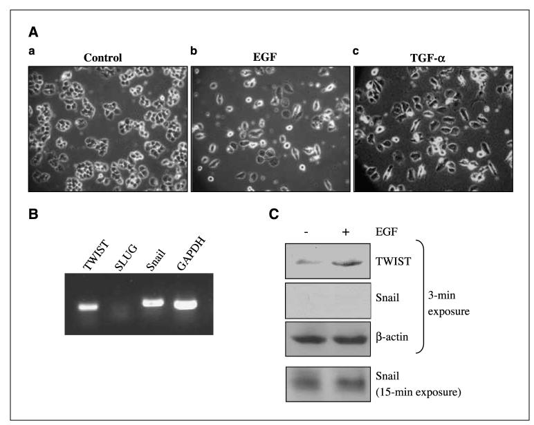
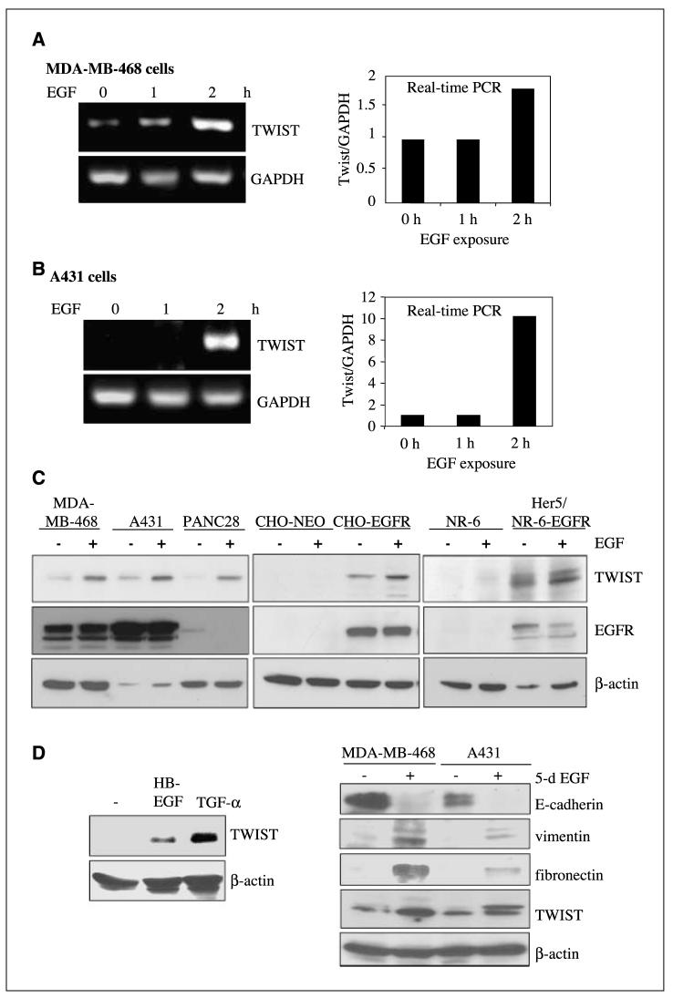
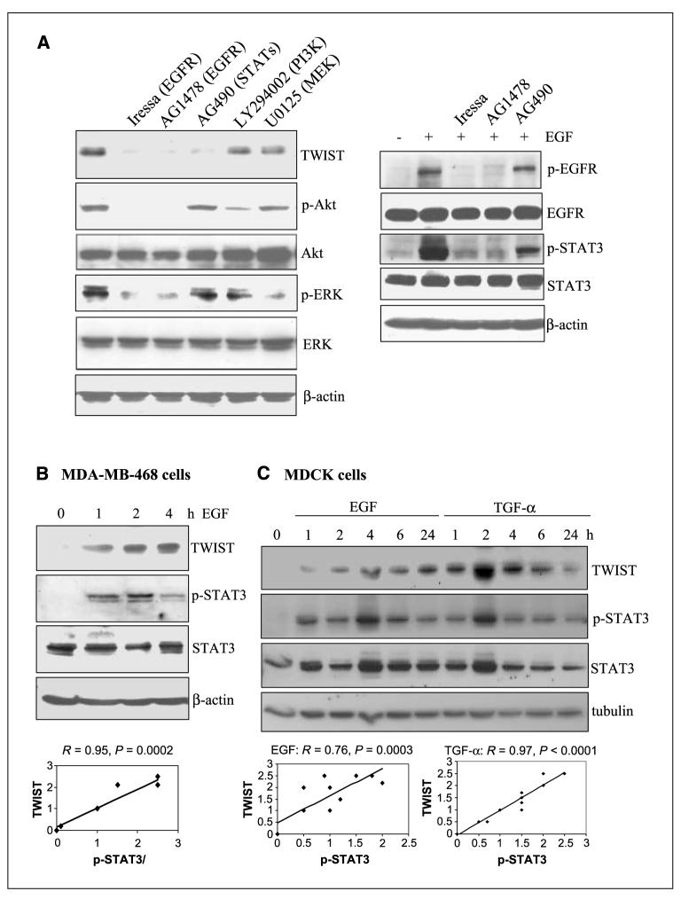
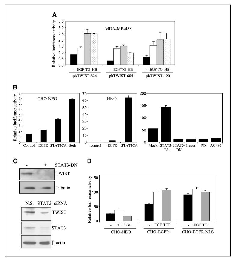
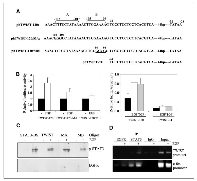
Aberrant epidermal growth factor receptor (EGFR) signaling is a major cause of tumor progression and metastasis; the underlying mechanisms, however, are not well understood. In particular, it remains elusive whether deregulated EGFR pathway is involved in epithelial-mesenchymal transition (EMT), an early event that occurs during metastasis of cancers of an epithelial origin. Here, we show that EGF induces EGFR-expressing cancer cells to undergo a transition from the epithelial to the spindle-like mesenchymal morphology. EGF reduced E-cadherin expression and increased that of mesenchymal proteins. In search of a downstream mediator that may account for EGF-induced EMT, we focused on transcription repressors of E-cadherin, TWIST, SLUG, and Snail and found that cancer cells express high levels of TWIST and that EGF enhances its expression. EGF significantly increases TWIST transcripts and protein in EGFR-expressing lines. Forced expression of EGFR reactivates TWIST expression in EGFR-null cells. TWIST expression is suppressed by EGFR and Janus-activated kinase (JAK)/signal transducer and activator of transcription 3 (STAT3) inhibitors, but not significantly by those targeting phosphoinositide-3 kinase and MEK/ERK. Furthermore, constitutively active STAT3 significantly activates the TWIST promoter, whereas the JAK/STAT3 inhibitor and dominant-negative STAT3 suppressed TWIST promoter. Deletion/mutation studies further show that a 26-bp promoter region contains putative STAT3 elements required for the EGF-responsiveness of the TWIST promoter. Chromatin immunoprecipitation assays further show that EGF induces binding of nuclear STAT3 to the TWIST promoter. Immunohistochemical analysis of 130 primary breast carcinomas indicates positive correlations between non-nuclear EGFR and TWIST and between phosphorylated STAT3 and TWIST. Together, we report here that EGF/EGFR signaling pathways induce cancer cell EMT via STAT3-mediated TWIST gene expression.


Figures

Figure 1
Figure 1
EGF exposure induced transition of the epithelial to the mesenchymal-like phenotype in cultured breast cancer cells. Human breast carcinoma MDA-MB-468 cells were used in these studies. Three independent experiments were done. Cancer cells were serum-starved for 24 h before treatments. A, EGF-treated MDA-MB-468 cells displayed mesenchymal morphology. Cells were treated with 0.5% FCS only (a), 0.5% FCS with 50 ng/mL EGF (b), and 0.5% FCS with 50 ng/mL TGF-α (c). Cell morphology was examined and photographed daily using a phase-contrast microscope. At day 5, unstimulated MDA-MB-468 cells retained their epithelial phenotype (a). In contrast, cells treated for 5 d with EGF (b) and TGF-α (c) displayed detached mesenchymal-like morphology. B, transcripts of EMT mediators, TWIST and Snail, were detected. Total RNA was isolated and subjected to RT-PCR to detect TWIST, SLUG, Snail gene transcripts. Expression of GAPDH serves as a loading control. C, TWIST, but not Snail, protein was expressed at high levels. Cancer cells were starved for 24 h and treated without and with EGF for 6 h; total cell lysates were extracted and subjected to Western blot analyses for expression of TWIST and Snail. Note the exposure time to detect the expression of Snail was 5 times longer than that of TWIST.
Figure 2
Figure 2
EGF stimulation induces expression of TWIST and mesenchymal markers, as well as reduction of an epithelial marker. A and B, EGF increases TWIST transcription in EGFR-expressing cancer cells. MDA-MB-468 (human breast carcinoma cells in A) and A431 (human epidermoid carcinoma cells in B) were serum-starved for 24 h, exposed to EGF (100 ng/mL) for 0, 1, and 2 h, and harvested. Total RNA was isolated and subjected to RT-PCR (left) and quantitative real-time PCR (right). Expression of GAPDH serves as a loading control. C, EGF stimulation induces expression of TWIST. All cells were serum-starved for 24 h and exposed to EGF (100 ng/mL) for 6 h, and total lysates were extracted. Expressions of TWIST, EGFR, and β-actin were examined using Western blot analyses. Left, EGF stimulation increased TWIST expression in EGFR-expressing cancer cells. A panel of EGFR-expressing cancer cell lines were used in these studies including MDA-MB-468 (human breast carcinoma), A431 (human epidermoid carcinoma), and PANC28 (human pancreatic cancer). Middle and right, forced EGFR expression in EGFR-null cells induce TWIST reexpression. CHO-NEO (middle) and NR-6 (rat fibroblasts; right) are EGFR-null, whereas CHO-EGFR and Her5 are stable lines which express EGFR. As expected, EGFR in the PANC28 cells seem to undergo down-regulation after EGF treatment, whereas those in EGFR-overexpressing MDA-MB-468 and A431 cells displayed reduced degradation (36). D, EGFR ligands HB-EGF and TGF-α activate TWIST gene expression (left). MDA-MB-468 cells were treated without or with HB-EGF (100 ng/mL) and TGF-α (100 ng/mL) for 6 h and harvested; total cell lysates were extracted and subjected to Western blot analyses. Right, Prolonged EGF treatment reduced expression of E-cadherin and increased that of vimentin, fibronectin, and TWIST. MDA-MB-468 and A431cells were treated without or with EGF (100 ng/mL) for 5 d and harvested; total cell lysates were extracted and subjected to Western blot analyses for expression of TWIST, the epithelial marker E-cadherin and mesenchymal markers, vimentin and fibronectin.
Figure 3
Figure 3
EGF-induced TWIST expression involves EGFR and STAT3. A, EGFR and STATs inhibitors suppressed TWIST expression. Serum-starved MDA-MB-468 cells were pretreated for 1 h without and with various inhibitors including Iressa (EGFR; 5 μmol/L), AG1478 (EGFR; 10 μmol/L), AG490 (JAKs/STATs; 10 μmol/L), LY294002 (PI3K/Akt; 20 μmol/L), and U0125 (MEK/ERK; 10 μmol/L). Cells were then stimulated with EGF (100 ng/mL) for 5 h, harvested, and subjected to Western blot analysis. Levels of TWIST were determined. Efficiency of various inhibitors was indicated by reduced phosphorylation of EGFR (for Iressa and AG1478), STAT3 (for AG490), Akt (for LY294002), and ERK (for U0125). B, time-dependent correlation between TWIST expression and STAT3 activation in breast cancer cells. Serum-starved MDA-MB-468 cells were treated with EGF (100 ng/mL) for 0, 1, 2, and 4 h and harvested for Western blot analysis. Levels of TWIST expression, total STAT3, and p-STAT3 at Y705 (p-STAT3) were determined. These experiments were repeated thrice, and bands were quantified using the NIH ImageJ software. Regression analysis was then done to determine the correlation between p-STAT3 and TWIST. C, time-dependent correlation between TWIST expression and STAT3 activation in normal epithelial cells. MDCK epithelial cells were serum-starved and stimulated with EGFR (100 ng/mL) and TGF-α (100 ng/mL) for 0, 1, 2, 4, 6, and 24 h. Harvested cells were examined, via Western blot analysis, for expression of TWIST, p-STAT3 (Y705), STAT3, and α-tubulin. These experiments were repeated thrice, and signals were quantified using ImageJ (NIH). Regression analysis was then conducted to determine the correlation between p-STAT3 and TWIST.
Figure 4
Figure 4
EGFR and STAT3 activate the human TWIST gene promoter. The human TWIST promoter-driven luciferase constructs were engineered to contain 824, 604, and 120 bp of the promoter and designated as phTWIST-824, phTWIST-604, and phTWIST-120, respectively. All transfection and determination of luciferase activity were carried out as previously described (24). Relative luciferase activity was derived from firefly luciferase activity after normalization against the activity of the transfection efficiency control, Recilla luciferase. All data represent the mean and SD from at least three independent experiments. A, EGFR activation by various ligands induces the TWIST gene promoter. MDA-MB-468 cells in six-well culture plates were transfected with phTWIST-824, phTWIST-604, and phTWIST-120. Recilla luciferase construct was cotransfected as transfection controls. After 24 h, transfected cells were serum-starved for 20 h and stimulated with 100 ng/mL EGF, TGF-α, and HB-EGF for 4 h. Harvested cells were lysed and subjected to luciferase assay. B, expression of EGFR and constitutive STAT3 (STAT3CA) activate the TWIST promoter. EGFR-null CHO-NEO cells (left) were cotransfected with phTWIST-120 and expression plasmids, pEGFR, pSTAT3CA, or combination. EGFR-negative NR-6 cells (middle), rat fibroblasts, were cotransfected with pEGFR and pSTAT3CA. After 48 h, transfected cells were lysed and luciferase activities determined. Right, CHO-EGFR cells were cotransfected with phTWIST-120 and indicated plasmids (pSTAT3CA and pSTAT3-DN). At 48 h, serum-starved cells were stimulated with EGF (100 ng/mL) for 4 h. Additionally, aliquots of CHO-EGFR cells were transfected with phTWIST-120, serum-starved, and pretreated with EGFR inhibitors (Iressa, 5 μmol/L; PD158780/PD, 10 μmol/L) and Jak/STAT3 inhibitor (AG490, 10 μmol/L) before 4 h of EGF stimulation. Control phTWIST-120 transfected cells were treated with EGF (Mock) for 4 h before analysis for luciferase activity. C, forced expression of dominant-negative STAT3 (STAT3-DN) and STAT3 small interfering RNA reduced TWIST expression. Left, MDA-MB-468 cells were transfected with control and STAT3-DN vectors and, 48 h later, harvested and subjected to Western blot analysis for TWIST and α-tubulin expression. Right, Forced expression of STAT3 small interfering RNA reduced TWIST expression. MDA-MB-468 cells were transfected with control small interfering RNA [nonspecific (N.S.), small interfering RNA, and STAT3 small interfering RNA]. After 48 h, transfected cells were harvested and subjected to Western blot analysis for STAT3, TWIST, and β-actin expression. D, involvement of nuclear EGFR in EGF-responsiveness of the human TWIST gene promoter. CHO-NEO, CHO-EGFR, CHO-EGFR-NLS cells were transfected with phTWIST-120, serum-starved at 24 h posttransfection, and treated with 100 ng/mL EGF and TGF-α for 4 h. Harvested cells were lysed and subjected to luciferase assay.
Figure 5
Figure 5
Identification of STAT3-targeted region within the human TWIST promoter. A, schematic illustration of the proximal region of the human TWIST promoter. The human TWIST proximal promoter contains two putative STAT3-binding elements. A TATAA box is located at nt −32 to −28, relative to the transcription start site. Site-directed mutagenesis was done to generate the phTWIST-120/MA mutant that contains multiple nucleotide substitutions (underlined) at nt −116 to −107 region and the phTWIST-120/MB mutant with nucleotide changes (underlined) at nt −99 to −96. The pTWIST-94 was additionally generated to remove the putative STAT3-binding sites and thus contains the minimal promoter up to −94 bp. B, mutation at the putative STAT3-binding site II significantly reduced the ability of the human TWIST promoter to respond to EGFR ligands. MDA-MB-468 cells were transfected with phTWIST-120, phTWIST-94, phTWIST-120/MA, and phTWIST-120/MB as previously described. After 48 h, serum-starved transfected cells were stimulated with EGF (100 ng/mL) for 4 h before determination of luciferase activities. All date represent the mean and SD from three independent experiments. C, biotinylated oligonucleotides precipitation assay. These studies were done to determine the degree of the binding of STAT3 to the TWIST promoter fragments. MDA-MB-468 cells untreated and treated with EGF (100 ng/mL) for 1 h were harvested, and nuclear lysates were extracted. Nuclear extracts were then subject to binding affinity evaluation to a number of biotinylated oligonucleotides, namely, STAT3-BS/APRE (30, 31), TWIST-120/TWIST, TWIST-120/MA, and TWIST-120/MB. Biotinylated oligos were then precipitated by avidin beads, washed and subjected to Western blot analysis for p-STAT3 and EGFR. D, nuclear STAT3, but not nuclear EGFR, binds to the human TWIST gene promoter. A431 cells were serum-starved and stimulated without and with EGF (100 ng/mL) for 30 min and subjected to the in vivo binding assay ChIP, as we previously described. Briefly, EGFR monoclonal antibody (Neomarkers, Ab13) and STAT3 polyclonal antibody (Santa Cruz, C-20) were used in immunoprecipitation. IgG was used as negative control for immunoprecipitation, whereas input chromatins were used as positive controls for PCR and for equal loading. The c-fos promoter was also amplified as a positive control for both EGFR and STAT3 binding.
Figure 6
Figure 6
Positive correlations between non-nuclear EGFR/p-STAT3 and TWIST in a cohort of primary breast carcinomas. The cohort of primary breast carcinoma specimens, previously stained for EGFR (26), was analyzed for p-STAT3 (Y705) and TWIST expression via immunochemical staining analysis. All slides were independently viewed and scored by two pathologists. When the scoring discrepancy is >10%, slides were reevaluated and reconciled by two pathologists on a two-headed microscope. All statistical analyses were done using Statistica 6.0 software. A, positive correlation between non-nuclear EGFR and TWIST. Levels of TWIST were correlated with those of non-nuclear EGFR (P = 0.01). Regression analysis was done in these analyses. B, lack of correlation between levels of nuclear EGFR and TWIST (P = 0.6). Regression analysis was similarly done as in A. C, expression of p-STAT3 correlates with those of TWIST and non-nuclear EGFR. Regression analysis was done to determine R and P values. D, representative tumors immunostained for EGFR, p-STAT3, and TWIST. Top, tumor was stained strongly for EGFR (left), p-STAT3 (middle), and TWIST (right). Bottom, tumor with negative/low expression for all three proteins.

Similar articles

Cited by

References

    1. Verbeek BS, Adriaansen-Slot SS, Vroom TM, Beckers T, Rijksen G. Overexpression of EGFR and c-erbB2 causes enhanced cell migration in human breast cancer cells and NIH3T3 fibroblasts. FEBS Lett. 1998;425:145–50. - PubMed
    1. Bruns CJ, Solorzano CC, Harbison MT, et al. Blockade of the epidermal growth factor receptor signaling by a novel tyrosine kinase inhibitor leads to apoptosis of endothelial cells and therapy of human pancreatic carcinoma. Cancer Res. 2000;60:2926–35. - PubMed
    1. Matsuo M, Sakurai H, Saiki I. ZD1839, a selective epidermal growth factor receptor tyrosine kinase inhibitor, shows antimetastatic activity using a hepatocellular carcinoma model. Mol Cancer Ther. 2003;2:557–61. - PubMed
    1. Weber KL, Doucet M, Price JE, et al. Blockade of epidermal growth factor receptor signaling leads to inhibition of renal cell carcinoma growth in the bone of nude mice. Cancer Res. 2003;63:2940–7. - PubMed
    1. Shintani S, Li C, Mihara M, Nakashiro K, Hamakawa H. Gefitinib (‘Iressa’), an epidermal growth factor receptor tyrosine kinase inhibitor, mediates the inhibition of lymph node metastasis in oral cancer cells. Cancer Lett. 2003;201:149–55. - PubMed

Publication types

MeSH terms