

Depletion of peripheral macrophages and brain microglia increases brain tumor titers of oncolytic viruses
- PMID: 17909049
- PMCID: PMC2850558
- DOI: 10.1158/0008-5472.CAN-07-1063
Depletion of peripheral macrophages and brain microglia increases brain tumor titers of oncolytic viruses
Erratum in
- Cancer Res. 2007 Nov 15;67(22):11092. Rachamimov, Anat Stemmer [corrected to Stemmer-Rachamimov, Anat O]
Abstract
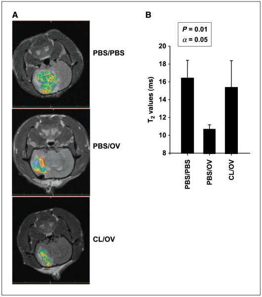
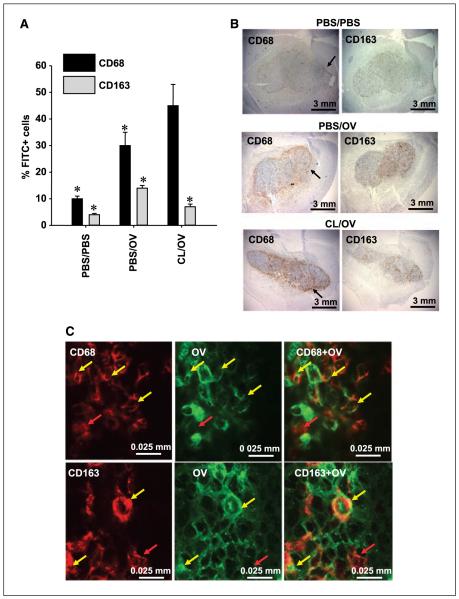
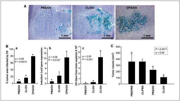
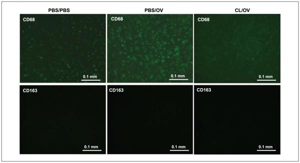
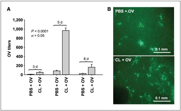
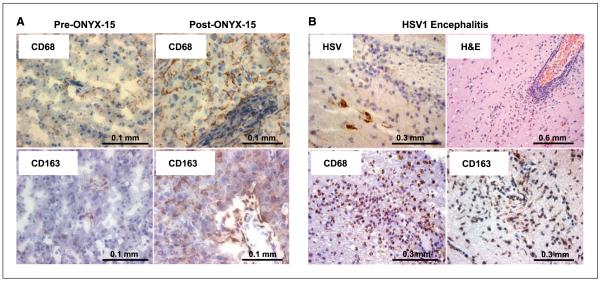
Clinical trials have proven oncolytic virotherapy to be safe but not effective. We have shown that oncolytic viruses (OV) injected into intracranial gliomas established in rodents are rapidly cleared, and this is associated with up-regulation of markers (CD68 and CD163) of cells of monocytic lineage (monocytes/microglia/macrophages). However, it is unclear whether these cells directly impede intratumoral persistence of OV through phagocytosis and whether they infiltrate the tumor from the blood or the brain parenchyma. To investigate this, we depleted phagocytes with clodronate liposomes (CL) in vivo through systemic delivery and ex vivo in brain slice models with gliomas. Interestingly, systemic CL depleted over 80% of peripheral CD163+ macrophages in animal spleen and peripheral blood, thereby decreasing intratumoral infiltration of these cells, but CD68+ cells were unchanged. Intratumoral viral titers increased 5-fold. In contrast, ex vivo CL depleted only CD68+ cells from brain slices, and intratumoral viral titers increased 10-fold. These data indicate that phagocytosis by both peripheral CD163+ and brain-resident CD68+ cells infiltrating tumor directly affects viral clearance from tumor. Thus, improved therapeutic efficacy may require modulation of these innate immune cells. In support of this new therapeutic paradigm, we observed intratumoral up-regulation of CD68+ and CD163+ cells following treatment with OV in a patient with glioblastoma.
Figures
Similar articles
-
Cyclophosphamide enhances glioma virotherapy by inhibiting innate immune responses.Proc Natl Acad Sci U S A. 2006 Aug 22;103(34):12873-8. doi: 10.1073/pnas.0605496103. Epub 2006 Aug 14. Proc Natl Acad Sci U S A. 2006. PMID: 16908838 Free PMC article.
-
Myxoma virus virotherapy for glioma in immunocompetent animal models: optimizing administration routes and synergy with rapamycin.Cancer Res. 2010 Jan 15;70(2):598-608. doi: 10.1158/0008-5472.CAN-09-1510. Epub 2010 Jan 12. Cancer Res. 2010. PMID: 20068158
-
Oncolytic Virotherapy Blockade by Microglia and Macrophages Requires STAT1/3.Cancer Res. 2018 Feb 1;78(3):718-730. doi: 10.1158/0008-5472.CAN-17-0599. Epub 2017 Nov 8. Cancer Res. 2018. PMID: 29118089
-
Immunosuppressive cells in oncolytic virotherapy for glioma: challenges and solutions.Front Cell Infect Microbiol. 2023 May 10;13:1141034. doi: 10.3389/fcimb.2023.1141034. eCollection 2023. Front Cell Infect Microbiol. 2023. PMID: 37234776 Free PMC article. Review.
-
Tumor-Associated Macrophages/Microglia in Glioblastoma Oncolytic Virotherapy: A Double-Edged Sword.Int J Mol Sci. 2022 Feb 4;23(3):1808. doi: 10.3390/ijms23031808. Int J Mol Sci. 2022. PMID: 35163730 Free PMC article. Review.
Cited by
-
Reciprocal Supportive Interplay between Glioblastoma and Tumor-Associated Macrophages.Cancers (Basel). 2014 Mar 26;6(2):723-40. doi: 10.3390/cancers6020723. Cancers (Basel). 2014. PMID: 24675569 Free PMC article.
-
Bevacizumab with angiostatin-armed oHSV increases antiangiogenesis and decreases bevacizumab-induced invasion in U87 glioma.Mol Ther. 2012 Jan;20(1):37-45. doi: 10.1038/mt.2011.187. Epub 2011 Sep 13. Mol Ther. 2012. PMID: 21915104 Free PMC article.
-
Non-secreting IL12 expressing oncolytic adenovirus Ad-TD-nsIL12 in recurrent high-grade glioma: a phase I trial.Nat Commun. 2024 Nov 8;15(1):9299. doi: 10.1038/s41467-024-53041-7. Nat Commun. 2024. PMID: 39516192 Free PMC article. Clinical Trial.
-
Advances in oncolytic virus therapy for glioma.Recent Pat CNS Drug Discov. 2009 Jan;4(1):1-13. doi: 10.2174/157488909787002573. Recent Pat CNS Drug Discov. 2009. PMID: 19149710 Free PMC article. Review.
-
Distinguishing inflammation from tumor and peritumoral edema by myeloperoxidase magnetic resonance imaging.Clin Cancer Res. 2011 Jul 1;17(13):4484-93. doi: 10.1158/1078-0432.CCR-11-0575. Epub 2011 May 10. Clin Cancer Res. 2011. PMID: 21558403 Free PMC article.
References
-
- Martuza RL, Malick A, Markert JM, Ruffner KL, Coen DM. Experimental therapy of human glioma by means of a genetically engineered virus mutant. Science. 1991;252:854–6. - PubMed
-
- Bischoff JR, Kirn DH, Williams A, et al. An adenovirus mutant that replicates selectively in p53-deficient human tumor cells. Science. 1996;274:373–6. - PubMed
-
- Alemany R, Balagué C, Curiel DT. Replicative adenoviruses for cancer therapy. Nat Biotechnol. 2000;18:723–7. - PubMed
-
- Alemany R, Gómez-Manzano C, Fueyo J. Oncolytic adenovirus for the treatment of cerebral tumors: past, present, and future [in Spanish] Neurologia. 2001;16:122–7. - PubMed
-
- Kirn D, Martuza RL, Zwiebel J. Replication-selective virotherapy for cancer: biological principles, risk management and future directions. Nat Med. 2001;7:781–7. - PubMed
Publication types
MeSH terms
Grants and funding
LinkOut - more resources
Full Text Sources
Other Literature Sources
Medical
Research Materials
