. 2007 Oct 1;67(19):9463-71.
doi: 10.1158/0008-5472.CAN-07-2034.

Autocrine glutamate signaling promotes glioma cell invasion

Affiliations

Autocrine glutamate signaling promotes glioma cell invasion

Susan A Lyons et al. Cancer Res. .

Erratum in

  • Cancer Res. 2007 Nov 1;67(21):10624

Abstract

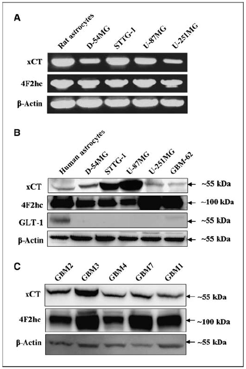
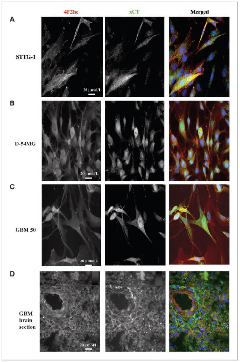
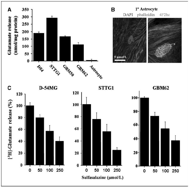
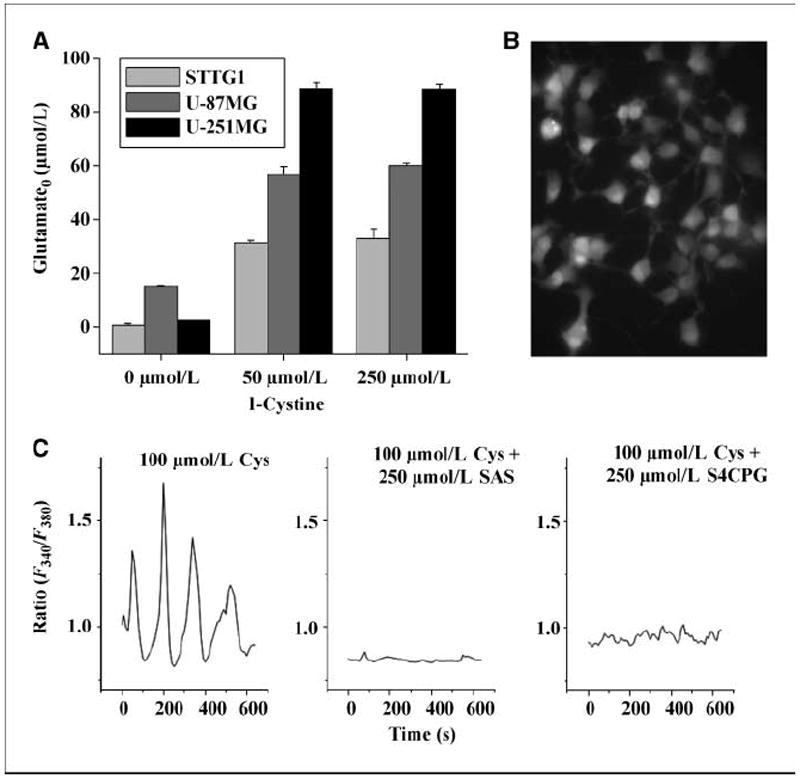
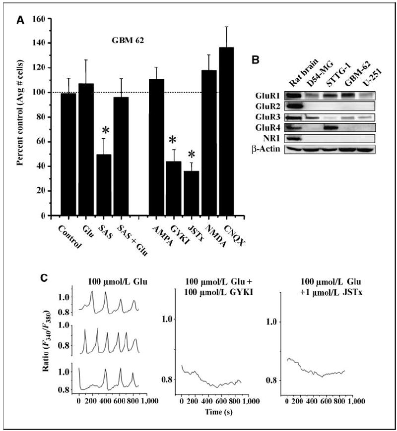
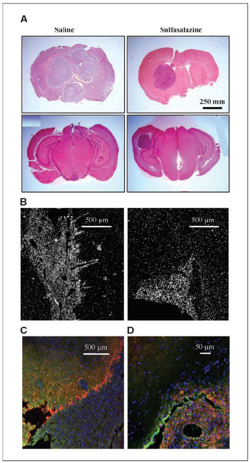
Malignant gliomas have been shown to release glutamate, which kills surrounding brain cells, creating room for tumor expansion. This glutamate release occurs primarily via system xC, a Na+-independent cystine-glutamate exchanger. We show here, in addition, that the released glutamate acts as an essential autocrine/paracrine signal that promotes cell invasion. Specifically, chemotactic invasion and scrape motility assays each show dose-dependent inhibition of cell migration when glutamate release was inhibited using either S-(4)-CPG or sulfasalazine, both potent blockers of system xC. This inhibition could be overcome by the addition of exogenous glutamate (100 micromol/L) in the continued presence of the inhibitors. Migration/invasion was also inhibited when Ca2+-permeable alpha-amino-3-hydroxy-5-methylisoxazole-4-propionic acid receptors (AMPA-R) were blocked using GYKI or Joro spider toxin, whereas CNQX was ineffective. Ca2+ imaging experiments show that the released glutamate activates Ca2+-permeable AMPA-R and induces intracellular Ca2+ oscillations that are essential for cell migration. Importantly, glioma cells release glutamate in sufficient quantities to activate AMPA-Rs on themselves or neighboring cells, thus acting in an autocrine and/or paracrine fashion. System xC and the appropriate AMPA-R subunits are expressed in all glioma cell lines, patient-derived glioma cells, and acute patient biopsies investigated. Furthermore, animal studies in which human gliomas were xenographed into scid mice show that chronic inhibition of system xC-mediated glutamate release leads to smaller and less invasive tumors compared with saline-treated controls. These data suggest that glioma invasion is effectively disrupted by inhibiting an autocrine glutamate signaling loop with a clinically approved candidate drug, sulfasalazine, already in hand.


Figures

Figure 1
Figure 1
Subunits of system xC are expressed in glioma cell lines and patient tumor samples. A, RT-PCR indicated that transcripts of catalytic subunit xCT and the regulatory subunit 4F2hc of system xC are expressed in primary cultures of rat astrocytes and all glioma cell lines tested; four lines are shown. B, protein expression of xCT and 4F2hc are shown in human astrocytes and five human glioma cell lines. This is compared with the glutamate transporter GLT-1, expressed robustly in mature astrocytes but less so in human fetal astrocytes (immature). C, protein expression of system xC subunits in five GBM patient tissues are shown from individuals aged 51 to 72 y. β-Actin is shown as a control for protein loading efficiency. The approximate molecular weights of the proteins are indicated to the right as each blot image was cropped for clarity.
Figure 2
Figure 2
Specificity of system xC subunits show antibody binding in the membranes of glioma cells. A, sister coverslips of glioma cell lines were stained with mouse anti-4F2hc and rabbit anti-xCT. Both fluorescence channels are shown separately in black and white until merged in the third color panels. A to D, merged images: red, xCT; green, 4F2hc; blue, DAPI, a nuclear stain. The GBM-stained patient section is shown in D, where xCT is green and 4F2hc is red. Merged images were taken at the exposure time appropriate for each wavelength. All images were taken at ×40 magnification.
Figure 3
Figure 3
Glutamate release by glioma cells is inhibited by sulfasalazine in a dose-dependent manner. A, glutamate released by four glioma cell lines were compared with primary cultured astrocytes using an enzyme-based bioluminescent signal normalized to protein. B, images of primary cultures of rat astrocytes show a control (left) and positive staining for 4F2hc on the right, labeling only the Golgi, in green. DAPI-labeled nuclei and phalloidin (a cytoskeleton protein conjugated with Alexa 546) are included. System xC is nonfunctional in astrocyte cultures. C, glutamate release is shown in the presence of increasing concentrations of sulfasalazine using [3H]glutamate-loaded glioma cell lines to measure release. Columns, percent control; bars, SE. All experiments were done with at least n = 3.
Figure 4
Figure 4
Glutamate released through system xC caused glioma Ca2+ oscillations. A, increasing amounts of the cystine were added to three glioma cell lines, thus inducing the release of glutamate in a dose-dependent manner as determined by the highly sensitive enzyme-based bioluminescent assay. B, FURA2-AM–loaded D54-MG cells are shown in a representative field before measuring calcium oscillations at ×40 magnification. C, three representative cells from three coverslips were imaged every 15 s in the presence of cystine or with cystine but in the presence of one of the two system xC inhibitors, sulfasalazine (SAS) or S-(4)-CPG.
Figure 5
Figure 5
Glutamate induces AMPA-R–dependent calcium oscillations in glioma cells. A, the invasion results of patient-derived glioma cells (GBM 62) are shown in the presence of sulfasalazine (250 μmol/L SAS) and SAS + glutamate (100 μmol/L). Using sister wells, in the second half of A, glioma cells were allowed to invade the filter pores in the presence of the AMPA-R agonist AMPA (100 μmol/L); AMPA-R antagonists GYKI (100 μmol/L) and Joro spider toxin (JSTx; 1 μmol/L); NMDA-R agonist NMDA (100 μmol/L); and AMPA/KA-R antagonist, CNQX (100 μmol/L). Each experiment had three similarly treated inserts where six fields per insert were imaged and data averaged. This was repeated thrice. Bars, SE * P ≤ 0.05. B, glioma cells express Ca2+-permeable AMPA-Rs as shown by Western blot analysis for four glioma cell lines with whole rat brain lysates as a positive control. Glutamate subunits of AMPA-Rs were probed with antibodies to GluR1-4. β-Actin is shown as a control for protein loading efficiency. C, representative traces from three glioma cells loaded with FURA2-AM showed oscillatory changes in [Ca2+]i in response to 100 μmol/L glutamate. Single representative traces are shown following simultaneous application of 100 μmol/L glutamate and the AMPA-R blockers GYKI (100 μmol/L) and Joro spider toxin showing that [Ca2+]i oscillations stimulated by glutamate are inhibited. Each experiment was repeated three independent times.
Figure 6
Figure 6
Glioma invasion of tumor growth in the mouse glioma model is inhibited by sulfasalazine. A, glioma xenographed mice brains were sectioned for H&E staining to identify tumor regions. Left, tumor formation in two saline-treated mice. Right, two SAS-treated brains. Top row, tumor formation at 25 d posttumor injection; bottom row, tumor formation at 21 d posttumor injection. B, fluorescent images of DAPI-labeled nuclei show densely populated tumor formation in brain sections of saline and SAS-treated mice. C, merged GLT-1–, GFAP-, and DAPI-stained brain slice shows astrocytes lining the tumor perimeter. D, merged system xC staining in tumor is shown in sulfasalazine-treated mouse brain section.

References

    1. Laerum OD, Bjerkvig R, Steinsvag S, de Ridder L. Invasiveness of primary brain tumors. Cancer Metastasis Rev. 1984;3:223–36. [review] [91 refs] - PubMed
    1. Takano T, Lin JH, Arcuino G, Gao Q, Yang J, Nedergaard M. Glutamate release promotes growth of malignant gliomas. Nat Med. 2001;7:1010–5. - PubMed
    1. Kaba SE, Kyritsis AP. Recognition and management of gliomas. Drugs. 1997;53:235–44. - PubMed
    1. Ye ZC, Sontheimer H. Glioma cells release excitotoxic concentrations of glutamate. Cancer Res. 1999;59:4383–91. - PubMed
    1. Ishiuchi S, Tsuzuki K, Yoshida Y, et al. Blockage of Ca(2+)-permeable AMPA receptors suppresses migration and induces apoptosis in human glioblastoma cells. Nat Med. 2002;8:971–8. - PubMed

Publication types

MeSH terms