

Effect of Alzheimer disease risk on brain function during self-appraisal in healthy middle-aged adults
- PMID: 17909128
- PMCID: PMC2650497
- DOI: 10.1001/archpsyc.64.10.1163
Effect of Alzheimer disease risk on brain function during self-appraisal in healthy middle-aged adults
Abstract
Context: Asymptomatic middle-aged adult children of patients with Alzheimer disease (AD) recently were found to exhibit functional magnetic resonance imaging (fMRI) deficits in the mesial temporal lobe during an encoding task. Whether this effect will be observed on other fMRI tasks is yet unknown. This study examines the neural substrates of self-appraisal (SA) in persons at risk for AD. Accurate appraisal of deficits is a problem for many patients with AD, and prior fMRI studies of healthy young adults indicate that brain areas vulnerable to AD such as the anterior mesial temporal lobe and posterior cingulate are involved during SA tasks.
Objective: To determine whether parental family history of AD (hereafter referred to as FH) or presence of the epsilon4 allele of the apolipoprotein E gene (APOE4) exerts independent effects on brain function during SA.
Design: Cross-sectional factorial design in which APOE4 status (present vs absent) was one factor and FH was the other. All participants received cognitive testing, genotyping, and an fMRI task that required subjective SA decisions regarding trait adjective words in comparison with semantic decisions about the same words.
Setting: An academic medical center with a research-dedicated 3.0-T MR imaging facility.
Participants: Cognitively normal middle-aged adults (n = 110), 51 with an FH and 59 without an FH.
Main outcome measure: Blood oxygen-dependent contrast measured using T2*-weighted echo-planar imaging.
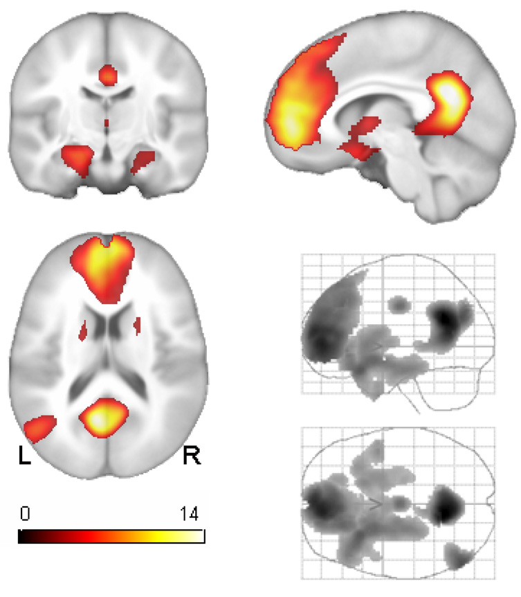
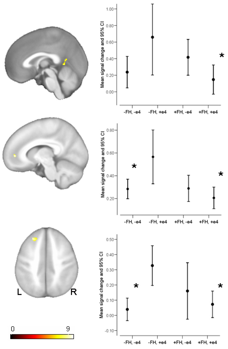
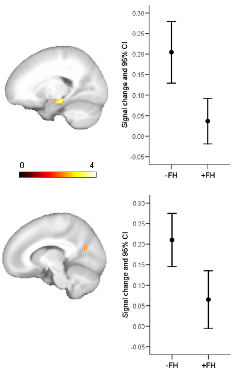
Results: Parental family history of AD and APOE4 status interacted in the posterior cingulate and left superior and medial frontal regions. There were main effects of FH (FH negative > FH positive) in the left hippocampus and ventral posterior cingulate. There were no main effects of APOE genotype.
Conclusions: Our results suggest that FH may affect brain function during subjective SA in regions commonly affected by AD. Although the participants in this study were asymptomatic and middle-aged, the findings suggest that there may be subtle alterations in brain function attributable to AD risk factors.
Figures
Similar articles
-
The influence of parental history of Alzheimer's disease and apolipoprotein E epsilon4 on the BOLD signal during recognition memory.Brain. 2009 Feb;132(Pt 2):383-91. doi: 10.1093/brain/awn254. Epub 2008 Oct 1. Brain. 2009. PMID: 18829694 Free PMC article.
-
fMRI activation during episodic encoding and metacognitive appraisal across the lifespan: risk factors for Alzheimer's disease.Neuropsychologia. 2008;46(6):1667-78. doi: 10.1016/j.neuropsychologia.2007.11.035. Epub 2007 Dec 17. Neuropsychologia. 2008. PMID: 18241895 Free PMC article.
-
The influence of Alzheimer disease family history and apolipoprotein E epsilon4 on mesial temporal lobe activation.J Neurosci. 2006 May 31;26(22):6069-76. doi: 10.1523/JNEUROSCI.0959-06.2006. J Neurosci. 2006. PMID: 16738250 Free PMC article.
-
Use of functional magnetic resonance imaging in the early identification of Alzheimer's disease.Neuropsychol Rev. 2007 Jun;17(2):127-43. doi: 10.1007/s11065-007-9025-y. Epub 2007 May 3. Neuropsychol Rev. 2007. PMID: 17476598 Free PMC article. Review.
-
Neuroimaging biomarkers for Alzheimer's disease in asymptomatic APOE4 carriers.Rev Neurol (Paris). 2013 Oct;169(10):729-36. doi: 10.1016/j.neurol.2013.07.025. Epub 2013 Sep 6. Rev Neurol (Paris). 2013. PMID: 24016463 Review.
Cited by
-
The effect of TOMM40 poly-T length on gray matter volume and cognition in middle-aged persons with APOE ε3/ε3 genotype.Alzheimers Dement. 2011 Jul;7(4):456-65. doi: 10.1016/j.jalz.2010.11.012. Alzheimers Dement. 2011. PMID: 21784354 Free PMC article.
-
Corticostriatal connectivity mediates the reciprocal relationship between parent-reported sleep duration and impulsivity in early adolescents.J Child Psychol Psychiatry. 2023 Nov;64(11):1545-1554. doi: 10.1111/jcpp.13843. Epub 2023 May 29. J Child Psychol Psychiatry. 2023. PMID: 37248201 Free PMC article.
-
White matter microstructure in late middle-age: Effects of apolipoprotein E4 and parental family history of Alzheimer's disease.Neuroimage Clin. 2014 Apr 21;4:730-42. doi: 10.1016/j.nicl.2014.04.008. eCollection 2014. Neuroimage Clin. 2014. PMID: 24936424 Free PMC article.
-
Failing compensatory mechanisms during working memory in older apolipoprotein E-epsilon4 healthy adults.Brain Imaging Behav. 2010 Jun;4(2):177-88. doi: 10.1007/s11682-010-9097-9. Brain Imaging Behav. 2010. PMID: 20502990 Free PMC article.
-
Increased functional brain response during word retrieval in cognitively intact older adults at genetic risk for Alzheimer's disease.Neuroimage. 2010 Jul 1;51(3):1222-33. doi: 10.1016/j.neuroimage.2010.03.021. Epub 2010 Mar 16. Neuroimage. 2010. PMID: 20298792 Free PMC article.
References
-
- Corder EH, Ghebremedhin E, Taylor MG, Thal DR, Ohm TG, Braak H. The biphasic relationship between regional brain senile plaque and neurofibrillary tangle distributions: modification by age, sex, and APOE polymorphism. Ann N Y Acad Sci. 2004 Jun;1019:24–28. - PubMed
-
- Braak E, Griffing K, Arai K, Bohl J, Bratzke H, Braak H. Neuropathology of Alzheimer's disease: what is new since A. Alzheimer? Eur Arch Psychiatry Clin Neurosci. 1999;249 Suppl 3:14–22. - PubMed
-
- Ohm TG, Muller H, Braak H, Bohl J. Close-meshed prevalence rates of different stages as a tool to uncover the rate of Alzheimer's disease-related neurofibrillary changes. Neuroscience. 1995 Jan;64(1):209–217. - PubMed
-
- Fratiglioni L, Ahlbom A, Viitanen M, Winblad B. Risk factors for late-onset Alzheimer's disease: a population-based, case-control study. Ann Neurol. 1993 Mar;33(3):258–266. - PubMed
Publication types
MeSH terms
Substances
Grants and funding
LinkOut - more resources
Full Text Sources
Medical
Miscellaneous
