. 2007 Oct 9;104(41):16287-92.
doi: 10.1073/pnas.0703508104. Epub 2007 Oct 1.

Kaposi's sarcoma herpesvirus-encoded latency-associated nuclear antigen stabilizes intracellular activated Notch by targeting the Sel10 protein

Affiliations

Kaposi's sarcoma herpesvirus-encoded latency-associated nuclear antigen stabilizes intracellular activated Notch by targeting the Sel10 protein

Ke Lan et al. Proc Natl Acad Sci U S A. .

Abstract

Deregulation of the evolutionarily conserved Notch signaling is highly correlated with oncogenesis. Intracellular activated Notch (ICN) is a protooncogene linked to the transcription activation of a number of cellular genes involved in cell cycle regulation, differentiation, and proliferation. Stability of ICN is tightly regulated by the Sel10-mediated ubiquitin-proteasome pathway. Sel10 can function as a negative regulator of Notch and exhibits activities of a tumor-suppressor protein. This article shows that the Kaposi's sarcoma-associated herpesvirus (KSHV) latency-associated nuclear antigen (LANA) directly interacts with Sel10 and forms a complex in KSHV-infected cells. This results in suppression of ICN ubiquitination and degradation. The carboxyl terminus of LANA interacts with the F-box and WD40 domains of Sel10 and competes with ICN for binding to Sel10. This elevated level of ICN is also critical for maintaining the enhanced proliferation of KSHV-infected tumor cells. These findings describe a mechanism by which the KSHV-encoded LANA protein regulates ubiquitination of ICN mediated by the F-box component of the E3 ligase Sel10, leading to proliferation of the virus-infected cells.


Conflict of interest statement

The authors declare no conflict of interest.

Figures

Fig. 1.
Fig. 1.
LANA associates with Sel10. (A and B) Immunoprecipitation analysis with mouse anti-Myc antibody or rabbit anti-GFP antibody showed that Sel10-GFP was directly immunoprecipitated with LANA-myc in 293T (A) and DG75 (B) cells. Fifteen million cells were cotransfected with 10 μg of Sel10-GFP and 10 μg of LANA-myc expression vectors or transfected with these vectors, respectively, 24 h after transfection; cell lysates were use for immunoprecipitate (IP) analysis. In, 5% total cell lysate input; Pc, preclear. (C) Endogenous LANA interacts with Sel10 in KSHV-infected primary effusion lymphoma cells BCBL1 and BC3. Immunoprecipitation analysis with polyclonal rabbit LANA antiserum showed that Sel10 was directly immunoprecipitated with LANA in BCBL1 and BC3 cells. For this experiment, 30 million cells were harvested for preparation of cell lysates, which were use for IP analysis. BJAB cells, which are KSHV-negative were used as a control. IN, 5% total lysate input; PC, preclear; IP, immunoprecipitate. (D) Immunofluorescence analysis showed that Sel10 was localized to the same nuclear compartment as LANA in different cells. (Top and Middle) DG75 and 293T cells were cotransfected with 10 μg each of Sel10-myc (Myc tagged) and pCDNA-LANA expression vectors, respectively; 24 h after transfection, cells were harvested for immunofluorescence analysis. Rabbit anti-LANA polyclonal antibody and mouse anti-Myc monoclonal antibody and corresponding secondary antibodies were used for this assay. (Bottom) BCBL1 cells were also used for immunofluorescence analysis. Human anti-LANA serum and rabbit anti-Sel10 polyclonal antibody and corresponding secondary antibodies were used for this assay. (E Top and Middle) C terminus LANA associates with Sel10 in vitro. The 35S-labeled products were incubated with GST as well as GST-Sel10 fusion protein. The pulldown products were electrophoresed on 10% SDS/PAGE gel, dried, and exposed to a PhosphorImager. Input controls of 10% of total LANA translation products. (Bottom) A schematic for the LANA clones used is given. NLS, nuclear localization signal; AD, acidic domain; LZ, leucine zipper; DBD, DNA-binding domain.
Fig. 2.
Fig. 2.
LANA inhibits degradation of ICN. (A) LANA rescues ICN degradation mediated by Sel10. In lanes 1–5, 5 μg of ICN expression vectors along with 0 μg, 2.5 μg, 5 μg, 10 μg, and 15 μg of Sel10 were transfected into 293T cells. In lanes 6-, 5 μg of ICN and 15 μg of Sel10 along with 2.5 μg, 5 μg, 10 μg, and 15 μg of LANA were transfected into 293 cells. Forty-eight hours after transfection, cells were harvested for preparation of lysates and examined by Western blotting. Each protein was checked by specific antibody individually, and actin was used for loading control. (B) LANA inhibits ubiquitination of ICN by Sel10. Transfection of 293T cells with the ICN (5 μg) together with HA-tagged ubiquitin (1.5 μg), Sel10 (10 μg), and 10 μg of LANA by using different combinations indicated by +/−. Forty-eight hours after transfection, cells were harvested for preparation of lysates and IP experiments. MG132 was added to the cell culture 3 h before harvesting. The cell lysates were immunoprecipitated by rabbit anti-ICN antibody and Western blotting for HA-tagged ubiquitin (Upper) as well as ICN (Lower).
Fig. 3.
Fig. 3.
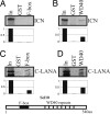
C terminus LANA interacts with both F-box and WD40 domain. ICN was in vitro-transcribed and translated. (A and B) The 35S-labeled products were incubated with GST as well as GST-F-box (A) or GST-WD40 fusion protein (B). (C and D) C terminus LANA was in vitro-transcribed and translated. The 35S-labeled products were incubated with GST as well as GST-F-box (C) or GST-WD40 fusion protein (D). The pulldown products were electrophoresed on 10% SDS/PAGE gel, dried, and exposed to a PhosphorImager. Input control is 10% of total translation product. A schematic diagram of Sel10 is shown below the blots. Sel10 is a 540-aa protein, and the F-box motif and WD40 repeated are indicated.
Fig. 4.
Fig. 4.
LANA/ICN competition for Sel10 binding. (A) Seven hundred sixty-two to 1,162-aa truncations of LANA and ICN were in vitro-transcribed and translated. The 35S-labeled products were incubated with GST-Sel10. A fixed amount of ICN and increasing amounts of LANA were used. Pulldown products were electrophoresed on 10% SDS/PAGE gels, dried, and exposed to a PhosphorImager. Input controls of 10% for LANA and for ICN were run as well. (Right) A fixed amount of ICN and increasing amounts of luciferase were used. Pulldown products were electrophoresed on 10% SDS/PAGE gels, dried, and exposed to a PhosphorImager. The quantification of each band is shown at the bottom. (B) LANA/ICN competition for Sel10 WD40 domain binding. Seven hundred sixty-two to 1,162-aa truncations of LANA and ICN were in vitro-transcribed and translated. The 35S-labeled products were incubated with GST-WD40. The procedure is the same as that mentioned in A.
Fig. 5.
Fig. 5.
Stabilization of ICN by LANA is associated with proliferation of KSHV-infected cells. (A) Western blotting showing expression of LANA and ICN in BCBL1, LANA siRNA-transfected BCBL1, and luciferase siRNA-transfected BCBL1 cells. Action level is as the loading control. (B) Western blotting to show expression level of LANA and ICN in BCBL1 cells, LANA siRNA-transfected BCBL1 cells, and LANA siRNA/Sel10 dominant-negative cotransfected BCBL1 cells. (C) CFSE staining showing the mitosis of cells. (Top) BCBL1 was transfected with LANA siRNA or Luciferase siRNA expression vectors and selected for stable clones. (Middle) BCBL1, LANA siRNA-transfected BCBL1, and LANA siRNA/Sel10 dominant-negative cotransfected BCBL1 cells were compared for mitosis. (Bottom) The standard proliferation curve of BCBL1 cells from 1st day to 7th day after CFSE staining. The negative control cell is CFSE nonstaining cells and the positive control cell is CFSE-staining but immediately fixed cell. (D) Mapping of the precise region of LANA responsible for Sel10 binding. A series of truncated LANA T1 (992–1,162), T2 (1,022–1,162), T3 (1,052–1,162), and T4 (1,082–1,162) were constructed, and in vitro binding assays were performed by using translated individual LANA- and GST-fused Sel10. (E) LANA lacking 1,052–1,082 aa were generated and used for in vitro binding assay with GST-Sel10. (F) Immunoprecipitation assays performed with mouse anti-Myc antibody and rabbit anti-GFP antibody showed that WT LANA but not the mutant type LANA (1,052–1,082 deletion) was directly immunoprecipitated with Sel10-GFP in 293T cells. Fifteen million cells were cotransfected with 10 μg of Sel10-GFP and 10 μg of WT LANA expression vector or mutant-type (MT) LANA expression vector. Twenty-four hours after transfection, cell lysates were used for IP analysis with GFP antibody. In, 5% total cell lysate input; Pc, preclear; IP, immunoprecipitate. (G) DG75 cells were -transfected with WT LANA and MT LANA and selected for stable clones. The individual stable cells expressing WT LANA or MT LANA were labeled with CFSE for proliferation assay. This figure shows that the proliferation of WT LANA-expressing cells is faster than that of MT-expressing cells. (H) Proposed model of ICN stability enhanced by KSHV LANA. ICN degradation is a tightly controlled ubiquitin–proteasome pathway mediated by Sel10 F-box protein. In KSHV-infected cells, LANA is expressed at a constant high level because LANA autoactivates its own promoter. In these cells, LANA competes with ICN for Sel10 binding, thus reducing ubiquitination and degradation of ICN. ICN is therefore stabilized in KSHV latently infected tumor cells.

Similar articles

Cited by

References

    1. Chang Y, Cesarman E, Pessin MS, Lee F, Culpepper J, Knowles DM, Moore PS. Science. 1994;266:1865–1869. - PubMed
    1. Russo JJ, Bohenzky RA, Chien MC, Chen J, Yan M, Maddalena D, Parry JP, Peruzzi D, Edelman IS, Chang Y, Moore PS. Proc Natl Acad Sci USA. 1996;93:14862–14867. - PMC - PubMed
    1. Boshoff C, Weiss RA. Philos Trans R Soc London Ser B. 2001;356:517–534. - PMC - PubMed
    1. Boshoff C, Chang Y. Annu Rev Med. 2001;52:453–470. - PubMed
    1. Brander C, Raje N, O'Connor PG, Davies F, Davis J, Chauhan D, Hideshima T, Martin J, Osmond D, Kedes DH, et al. Blood. 2002;100:698–700. - PubMed

Publication types

MeSH terms