Nipping at cardiac remodeling
- PMID: 17909620
- PMCID: PMC1994642
- DOI: 10.1172/JCI33706
Nipping at cardiac remodeling
Abstract
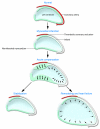
Much of the mortality following myocardial infarction results from remodeling of the heart after the acute ischemic event. Cardiomyocyte apoptosis has been thought to play a key role in this remodeling process. In this issue of the JCI, Diwan and colleagues present evidence that Bnip3, a proapoptotic Bcl2 family protein, mediates cardiac enlargement, reshaping, and dysfunction in mice without influencing infarct size.
Figures

Comment on
-
Inhibition of ischemic cardiomyocyte apoptosis through targeted ablation of Bnip3 restrains postinfarction remodeling in mice.J Clin Invest. 2007 Oct;117(10):2825-33. doi: 10.1172/JCI32490. J Clin Invest. 2007. PMID: 17909626 Free PMC article.
References
-
- Kajstura J., et al. Apoptotic and necrotic myocyte cell deaths are independent contributing variables of infarct size in rats. Lab. Invest. 1996;74:86–107. - PubMed
-
- Sutton M.G., Sharpe N. Left ventricular remodeling after myocardial infarction: pathophysiology and therapy. Circulation. 2000;101:2981–2988. - PubMed
-
- Frangogiannis N.G. The mechanistic basis of infarct healing. Antioxid. Redox Signal. 2006;8:1907–1939. - PubMed
-
- Mani K., Kitsis R.N. Myocyte apoptosis: programming ventricular remodeling. J. Am. Coll. Cardiol. 2003;41:761–764. - PubMed

