. 2007 Oct;117(10):2825-33.
doi: 10.1172/JCI32490.

Inhibition of ischemic cardiomyocyte apoptosis through targeted ablation of Bnip3 restrains postinfarction remodeling in mice

Affiliations

Inhibition of ischemic cardiomyocyte apoptosis through targeted ablation of Bnip3 restrains postinfarction remodeling in mice

Abhinav Diwan et al. J Clin Invest. 2007 Oct.

Abstract

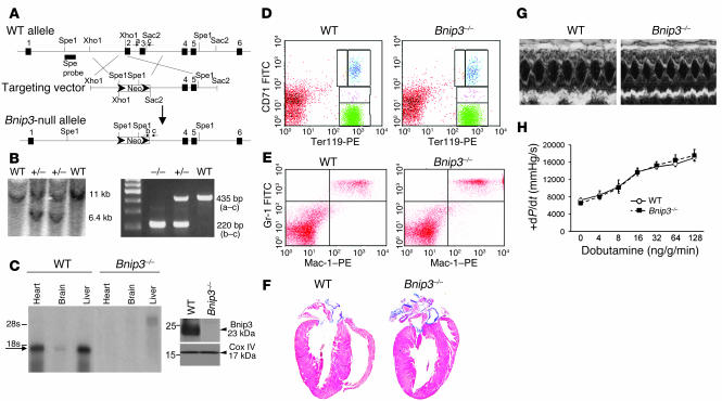
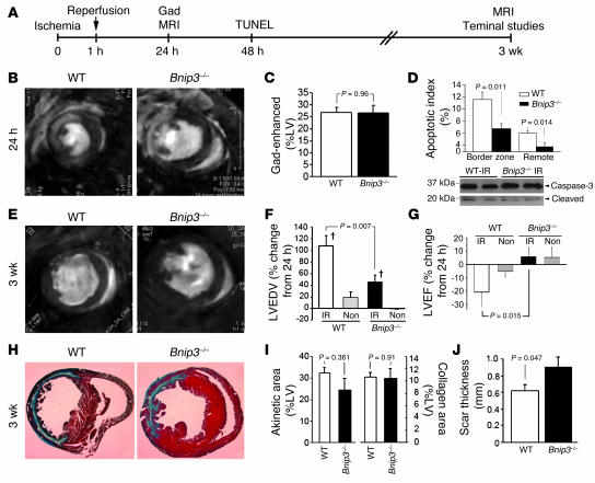
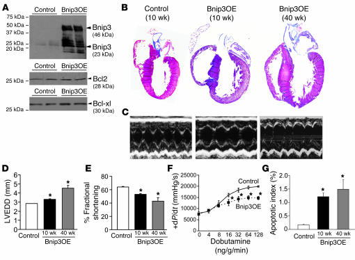
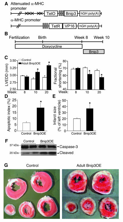
Following myocardial infarction, nonischemic myocyte death results in infarct expansion, myocardial loss, and ventricular dysfunction. Here, we demonstrate that a specific proapoptotic gene, Bnip3, minimizes ventricular remodeling in the mouse, despite having no effect on early or late infarct size. We evaluated the effects of ablating Bnip3 on cardiomyocyte death, infarct size, and ventricular remodeling after surgical ischemia/reperfusion (IR) injury in mice. Immediately following IR, no significant differences were observed between Bnip3(-/-) and WT mice. However, at 2 days after IR, apoptosis was diminished in Bnip3(-/-) periinfarct and remote myocardium, and at 3 weeks after IR, Bnip3(-/-) mice exhibited preserved LV systolic performance, diminished LV dilation, and decreased ventricular sphericalization. These results suggest myocardial salvage by inhibition of apoptosis. Forced cardiac expression of Bnip3 increased cardiomyocyte apoptosis in unstressed mice, causing progressive LV dilation and diminished systolic function. Conditional Bnip3 overexpression prior to coronary ligation increased apoptosis and infarct size. These studies identify postischemic apoptosis by myocardial Bnip3 as a major determinant of ventricular remodeling in the infarcted heart, suggesting that Bnip3 may be an attractive therapeutic target.


Figures

Figure 1
Figure 1. Generation and baseline characteristics of Bnip3-null mice.
(A) Schematic of Bnip3 deletion strategy depicting locations of exons 1–6 (black boxes), restriction sites, probe used for Southern blotting (Spe probe), and PCR primers (a–c). (B) Southern blot (left) and PCR (right) screening of Bnip3-targeted mice. (C) Bnip3 multiple tissue Northern blot (left) and immunoblot analysis of mitochondria-enriched myocardial protein (right). (D) Fluorescence cytometry of erythroid Ter119 and CD71 expression in splenocytes. Yellow represents proerythroblasts; blue, basophilic erythroblasts; pink, chromatophilic erythroblasts; green, orthochromatic erythroblasts. (E) Fluorescence cytometry of Gr-1 (granulocyte) and Mac-1 (macrophage) expression in bone marrow cells. (F) Masson trichrome–stained coronal heart sections. (G) M-mode echocardiograms; short-axis view of LV. (H) Invasive hemodynamic analysis of LV contractility at baseline and in response to β-adrenergic agonist dobutamine (n = 4, WT; n = 8, Bnip3–/–).
Figure 2
Figure 2. Effects of Bnip3 ablation on in vivo myocardial I/R injury.
(A) Schematic depiction of experimental design for in vivo IR studies. Gad, gadolinium. (B) Representative gadolinium-enhanced (white) MRI mid-ventricle end-diastolic images 24 hours after IR. (C) Quantitative infarct size of grouped data (n = 8–9). (D) Top: Apoptotic indices (TUNEL) 48 hours after IR (n = 6, WT; n = 7, Bnip3–/–). Bottom: Representative caspase-3 cleavage studies. (E) Representative mid-ventricle end-diastolic MRI images 3 weeks after IR. Quantitative change in MRI-derived LV end-diastolic volume (LVEDV) (F) and.LV ejection fraction (G) from 24 hours to 3 weeks after IR. P < 0.05 vs. nonoperated group (Non). (H) Masson trichrome–stained transverse heart sections (original magnification, ×5) 3 weeks after IR. (I) Grouped data for MRI-derived akinetic area (left) and histological collagen scar content (right) of LVs 3 weeks after IR. (J) Scar thickness 3 weeks after IR.
Figure 3
Figure 3. Studies of cardiac Bnip3 overexpression from the neonatal period.
(A) Representative Western blots of myocardial Bnip3, Bcl2, and Bcl-xl in mice with neonatal Bnip3 overexpression (Bnip3OE) and controls. (B) Masson trichrome–stained coronal heart sections. Original magnification, ×2. (C) Short-axis M-mode echocardiograms. (D and E) Echocardiographically determined LV end-diastolic diameter (LVEDD) and fractional shortening as a function of age (n = 7 controls, n = 5 Bnip3 OE at 10 and 40 weeks). (F) Invasively determined LV contractility at baseline and with β-adrenergic stimulation at 10 weeks (n = 3/group). (G) TUNEL-determined apoptotic indices (n = 4/group). *P < 0.05 vs. controls.
Figure 4
Figure 4. Studies of conditional Bnip3 overexpression in adult mice.
(A) Double conditional transgenesis strategy (see text for details). (B) Schematic of experimental design. (C) Echocardiographically determined LV end-diastolic diameter (left) and LV fractional shortening (right) before Bnip3 induction (week 8) and 2 weeks (week 10) and 12 weeks after induction (week 20); n = 10 pairs at 8 and 10 weeks and n = 5 pairs at 20 weeks. (D) TUNEL-derived apoptotic indices in infarcted mice. (E) Quantitative infarct size after 48 hours (n = 5–6/group). (F).Caspase-3 cleavage studies. (G) Triphenyltetrazolium chloride–stained cardiac cross-sections 48 hours after coronary occlusion. Original magnification, ×1. *P < 0.05 vs. controls.

Comment in

  • Nipping at cardiac remodeling.
    Whelan RS, Mani K, Kitsis RN. Whelan RS, et al. J Clin Invest. 2007 Oct;117(10):2751-3. doi: 10.1172/JCI33706. J Clin Invest. 2007. PMID: 17909620 Free PMC article.

References

    1. Zheng Z.J., Croft J.B., Giles W.H., Mensah G.A. Sudden cardiac death in the United States, 1989 to 1998. Circulation. 2001;104:2158–2163. - PubMed
    1. Thom T., et al. Heart disease and stroke statistics — 2006 update: a report from the American Heart Association Statistics Committee and Stroke Statistics Subcommittee. Circulation. 2006;113:e85–e151. - PubMed
    1. Gibson C.M. NRMI and current treatment patterns for ST-elevation myocardial infarction. Am. Heart J. 2004;148:S29–S33. - PubMed
    1. Kober L., et al. A clinical trial of the angiotensin-converting-enzyme inhibitor trandolapril in patients with left ventricular dysfunction after myocardial infarction. Trandolapril Cardiac Evaluation (TRACE) Study Group. N. Engl. J. Med. 1995;333:1670–1676. - PubMed
    1. Antman E.M., et al. ACC/AHA guidelines for the management of patients with ST-elevation myocardial infarction — executive summary: a report of the American College of Cardiology/American Heart Association Task Force on Practice Guidelines (Writing Committee to Revise the 1999 Guidelines for the Management of Patients With Acute Myocardial Infarction). Circulation. 2004;110:588–636. - PubMed

Publication types

MeSH terms