. 2007 Oct;117(10):2920-8.
doi: 10.1172/JCI31692.

CX3CR1-dependent subretinal microglia cell accumulation is associated with cardinal features of age-related macular degeneration

Affiliations

CX3CR1-dependent subretinal microglia cell accumulation is associated with cardinal features of age-related macular degeneration

Christophe Combadière et al. J Clin Invest. 2007 Oct.

Abstract

The role of retinal microglial cells (MCs) in age-related macular degeneration (AMD) is unclear. Here we demonstrated that all retinal MCs express CX3C chemokine receptor 1 (CX3CR1) and that homozygosity for the CX3CR1 M280 allele, which is associated with impaired cell migration, increases the risk of AMD. In humans with AMD, MCs accumulated in the subretinal space at sites of retinal degeneration and choroidal neovascularization (CNV). In CX3CR1-deficient mice, MCs accumulated subretinally with age and albino background and after laser impact preceding retinal degeneration. Raising the albino mice in the dark prevented both events. The appearance of lipid-bloated subretinal MCs was drusen-like on funduscopy of senescent mice, and CX3CR1-dependent MC accumulation was associated with an exacerbation of experimental CNV. These results show that CX3CR1-dependent accumulation of subretinal MCs evokes cardinal features of AMD. These findings reveal what we believe to be a novel pathogenic process with important implications for the development of new therapies for AMD.


Figures
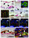
Figure 1
Figure 1. CX3CR1 expression in AMD.
(A) CX3CR1 (fast red labeling) was expressed in the inner retina (arrows); no CX3CR1-positive cells were found in the outer nuclear layer (ONL), photoreceptor OSs, or RPE. Inset: Confocal images of the nerve fiber layer of double-labeled retinal flatmounts showed the ramified morphology of cells expressing CX3CR1 (green) and their close proximity to the retinal vasculature (Ulex red) in a healthy eye. (B) In affected zones of the macula of age-matched subjects with AMD, additional CX3CR1-positive cells (arrows) were found in and below the outer nuclear layer that contains the photoreceptors. Inset: These cells were large and bloated (green, CX3CR1; blue, DAPI). (CE) CX3CR1-expressing cells (C) were ramified cells that also labeled positive for the MC marker CD18 (red, D); merged image is shown in E. (F) Drusen contained acellular CX3CR1 deposits (arrows). (G) CX3CR1-positive cells were in close physical contact with CNV (arrows). Results are representative for immunohistochemistry from 4 eyes with AMD and 3 age-matched control eyes. INL, inner nuclear layer; IPL, inner plexiform layer. Scale bar: 50 μm (AG and A, inset); 20 μm (B, inset).
Figure 2
Figure 2. The M280 polymorphism leads to CX3CR1 dysfunction and impaired migration.
(A) Representative CCL2-dependent migration through CX3CL1-coated filters of monocytes from individuals homozygous for the 2 extreme haplotypes of CX3CR1 (VT and IM). Flow cytometry was used to count the number of CD14-positive cells that migrated into the lower chamber. Each data point is the mean ± SEM of 3 different determinations. (B) Genetic variations of CX3CR1-impaired CCL2-dependent migration through filters coated with CX3CL1. Monocytes from individuals homozygous for CX3CR1-IM variants (n = 5) have less CCL2-dependent chemotactic ability in the presence of a CX3CL1-coated filter than do monocytes from individuals homozygous for CX3CR1-VT variants (n = 8). Migration ability is expressed as the ratio of the chemotactic index (CI) in response to CCL2 of monocytes migrating through a filter with or without a CX3CL1 coating (chemotactic index without CX3CL1 relative to chemotactic index with CX3CL1). ***P = 0.0006.
Figure 3
Figure 3. SrMCs accumulate in CX3CR1–/– C57BL/6 mice with age and lead to retinal degeneration.
(A) Sections from an 18-month-old CX3CR1+/GFP mouse showed GFP-positive cells in close proximity to Griffonia simplicifolia–positive (red) retinal endothelial cells. (B) Sections from an 18-month-old CX3CR1GFP/GFP mouse revealed the same distribution of GFP-positive cells in the inner retina but showed additional subretinal GFP-positive cells juxtaposed to the RPE cell layer (arrows). (C and D) Confocal images of the outer plexiform layer revealed that the entire network of ramified MCs was GFP positive and similarly dense in CX3CR1+/GFP (C) and CX3CR1GFP/GFP mice (D). (E and F) RPE flatmounts of aged mice showed a strong accumulation of SrMCs in CX3CR1+/GFP (E) and CX3CR1GFP/GFP mice (F). (G) Quantification of subretinal GFP-positive cells on RPE flatmounts revealed that SrMCs accumulated progressively in CX3CR1GFP/GFP mice and were significantly more numerous than in CX3CR1+/GFP mice at all time points. (H and I) Toluidine blue–stained epoxy retinal semithin sections showed degeneration of photoreceptors in 18-month-old CX3CR1–/– (I) mice compared with CX3CR1+/+ (H) mice. (J) Measurements of photoreceptor cell layer thickness showed significant thinning of the photoreceptor cell layer in CX3CR1–/– mice compared with CX3CR1+/+ mice. Experiments were performed on 4–8 eyes from different mice per group. *P < 0.05. Ch, choroid. ROS, rod OS. Scale bars: 50 μm.
Figure 4
Figure 4. SrMC accumulation induces retinal degeneration in albino CX3CR1–/– mice.
(A and B) RPE flatmounts of albino CX3CR1+/+ BALB/c (A) and CX3CR1–/– BALB/c mice (B) showed more numerous CD11b-positive (green) SrMC abutting the RPE (Phalloidin, red) in CX3CR1-deficient animals. (C) Quantification of subretinal CD11b-positive cells on RPE flatmounts revealed a significantly higher density of MCs in CX3CR1–/– mice at 1 and 2 months of age raised in ambient light conditions. CX3CR1–/– BALB/c mice raised in complete darkness showed significantly fewer SrMC than ambient light–raised CX3CR1–/– BALB/c mice. (DF) Toluidine blue–stained epoxy retinal semithin sections showed complete degeneration of all photoreceptors in albino CX3CR1–/– BALB/c mice (E) at 4 months of age compared with CX3CR1+/+ BALB/c mice (D). This degeneration was prevented in CX3CR1–/– BALB/c mice raised in darkness (F). (G) Measurements of photoreceptor cell layer thickness showed significant and progressive degeneration in albino CX3CR1–/– BALB/c mice, which was completely reversed by raising CX3CR1–/– BALB/c mice in darkness. Experiments were performed on 8–10 eyes from different mice per group. *P < 0.05. Scale bars: 50 μm.
Figure 5
Figure 5. Drusen observed in CX3CR1 knockout animals are bloated SrMCs.
(AD) Comparison of fundus photos and micrographs of a RPE flatmount in tangential light of 1-year-old CX3CR1+/+ (A and C) and CX3CR1–/– mice (B and D) revealed multiple drusen in CX3CR1–/– mice. (EG) Higher-magnification image of a drusen (arrow) in a CX3CR1GFP/GFP mouse in tangential light (E) superimposed with GFP-positive (green; red, Phalloidin; blue, DAPI) subretinal ramified cells (F) on RPE flatmounts. Merge of tangential light and DAPI is shown in G. (H and I) GFP-positive subretinal ramified cells (H) on CX3CR1GFP/GFP RPE flatmounts were positive for the MC marker 5D4 (red, I). (J and K) Histological sections of historesin-embedded 12-month-old CX3CR1+/+ (J) and CX3CR1–/– (K) eyes showed subretinal cells abutting the RPE in CX3CR1–/– (K, arrow) but not in CX3CR1+/+ mice. (L) Electron microscopy of the RPE/OS interface in CX3CR1–/– mice revealed subretinal cells (asterisk) with multiple phagosomes juxtaposed to the RPE cell layer. (M) Higher magnification of these subretinal cells revealed multiple lipid deposits and typical remnants of rod OSs in phagosomes (arrow). (N) Subretinal CD11b-positive (green) dendritic cells contained rhodopsin-positive inclusions (red) in confocal micrographs of RPE flatmounts from CX3CR1–/– mice. Results are representative of at least 3 independent experiments. Scale bars: 500 μm (AD), 50 μm (EG), 20 μm (HK and N), 1 μm (L and M).
Figure 6
Figure 6. SrMCs accumulate after laser injury, exacerbate neovascularization, and induce adjacent retinal degeneration.
(A and B) At 3 months of age, CX3CR1+/GFP mice displayed some GFP-positive (green) SrMCs adjacent to the CNV (red, Griffonia simplicifolia) 14 days after laser injury (A), and CX3CR1GFP/GFP mice showed strong SrMC accumulation (B). (C) CX3CR1+/GFP mice showed a transient increase of SrMCs after laser impact, peaking at day 7; however, SrMCs in CX3CR1GFP/GFP mice were significantly more numerous at that time and continued to accumulate. (DF) Immunohistochemistry at day 7 revealed VEGF expression (green) in F4/80-positive activated CX3CR1+/+ MCs (red) in the subretinal space. Nuclei were stained with DAPI (blue). (G and H) Micrographs of fluorescein dextran (green) perfused RPE/choroidal flatmounts double labeled with endothelial cell marker Griffonia simplicifolia (red) 14 days after laser impact in CX3CR1+/+ (G) and CX3CR1–/– (H) mice showed representative CNV. (I) Quantification showed exacerbated neovascularization in the knockout strains. (J) Eyes of CX3CR1+/+ mice (12 months old) showed a normal photoreceptor cell layer 200 μm adjacent to the impact 21 days after laser treatment. (K) At this stage CX3CR1–/– mice displayed SrMCs and a thinned and irregular photoreceptor cell layer. (L) Laser-dependent retinal degeneration (calculated as described in Methods) was significantly more pronounced in CX3CR1–/– mice only at 100 and 200 μm. Experiments were performed on 8–10 eyes from different mice per group. *P < 0.05. Scale bars: 50 μm (A, B, G, H, J, and K), 10 μm (DF).

Comment in

Similar articles

Cited by

References

    1. Friedman D.S., et al. Prevalence of age-related macular degeneration in the United States. Arch. Ophthalmol. 2004;122:564–572. - PubMed
    1. Sarks S.H. Ageing and degeneration in the macular region: a clinico-pathological study. Br. J. Ophthalmol. 1976;60:324–341. - PMC - PubMed
    1. Bird A.C., et al. An international classification and grading system for age-related maculopathy and age-related macular degeneration. The International ARM Epidemiological Study Group. Surv. Ophthalmol. 1995;39:367–374. - PubMed
    1. Farkas T.G., Sylvester V., Archer D., Altona M. The histochemistry of drusen. Am. J. Ophthalmol. 1971;71:1206–1215. - PubMed
    1. Hageman G.S., et al. An integrated hypothesis that considers drusen as biomarkers of immune-mediated processes at the RPE-Bruch’s membrane interface in aging and age-related macular degeneration. Prog. Retin. Eye Res. 2001;20:705–732. - PubMed

Publication types

MeSH terms