. 2007 Oct;117(10):2929-40.
doi: 10.1172/JCI31854.

Pten controls lung morphogenesis, bronchioalveolar stem cells, and onset of lung adenocarcinomas in mice

Affiliations

Pten controls lung morphogenesis, bronchioalveolar stem cells, and onset of lung adenocarcinomas in mice

Shigehisa Yanagi et al. J Clin Invest. 2007 Oct.

Abstract

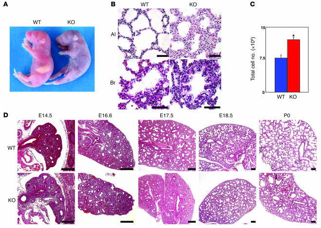
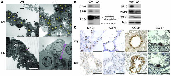
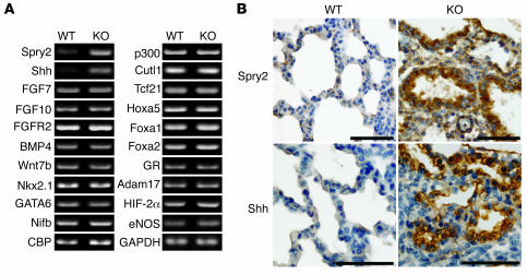
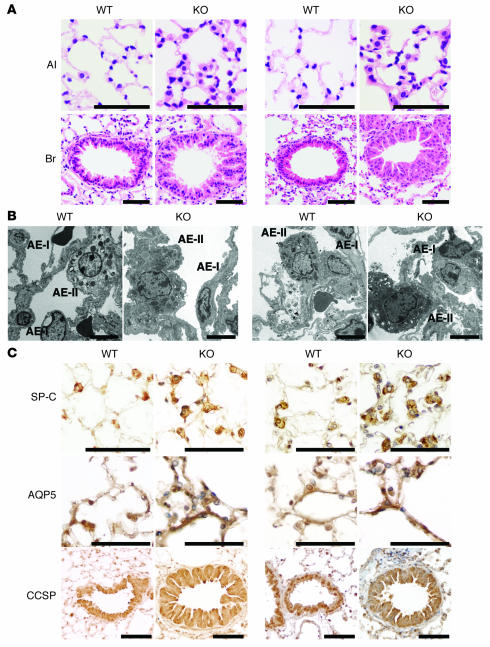
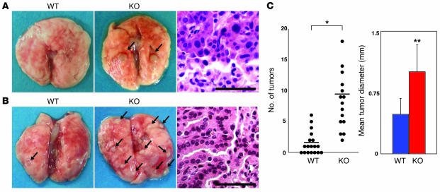
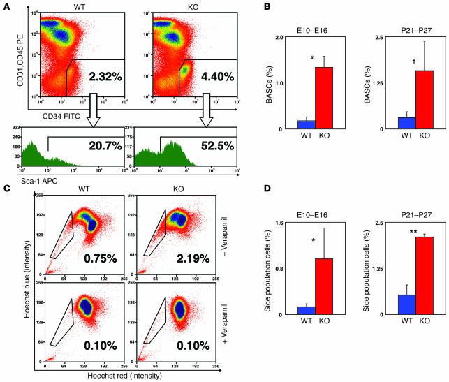
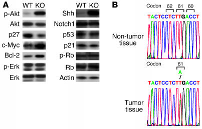
PTEN is a tumor suppressor gene mutated in many human cancers. We generated a bronchioalveolar epithelium-specific null mutation of Pten in mice [SP-C-rtTA/(tetO)(7)-Cre/Pten(flox/flox) (SOPten(flox/flox)) mice] that was under the control of doxycycline. Ninety percent of SOPten(flox/flox) mice that received doxycycline in utero [SOPten(flox/flox)(E10-16) mice] died of hypoxia soon after birth. Surviving SOPten(flox/flox)(E10-16) mice and mice that received doxycycline postnatally [SOPten(flox/flox)(P21-27) mice] developed spontaneous lung adenocarcinomas. Urethane treatment accelerated number and size of lung tumors developing in SOPten(flox/flox) mice of both ages. Histological and biochemical examinations of the lungs of SOPten(flox/flox)(E10-16) mice revealed hyperplasia of bronchioalveolar epithelial cells and myofibroblast precursors, enlarged alveolar epithelial cells, and impaired production of surfactant proteins. Numbers of bronchioalveolar stem cells (BASCs), putative initiators of lung adenocarcinomas, were increased. Lungs of SOPten(flox/flox)(E10-16) mice showed increased expression of Spry2, which inhibits the maturation of alveolar epithelial cells. Levels of Akt, c-Myc, Bcl-2, and Shh were also elevated in SOPten(flox/flox)(E10-16) and SOPten(flox/flox)(P21-27) lungs. Furthermore, K-ras was frequently mutated in adenocarcinomas observed in SOPten(flox/flox)(P21-27) lungs. These results indicate that Pten is essential for both normal lung morphogenesis and the prevention of lung carcinogenesis, possibly because this tumor suppressor is required for BASC homeostasis.


Figures

Figure 1
Figure 1. Abnormal lung morphogenesis and lung epithelial cell hyperplasia in SOPtenflox/flox(E10–E16) mice.
(A) Gross appearance of representative neonates. Skin color is normal in WT(E10–E16) neonates but cyanotic in SOPtenflox/flox(E10–E16) (KO) littermates. (B) Histologic analysis of neonatal lungs. Left: normal alveolar (Al) and bronchiolar (Br) epithelial cells from a WT(E10–E16) neonate. Right: epithelial cell hyperplasia in alveoli and a bronchiole from a SOPtenflox/flox(E10–E16) neonate. Scale bars: 50 μm. (C) Increased total cell numbers in lungs. Total cells from WT(E10–E16) and SOPtenflox/flox(E10–E16) lungs at P0 were counted. Data are expressed as the mean total lung cells ± SD for 4 mice/group. *P < 0.05, Student’s t test. (D) Histological analysis of lungs at various embryonic stages. Lung sections were prepared from SOPtenflox/flox(E10–E16) mice at the indicated gestational stages and stained with H&E. Representative sections from WT(E10–E16) and SOPtenflox/flox(E10–E16) embryos are shown. No differences were detected between the WT and mutant embryos at E14.5 or E16.6, but dramatic differences were visible from E17.5 onward. WT(E10–E16) lungs showed dilatation of distal tubules and mesenchyme thinning at E17.5, with progression of septation from E17.5 to P0. SOPtenflox/flox(E10–E16) lungs showed fewer saccular structures during this period. Scale bars: 200 μm.
Figure 2
Figure 2. Abnormal thickness of the blood-air barrier and impaired alveolar epithelial cell differentiation in SOPtenflox/flox(E10–E16) lungs.
(A) Transmission electron micrographs of the lung septa of E19.5 embryos shown at low (LM) and high (HM) magnification. WT(E10–E16) lung showed AE-I and AE-II cells plus 2 layers of capillaries (asterisks) separated by mesenchymal cells (M; upper left panel). The AE-II cells contained many lamellar bodies (white arrowheads) and apical microvilli (lower left panel). SPs (arrow) were visible in the saccular spaces (upper left panel). Black arrowheads (left panels) indicate the thin, normal blood-air barrier, composed of AE-I cells and capillary endothelial cells, in the WT lung. In the SOPtenflox/flox(E10–E16) lung, the septa were thick with increased mesenchymal cells (upper right panel). Increased numbers of undifferentiated cuboidal epithelial cells of enlarged size were present (CC; upper right panel). The blood-air barrier (red bars) was significantly thicker in SOPtenflox/flox lungs than in WT lungs (lower panels). Scale bars: 10 μm (LM); 5 μm (HM). (B and C) Altered marker protein expression. (B) Western blotting of SP-A, -B, -C, -D, AQP5, and CCSP proteins in extracts of whole lungs taken from WT(E10–E16) and SOPtenflox/flox(E10–E16) mice at E19.5. Actin was used as a loading control. Data shown are representative of 3 trials. (C) IHC analysis of SP-C, AQP5, CCSP and CGRP. SP-C and AQP5 immunostaining was intense in cuboidal AE-II and flat AE-I cells, respectively, in WT(E10–E16) lungs at E19.5 but dramatically decreased in SOPtenflox/flox(E10–E16) lung at E19.5. Scale bars: 50 μm.
Figure 3
Figure 3. Altered gene expression profile during lung morphogenesis and distal epithelial cell differentiation in SOPtenflox/flox(E10–E16) lungs.
(A) RT-PCR analyses of the mRNA expression of the indicated genes in lungs of WT(E10–E16) and SOPtenflox/flox(E10–E16) mice at E19.5. Spry2 and Shh are differentially expressed. GAPDH was used as a loading control. Data shown are representative of 3 trials. (B) IHC analysis of Spry2 and Shh expression in lungs of WT(E10–E16) and SOPtenflox/flox(E10–E16) mice at E19.5. A dramatic upregulation of Spry2 and Shh in the alveolar epithelium in SOPtenflox/flox(E10–E16) lungs was observed. Scale bars: 50 μm.
Figure 4
Figure 4. Bronchiolar and alveolar epithelial cell hyperplasia and increased cell size in SOPtenflox/flox mice that received doxycycline postnatally.
(A) Histologic analyses of lungs from WT(P21–P27) and SOPtenflox/flox(P21–P27) and WT(P84–P90) and SOPtenflox/flox(P84–P90) mice. Mild epithelial cell hyperplasia in alveoli and a bronchiole could be observed at both time points. Scale bars: 50 μm. (B) Transmission electron micrographs of the lung septa from WT(P21–P27) and SOPtenflox/flox(P21–P27) and WT(P84–P90) and SOPtenflox/flox(P84–P90) mice. The increased sizes of AE-I and AE-II cells can be seen in SOPtenflox/flox lungs at both time points. Lamellar bodies and apical microvilli, which are signature structures of AE-II cells, are visible within the AE-II cells of both WT and SOPtenflox/flox lungs. Scale bars: 5 μm. (C) IHC analysis of SP-C, AQP5, and CCSP expression in lungs of WT(P21–P27) and SOPtenflox/flox(P21–P27) mice (left panels) and WT(P84–P90) and SOPtenflox/flox(P84–P90) mice (right panels). Although the numbers of AE-I and AE-II cells were increased in lungs of SOPtenflox/flox(P21–P27) and SOPtenflox/flox(P84–P90) mice, no significant differences between WT and SOPtenflox/flox mice were observed in the staining intensity of SP-C, AQP5, or CCSP at either time point. Scale bars: 50 μm.
Figure 5
Figure 5. Spontaneous lung adenocarcinomas and urethane-induced lung tumors in SOPtenflox/flox mice that received doxycycline postnatally.
(A) Gross and histological analyses of spontaneous adenocarcinomas. Left and middle: gross appearance of a healthy lung in a 48-week-old WT(P21–P27) mouse and representative lung adenocarcinomas (arrow) in a lung of a 48-week-old SOPtenflox/flox(P21–P27) mouse. Right: histology of a spontaneous lung adenocarcinoma in the lung of a 48-week-old SOPtenflox/flox(P21–P27) mouse, representative of adenocarcinomas observed in 13/15 of these mice. Scale bar: 50 μm. (B) Gross and histological analyses of urethane-induced tumors. WT(P21–P27) (n = 18) and SOPtenflox/flox(P21–P27) mice (n = 15) were injected i.p. with urethane. After 20 weeks, the number of tumors and their diameters were measured from the lung surface. Left and middle: gross appearance of urethane-induced lung tumors (arrows) in WT(P21–P27) and SOPtenflox/flox(P21–P27) mice. Right: histology of a lung adenoma representative of those observed in both WT(P21–P27) and SOPtenflox/flox(P21–P27) mice. Scale bar: 50 μm. (C) Numbers and mean size of urethane-induced tumors in WT(P21–P27) and SOPtenflox/flox(P21–P27) mice. Horizontal bars in the left panel represent the mean tumor numbers for the WT(P21–P27) and SOPtenflox/flox(P21–P27) groups. The right panel shows the mean diameter ± SD of tumors in WT(P21–P27) and SOPtenflox/flox(P21–P27) lungs. *P < 0.001; **P < 0.005, Welch’s t test.
Figure 6
Figure 6. Pten deficiency induces significant increases in numbers of BASCs and side population cells.
(A) Increased numbers of BASCs in lungs of SOPtenflox/flox(P21–P27) mice. Representative flow cytometric profiles of BASCs in lungs of 8-week-old WT(P21–P27) and SOPtenflox/flox(P21–P27) mice. Upper panels show the percentages of CD34+CD45CD31 cells in the total lung cell population. Lower histograms show the percentages of Sca-1+ cells in the CD34+CD45CD31 population. (B) Bar graph representation of the percentages of BASCs in total lung cells of WT(E10–E16) and SOPtenflox/flox(E10–E16) mice and WT(P21–P27) and SOPtenflox/flox(P21–P27) mice. Data are expressed as the mean percentage ± SD for 4 mice/group. #P = 0.011; P = 0.015. (C) Increased numbers of side population cells in SOPtenflox/flox(P21–P27) lungs. Representative flow cytometric profiles of side population cells in lungs of 8-week-old WT(P21–P27) and SOPtenflox/flox(P21–P27) mice. Side population cells were identified by staining with 5 μg/ml Hoechst 33342 alone (upper panels) or in combination with 50 μM verapamil, which is an inhibitor of ATP-binding cassette transporters and blocks the dye efflux (lower panels). The percentages of side population cells (boxed ares) in the total lung cells are indicated in each panel. (D) Bar graph representation of the percentages of side population cells in total lung cells of WT(E10–E16) and SOPtenflox/flox(E10–E16) and WT(P21–P27) and SOPtenflox/flox(P21–P27) mice. Data are expressed as the mean percentage of side population cells ± SD for 3 mice/group. *P < 0.05; **P = 0.001. For B and D, statistical differences were determined using the Student’s t test. Data shown are representative of at least 4 trials.
Figure 7
Figure 7. Altered cancer-related molecules in SOPtenflox/flox(P21–P27) lungs and identification of K-ras mutations in spontaneous lung adenocarcinomas of SOPtenflox/flox(P21–P27) mice.
(A) Altered cancer-related molecules in SOPtenflox/flox(P21–P27) lungs. The phosphorylated forms of Akt, Erk1/2 and Rb and the expression of p27, c-Myc, Bcl-2, Notch1, Shh, p53 and p21 were detected by immunoblotting of lysates of whole lung tissue from 8-week-old WT(P21–P27) and SOPtenflox/flox(P21–P27) mice. Total Akt, total Erk, total Rb, and actin were evaluated as controls. Data shown are representative of 3 trials. (B) Identification of K-ras mutations in spontaneous lung adenocarcinomas of SOPtenflox/flox(P21–P27) mice. Top: Normal nucleotide sequence of codons 60–62 of the K-ras gene in the genomic DNA of non-tumorous tissue taken from SOPtenflox/flox(P21–P27) lungs. The antisense strand is shown for exon 2. Bottom: Detection of a T-to-A transversion at the second position of codon 61 in the genomic DNA of a spontaneous adenocarcinoma in SOPtenflox/flox(P21–P27) lung tissue. Data shown are representative of 3 trials.

Similar articles

Cited by

References

    1. Jemal A., Thomas A., Murray T., Thun M. Cancer statistics, 2002. CA Cancer J. Clin. 2002;52:23–47. - PubMed
    1. Giangreco A., Groot K.R., Janes S.M. Lung cancer and lung stem cells: strange bedfellows? Am. J. Respir. Crit. Care Med. 2007;175:547–553. - PubMed
    1. Minna J.D., Roth J.A., Gazdar A.F. Focus on lung cancer. Cancer Cell. 2002;1:49–52. - PubMed
    1. Reya T., Morrison S.J., Clarke M.F., Weissman I.L. Stem cells, cancer, and cancer stem cells. Nature. 2001;414:105–111. - PubMed
    1. Kim C.F., et al. Identification of bronchioalveolar stem cells in normal lung and lung cancer. Cell. 2005;121:823–835. - PubMed

Publication types

MeSH terms