

Lipopolysaccharide is a frequent and significant contaminant in microglia-activating factors
- PMID: 17910052
- PMCID: PMC2926344
- DOI: 10.1002/glia.20585
Lipopolysaccharide is a frequent and significant contaminant in microglia-activating factors
Abstract
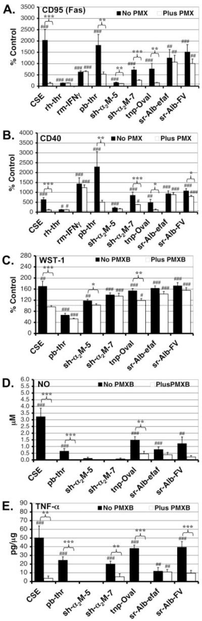
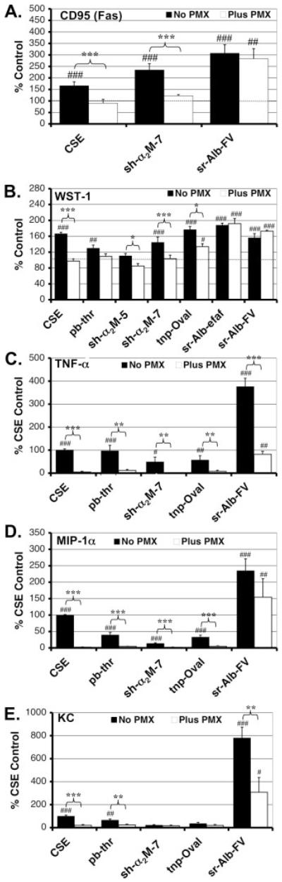
Lipopolysaccharide (LPS/endotoxin) is a potent immunologic stimulant. Many commercial-grade reagents used in research are not screened for LPS contamination. LPS induces a wide spectrum of proinflammatory responses in microglia, the immune cells of the brain. Recent studies have demonstrated that a broad range of endogenous factors including plasma-derived proteins and bioactive phospholipids can also activate microglia. However, few of these studies have reported either the LPS levels found in the preparations used or the effect of LPS inhibitors such as polymyxin B (PMX) on factor-induced responses. Here, we used the Limulus amoebocyte lysate assay to screen a broad range of commercial- and pharmaceutical-grade proteins, peptides, lipids, and inhibitors commonly used in microglia research for contamination with LPS. We then characterized the ability of PMX to alter a representative set of factor-induced microglial activation parameters including surface antigen expression, metabolic activity/proliferation, and NO/cytokine/chemokine release in both the N9 microglial cell line and primary microglia. Significant levels of LPS contamination were detected in a number of commercial-grade plasma/serum- and nonplasma/serum-derived proteins, phospholipids, and synthetic peptide preparations, but not in pharmaceutical-grade recombinant proteins or pharmacological inhibitors. PMX had a significant inhibitory effect on the microglia-activating potential of a number of commercial-, but not pharmaceutical-grade, protein preparations. Novel PMX-resistant responses to alpha(2)-macroglobulin and albumin were incidentally observed. Our results indicate that LPS is a frequent and significant contaminant in commercial-grade preparations of previously reported microglia-activating factors. Careful attention to LPS levels and appropriate controls are necessary for future studies in the neuroinflammation field.
(c) 2007 Wiley-Liss, Inc.
Figures
Similar articles
-
Unraveling thrombin's true microglia-activating potential: markedly disparate profiles of pharmaceutical-grade and commercial-grade thrombin preparations.J Neurochem. 2005 Nov;95(4):1177-87. doi: 10.1111/j.1471-4159.2005.03499.x. J Neurochem. 2005. PMID: 16271051
-
Suppressive effects of levobupivacaine on endotoxin-induced microglial activation.J Surg Res. 2013 Oct;184(2):989-96. doi: 10.1016/j.jss.2013.03.074. Epub 2013 Apr 10. J Surg Res. 2013. PMID: 23590869
-
Novel celecoxib analogues inhibit glial production of prostaglandin E2, nitric oxide, and oxygen radicals reverting the neuroinflammatory responses induced by misfolded prion protein fragment 90-231 or lipopolysaccharide.Pharmacol Res. 2016 Nov;113(Pt A):500-514. doi: 10.1016/j.phrs.2016.09.010. Epub 2016 Sep 22. Pharmacol Res. 2016. PMID: 27667770
-
Differential expression of chemokines and chemokine receptors during microglial activation and inhibition.J Neuroimmunol. 2004 Apr;149(1-2):1-9. doi: 10.1016/j.jneuroim.2003.11.012. J Neuroimmunol. 2004. PMID: 15020059
-
Activation of microglial cells by thrombin: past, present, and future.Semin Thromb Hemost. 2006 Apr;32 Suppl 1:69-76. doi: 10.1055/s-2006-939556. Semin Thromb Hemost. 2006. PMID: 16673268 Review.
Cited by
-
Association between periodontitis and bipolar disorder: A nationwide cohort study.Medicine (Baltimore). 2020 Jul 31;99(31):e21423. doi: 10.1097/MD.0000000000021423. Medicine (Baltimore). 2020. PMID: 32756145 Free PMC article.
-
Thrombin-induced regulation of CD95(Fas) expression in the N9 microglial cell line: evidence for involvement of proteinase-activated receptor(1) and extracellular signal-regulated kinase 1/2.Neurochem Res. 2009 Mar;34(3):445-52. doi: 10.1007/s11064-008-9803-9. Epub 2008 Aug 7. Neurochem Res. 2009. PMID: 18686031
-
Brain resident microglia in Alzheimer's disease: foe or friends.Inflammopharmacology. 2024 Oct;32(5):2781-2800. doi: 10.1007/s10787-024-01550-8. Epub 2024 Aug 21. Inflammopharmacology. 2024. PMID: 39167311 Review.
-
Bacterial vaginosis toxins impair sperm capacitation and fertilization.Hum Reprod. 2025 Sep 1;40(9):1720-1734. doi: 10.1093/humrep/deaf132. Hum Reprod. 2025. PMID: 40652342 Free PMC article.
-
Saturated fatty acids produce an inflammatory response predominantly through the activation of TLR4 signaling in hypothalamus: implications for the pathogenesis of obesity.J Neurosci. 2009 Jan 14;29(2):359-70. doi: 10.1523/JNEUROSCI.2760-08.2009. J Neurosci. 2009. PMID: 19144836 Free PMC article.
References
-
- Badie B, Schartner J, Vorpahl J, Preston K. Interferon-gamma induces apoptosis and augments the expression of Fas and Fas ligand by microglia in vitro. Exp Neurol. 2000;162:290–296. - PubMed
-
- Balcaitis S, Xie Y, Weinstein JR, Andersen H, Hanisch UK, Ransom BR, Möller T. Expression of proteinase-activated receptors in mouse microglial cells. Neuroreport. 2003;14:2373–2377. - PubMed
-
- Bausinger H, Lipsker D, Ziylan U, Manie S, Briand JP, Cazenave JP, Muller S, Haeuw JF, Ravanat C, de la Salle H, Hanau D. Endotoxin-free heat-shock protein 70 fails to induce APC activation. Eur J Immunol. 2002;32:3708–3713. - PubMed
-
- Bianco F, Ceruti S, Colombo A, Fumagalli M, Ferrari D, Pizzirani C, Matteoli M, Di Virgilio F, Abbracchio MP, Verderio C. A role for P2x in microglia proliferation. J Neurochem. 2006;99:745–758. - PubMed
Publication types
MeSH terms
Substances
Grants and funding
LinkOut - more resources
Full Text Sources
