. 2007 Oct 2:4:96.
doi: 10.1186/1743-422X-4-96.

A pandemic strain of calicivirus threatens rabbit industries in the Americas

Affiliations

A pandemic strain of calicivirus threatens rabbit industries in the Americas

Michael T McIntosh et al. Virol J. .

Abstract

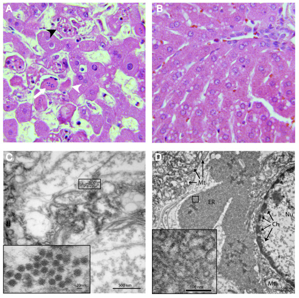
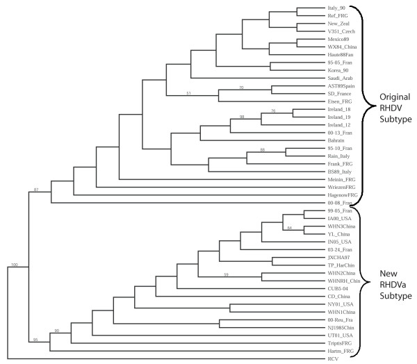
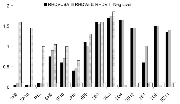
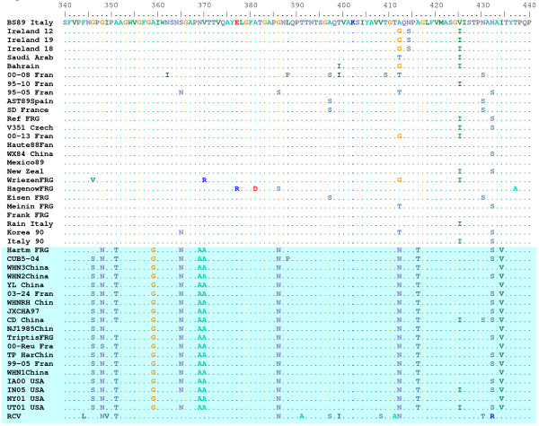
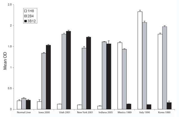
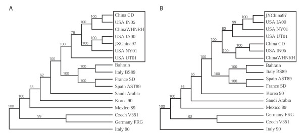
Rabbit Hemorrhagic Disease (RHD) is a severe acute viral disease specifically affecting the European rabbit Oryctolagus cuniculus. As the European rabbit is the predominant species of domestic rabbit throughout the world, RHD contributes towards significant losses to rabbit farming industries and endangers wild populations of rabbits in Europe and other predatory animals in Europe that depend upon rabbits as a food source. Rabbit Hemorrhagic Disease virus (RHDV) - a Lagovirus belonging to the family Caliciviridae is the etiological agent of RHD. Typically, RHD presents with sudden death in 70% to 95% of infected animals. There have been four separate incursions of RHDV in the USA, the most recent of which occurred in the state of Indiana in June of 2005. Animal inoculation studies confirmed the pathogenicity of the Indiana 2005 isolate, which caused acute death and pathological changes characterized by acute diffuse severe liver necrosis and pulmonary hemorrhages. Complete viral genome sequences of all USA outbreak isolates were determined and comparative genomics revealed that each outbreak was the result of a separate introduction of virus rather than from a single virus lineage. All of the USA isolates clustered with RHDV genomes from China, and phylogenetic analysis of the major capsid protein (VP60) revealed that they were related to a pandemic antigenic variant strain known as RHDVa. Rapid spread of the RHDVa pandemic suggests a selective advantage for this new subtype. Given its rapid spread, pathogenic nature, and potential to further evolve, possibly broadening its host range to include other genera native to the Americas, RHDVa should be regarded as a threat.


Figures

Figure 1
Figure 1
Histopathology and cytopathology associated with IN-05 RHDVa infection. A. A liver section from one of the fatally infected rabbits (day 2 post infection) is shown (H&E stain, 40× objective). Note the acute hepatocellular necrosis characterized by destruction and disassociation of hepatocytes, loss of cellular organization, and evidence of acidophilic bodies (white arrow head), karyorrhexis (white arrow), and necrotic or apoptotic hepatocytes (black arrow head). B. A liver section from the surviving infected rabbit (day 21 post infection) exhibited normal liver morphology (H&E staine, 40× objective). C. Transmission electron micrograph showing the ultrastructure of a hepatocyte from a fatally infected rabbit revealed the presence of 26.5 nm +/- 1.9 diameter viral particles with morphology characteristic of caliciviruses. D. An example of ultrastructural changes to a hepatocyte from one of the fatally infected rabbits. Note the margination of chromatin (Ch) in the nucleus (Nu), and disruption of cristae in mitochondria (Mt). Often, an abnormal condensation of the endoplasmic reticulum (ER) was observed. The inset shows an abnormally dense reticular network.
Figure 2
Figure 2
Relationship of VP60 capsid proteins among diverse isolates of RHDV. The predicted amino acid sequences of 45 RHDV isolates were aligned in CLUSTAL W. One thousand bootstrap replicates were subjected to protein distance and UPGMA methods and the consensus phylogenetic tree is shown. The VP60 region of a non-pathogenic rabbit calicivirus (RCV) was used as an outgroup. Two clades, one representing the original RHDV serotype and a second representing the new RHDVa subtype were identified. Bootstrap values greater than 50% are displayed above the tree branches.
Figure 3
Figure 3
Epitope profile of the first U.S. outbreak isolate RHDV IA-00. The RHDV IA-00 isolate was subtyped by antigen capture ELISA using a panel of monoclonal antibodies. Previous studies and communication from Lorenzo Capucci [35] have determined that monoclonal antibodies 1H8, 2A10, and 1H3 recognize the original serotype of RHDV while antibodies 3D4, 3B12, 2E1, 3D6, and 5D11 recognize RHDVa-specific epitopes. Additional monoclonal antibodies used (6H6, 1F10, 3H6, 6F9, 2B4, and 2G3) were not subtype-specific. The IA-00 isolate (black bars) correlated in antibody recognition profile to a prototype RHDVa strain, Pavia 1997 (grey bars). The Brescia 1989 strain (stippled bars) was used as an original RHDV serotype virus control. Normal liver from an uninfected rabbit served as a negative control (white bars).
Figure 4
Figure 4
RHDVa-specific epitope between residues 340 and 440 of the VP60 capsid protein. A portion of the CLUSTAL W alignment of the VP60 sequence for 45 isolates of RHDV and 1 isolate of a non-pathogenic rabbit calicivirus (RCV) is shown. The top reference sequence for the alignment came from the Brescia 1989 strain (BS89 Italy) and identical amino acids were indicated by a dot. Note the large number of shared amino acid substitutions within the RHDVa clade (shaded blue).
Figure 5
Figure 5
Type-specific antigenicity of the U.S. isolates of RHDV. Liver homogenates from experimentally infected animals were tested by antigen-capture ELISA using type-specific HRP-conjugated monoclonal antibodies (MAb). MAb 1H8 is specific for the original RHDV serotype, MAb 3B12 is specific for the new RHDVa pandemic strain, and MAb 2B4 recognizes a shared epitope. The four U.S. RHDV isolates, Mexico 1989 isolate, an Italian isolate, and Korean isolate were compared in comparison with a control liver homogenate derived from an uninfected rabbit (Normal Liver). All U.S. isolates were recognized by MAb 3B12 as belonging to the RHDVa pandemic strain.
Figure 6
Figure 6
Relationship of U.S. isolates to genomes of other RHDV isolates. A. Genomes of RHDV isolates including the four U.S. isolates were aligned in CLUSTAL W and 1000 bootstrap replicates were subjected to DNA Distance and Neighbor Joining methods. A consensus tree is shown with bootstrap values greater than 50% placed above tree branches. The U.S. isolates all branched (100% of the time) with a distinct clade of RHDVa isolates from China (box). B. Analysis was repeated as shown in panel A. except that the VP60 coding regions were removed from the genomic sequences. All U.S. isolates continued to branch with the RHDVa isolates from China, despite removal of the RHDVa epitope.

Similar articles

Cited by

References

    1. Marcato PS, Benazzi C, Vecchi G, Galeotti M, Della Salda L, Sarli G, Lucidi P. Clinical and pathological features of viral haemorrhagic disease of rabbits and the European brown hare syndrome. Rev Sci Tech. 1991;10:371–392. - PubMed
    1. Capucci L, Scicluna MT, Lavazza A. Diagnosis of viral haemorrhagic disease of rabbits and the European brown hare syndrome. Rev Sci Tech. 1991;10:347–370. - PubMed
    1. Ferreira PG, Costa-e-Silva A, Monteiro E, Oliveira MJ, Aguas AP. Transient decrease in blood heterophils and sustained liver damage caused by calicivirus infection of young rabbits that are naturally resistant to rabbit haemorrhagic disease. Res Vet Sci. 2004;76:83–94. doi: 10.1016/j.rvsc.2003.08.003. - DOI - PubMed
    1. Arguello Villares JL. Viral haemorrhagic disease of rabbits: vaccination and immune response. Rev Sci Tech. 1991;10:459–480. - PubMed
    1. Meyers G, Wirblich C, Thiel HJ. Rabbit hemorrhagic disease virus – molecular cloning and nucleotide sequencing of a calicivirus genome. Virology. 1991;184:664–676. doi: 10.1016/0042-6822(91)90436-F. - DOI - PubMed

Publication types

MeSH terms

Substances