

doi: 10.1016/j.febslet.2007.09.030.
Epub 2007 Sep 24.
Interaction of a fragment of the cannabinoid CB1 receptor C-terminus with arrestin-2
Affiliations
- PMID: 17910957
- PMCID: PMC2151313
- DOI: 10.1016/j.febslet.2007.09.030
Item in Clipboard
Interaction of a fragment of the cannabinoid CB1 receptor C-terminus with arrestin-2
FEBS Lett.
.
Abstract
Desensitization of the cannabinoid CB1 receptor is mediated by the interaction with arrestin. In this study, we report the structural changes of a synthetic diphosphorylated peptide corresponding to residues 419-439 of the CB1 C-terminus upon binding to arrestin-2. This segment is pivotal to the desensitization of CB1. Using high-resolution proton NMR, we observe two helical segments in the bound peptide that are separated by the presence a glycine residue. The binding we observe is with a diphoshorylated peptide, whereas a previous study reported binding of a highly phosphorylated rhodopsin fragment to visual arrestin. The arrestin bound conformations of the peptides are compared.
Figures

The amino acid sequences and numbering of CB1419-438 and Rh330-348. The phosphorylated residues are shown in italics and underlined.
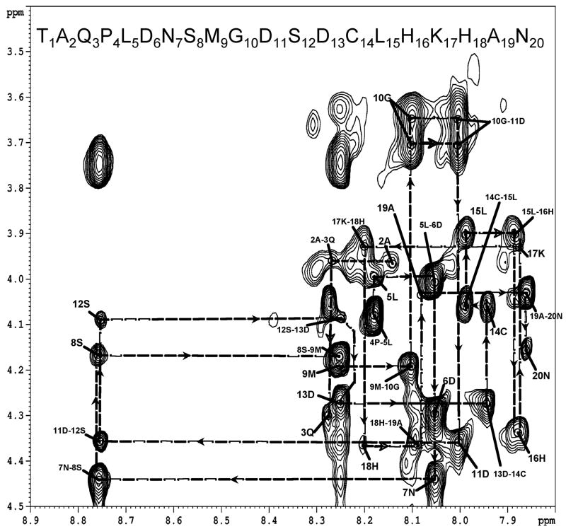
Sequential assignments in the fingerprint region of the NOESY spectrum of the CB1419-438 - arrestin-2 complex. The one letter codes of amino acids and numbering of the peptide from 1–20 is used for the sake of clarity. (See Figure 1 for numbering) Labels such as “5L” indicate the NH-α intra-residue NOEs for each residue while labels such as “5L-6D” indicate the ai-NHi+1 inter-residue NOE’s.
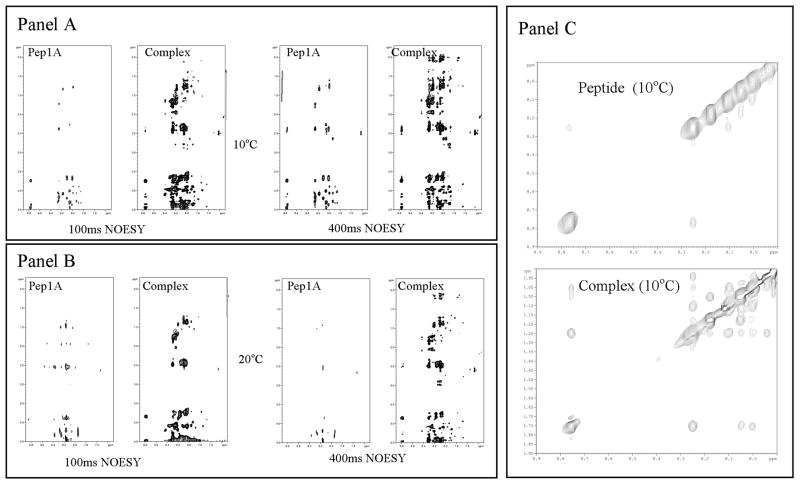
Panel A) NOESY spectra of free CB1419-438 (peptide) and CB1419-438 -arrestin2 mixtures (complex) at 10°C and at two different mixing times. Panel B) NOESY spectra of free CB1419-438 and CB1419-438 -arrestin2 mixtures (complex) at 20°C and at two different mixing times. Panel C) NH-NH region of NOESY spectra of free CB1419-438 (peptide) and CB1419-438 -arrestin2 mixture (complex) at 10°C.

Summary of inter-resdue NOEs and structural statistics of the ordered regions of the peptide (423–433) generated using PROCHECK. Approximately 90% of these residues occur in favored and allowed regions of the ramachandran plot with less than 5% occurring in disallowed regions. The residues that fall in the allowed or generously allowed regions were the residues that borderer the ordered helices and fell within the flexible region near the glycine.

NMR derived structures of CB1419-438 fragment bound to arrestin-2. (A) The low energy structures are aligned from residue 422–427 (B) The same set of structures are aligned from residue 429–433 (C) The ordered regions are shown together to emphasize the flexible GLY428 present in between the two ordered regions.

Comparison of (A) a minimimum energy conformer of diphosphorylated CB1419-438 fragment bound to arrestin and (B) the seven phosphorylated Rh330-348 fragment bound to visual arrestin. This later structure was downloaded from the protein data bank (www.rcsb.org.1NZS.pdb ) and oriented to highlight the similarity of the C-terminal end with CB1419-438. The phosphorylated residues are highlighted by CPK representations.
References
-
- Lohse MJ, Benovic JL, Codina J, Caron MG, Lefkowitz RJ. beta-Arrestin: a protein that regulates beta-adrenergic receptor function. Science. 1990;248:1547–1550. - PubMed
-
- Okada T, Le Trong I, Fox BA, Behnke CA, Stenkamp RE, Palczewski K. X-Ray diffraction analysis of three-dimensional crystals of bovine rhodopsin obtained from mixed micelles. J Struct Biol. 2000;130:73–80. - PubMed
-
- Palczewski K, et al. Crystal structure of rhodopsin: A G protein-coupled receptor. Science. 2000;289:739–745. - PubMed
-
- Yeagle PL, Albert AD. G-protein coupled receptor structure. Biochim Biophys Acta. 2007;1768:808–824. - PubMed
-
- Klabunde T, Hessler G. Drug design strategies for targeting G-protein-coupled receptors. Chembiochem. 2002;3:928–944. - PubMed
Publication types
MeSH terms
Substances
Grants and funding
LinkOut - more resources
Full Text Sources
