. 2007 Oct 9;104(41):16170-5.
doi: 10.1073/pnas.0703942104. Epub 2007 Oct 2.

Tumor protein 53-induced nuclear protein 1 expression is repressed by miR-155, and its restoration inhibits pancreatic tumor development

Affiliations

Tumor protein 53-induced nuclear protein 1 expression is repressed by miR-155, and its restoration inhibits pancreatic tumor development

Meritxell Gironella et al. Proc Natl Acad Sci U S A. .

Abstract

Pancreatic cancer is a disease with an extremely poor prognosis. Tumor protein 53-induced nuclear protein 1 (TP53INP1) is a proapoptotic stress-induced p53 target gene. In this article, we show by immunohistochemical analysis that TP53INP1 expression is dramatically reduced in pancreatic ductal adenocarcinoma (PDAC) and this decrease occurs early during pancreatic cancer development. TP53INP1 reexpression in the pancreatic cancer-derived cell line MiaPaCa2 strongly reduced its capacity to form s.c., i.p., and intrapancreatic tumors in nude mice. This anti-tumoral capacity is, at least in part, due to the induction of caspase 3-mediated apoptosis. In addition, TP53INP1(-/-) mouse embryonic fibroblasts (MEFs) transformed with a retrovirus expressing E1A/ras(V12) oncoproteins developed bigger tumors than TP53INP1(+/+) transformed MEFs or TP53INP1(-/-) transformed MEFs with restored TP53INP1 expression. Finally, TP53INP1 expression is repressed by the oncogenic micro RNA miR-155, which is overexpressed in PDAC cells. TP53INP1 is a previously unknown miR-155 target presenting anti-tumoral activity.


Conflict of interest statement

The authors declare no conflict of interest.

Figures

Fig. 1.
Fig. 1.
TP53INP1 expression is lost in PDAC and metastasis. Shown is TP53INP1-positive immunostaining in epithelial cells of large ducts from normal pancreatic tissue (A), mucinous cystadenoma (B), intraductal papillary mucinous neoplasm (C), and in chronic pancreatitis (D). TP53INP1 protein was not detected in PDAC (E) and in liver metastasis (F). (G–I). TP53INP1 positive staining in early lesions, PanIN-1A (red arrow) negative staining in PanIN-1B, PanIN-2 and PanIN-3 (black arrow).
Fig. 2.
Fig. 2.
TP53INP1-inducible expression in MiaPaCa2 cells and inhibition of s.c. and i.p. tumor growth. (A) TP53INP1- or EGFP-inducible MiaPaCa2 cells were cultured in the presence of PonA 10 μM or vehicle as control. Sixteen hours later, cells were tested for TP53INP1-EGFP or EGFP expression with an anti-EGFP antibody. (B) Cells were injected into the pancreas of mice, and 2 weeks later PonA-releasing pellets were implanted. After 48 h, TP53INP1 induction was visualized by green epifluorescence. (C) TP53INP1-inducible MiaPaCa2 cells were incubated with increasing concentrations of PonA for 48 h (Upper). Cells were treated for the indicated times with PonA 10 μM and TP53INP1 expression was analyzed by Western blot (Lower). β-tubulin level was used as loading control. (D) Ten millions of TP53INP1-inducible MiaPaCa2 cells were s.c. injected in mice carrying PonA (n = 6) or vehicle (n = 6) releasing pellets. Tumoral volume was weekly determined as described in SI Materials and Methods. (E) Histological analysis of s.c. and i.p. tumors from vehicle-treated mice. (F) Ten millions of TP53INP1-inducible MiaPaCa2 cells were i.p. injected in mice carrying PonA (n = 6) or vehicle (n = 6) releasing pellets. Representative photographs are shown. Values are represented as mean ± SE. *, P < 0.05.
Fig. 3.
Fig. 3.
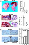
TP53INP1 restoration inhibits tumoral growth in a pancreatic cancer model in vivo. Eighteen millions of inducible MiaPaCa2 cells were injected into the pancreas of nude mice (n = 7 for each group) one day after s.c. implantation of pellets releasing vehicle as control (V) or PonA (P). Assessment of pancreatic tumor growth and spreading was done 19 days after cells injection. (A) (Left and Center) Representative photographs of pancreatic tumors. (Right) Intrapancreatic tumoral volume (tumor sizes were measured as described in SI Materials and Methods). (B) (Left) Representative images showing the spreading of pancreatic tumors in PonA and vehicle treated animals (Sp, spleen; Pa, pancreas). (Center) Representative images of HES-stained pancreas and spleen from vehicle and PonA-treated animals. Tumor surface is outlined in black and indicated by arrows. (Right) Representation of the proximal extension index. The index was determined as described in SI Materials and Methods. (C) (Left and Center) Representative images of pancreatic tumors active caspase-3 immunohistochemistry on vehicle and PonA-treated animals with TP53INP1-inducilbe MiaPaCa2 or EGFP-inducible MiaPaCa2 tumors. (Right) The percentage of tumor surface containing active caspase-3. Values are represented as mean ± SE. *, P < 0.05.
Fig. 4.
Fig. 4.
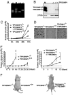
TP53INP1 reexpression reduces the growth rate of E1A/ras-TP53INP1−/− MEFs in vitro and in vivo. (A) TP53INP1 genotyping of primary MEFs determined by PCR. (B) Western blot showing the TP53INP1 restoration in E1A/ras-TP53INP1−/− MEFs. TP53INP1 restoration was obtained by transduction of E1A/ras-TP53INP1−/− MEFs with a retrovirus expressing TP53INP1 (TP53INP1−/− plus TP53INP1). β-tubulin (β-tub) was used as control. Total cellular extract containing TP53INP1α and β transfected proteins were migrated in parallel (M) (C) Growth curves for E1A/ras-MEFs. (D) Growth in soft-agar for E1A/ras-MEFs. (E) Subcutaneous tumoral growth in nude mice. One million cells were s.c. injected. Tumoral volume was determined as described in SI Materials and Methods. (Left) Tumoral growth for E1A/ras-TP53INP1−/− MEFs (n = 6) versus E1A/ras-TP53INP1+/+ MEFs (n = 6). The arrow corresponds to the excision of indicated tumors. (Right) Tumoral growth for E1A/ras-TP53INP1−/− MEFs (n = 6) versus E1A/ras-TP53INP1−/− MEFs reexpressing TP53INP1 (n = 5). (F) Representative images of s.c. tumors. Values are represented as mean ± SE. *, P < 0.05.
Fig. 5.
Fig. 5.
TP53INP1 is a miR-155 target. (A) Data represent mRNA and miR-155 level ratios between PDAC and peritumoral pancreas (PTP). TP53INP1 immunohistochemical analysis are presented below the graph: +, positive staining; −, negative staining. (B) Alignment of mouse, rat and human miR-155 and the TP53INP1 3′ UTR in different species. (C) Representation of the pMIR-TP53INP1 vectors used in the Luciferase assay. (D) 293T cells were cotransfected with pMIR-TP53INP1 3′ UTR constructs in the 5′→3′ or 3′→5′ orientation, and the indicated concentrations of miR-155 or miR-control. Luciferase activity was measured, 48 h after transfection. (E) MCF7 cells were transfected with 50 nM miR-155 or miR-control. After transfection with a p53-expressing vector and gamma irradiation (30 Gy), TP53INP1 level was evaluated by Western blot and RT-PCR. β-tubulin level was used as control. Values are represented as mean ± SE. *, P < 0.05. (F) Capan2 cells were transfected with Anti-miR-155 or Anti-miR-control and gamma-irradiated (30 Gy), TP53INP1 level was evaluated by Western blot. β-tubulin level was used as control. Capan2 cells transfected with a plasmid allowing the expression of TP53INP1 were migrated in parallel as molecular weight control (M). (G) Apoptosis was measured in Capan2 cells 24 h after transfection with Anti-miR-155 or Anti-miR-control.

References

    1. Hezel AF, Kimmelman AC, Stanger BZ, Bardeesy N, DePinho RA. Genes Dev. 2006;20:1218–1249. - PubMed
    1. Tomasini R, Samir AA, Vaccaro MI, Pebusque MJ, Dagorn JC, Iovanna JL, Dusetti NJ. J Biol Chem. 2001;276:44185–44192. - PubMed
    1. Tomasini R, Samir AA, Pebusque MJ, Calvo EL, Totaro S, Dagorn JC, Dusetti NJ, Iovanna JL. Eur J Cell Biol. 2002;81:294–301. - PubMed
    1. Tomasini R, Samir AA, Carrier A, Isnardon D, Cecchinelli B, Soddu S, Malissen B, Dagorn JC, Iovanna JL, Dusetti NJ. J Biol Chem. 2003;278:37722–37729. - PubMed
    1. Yoshida K, Liu H, Miki Y. J Biol Chem. 2006;281:5734–5740. - PubMed

Publication types

MeSH terms