. 2008 Jan 15;111(2):633-42.
doi: 10.1182/blood-2007-08-107359. Epub 2007 Oct 2.

ALK5- and TGFBR2-independent role of ALK1 in the pathogenesis of hereditary hemorrhagic telangiectasia type 2

Affiliations

ALK5- and TGFBR2-independent role of ALK1 in the pathogenesis of hereditary hemorrhagic telangiectasia type 2

Sung O Park et al. Blood. .

Abstract

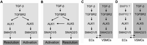
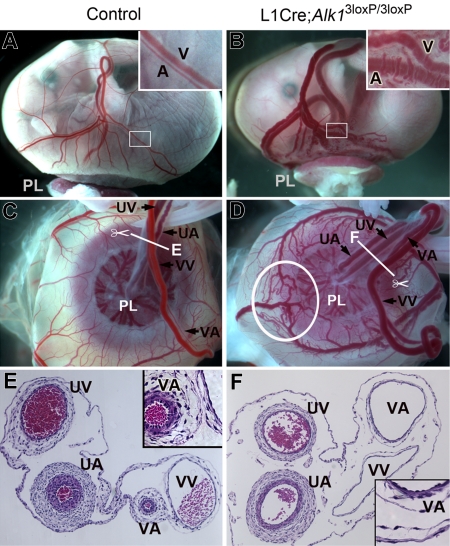
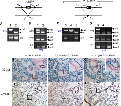
ALK1 belongs to the type I receptor family for transforming growth factor-beta family ligands. Heterozygous ALK1 mutations cause hereditary hemorrhagic telangiectasia type 2 (HHT2), a multisystemic vascular disorder. Based largely on in vitro studies, TGF-beta1 has been considered as the most likely ALK1 ligand related to HHT, yet the identity of the physiologic ALK1 ligand remains controversial. In cultured endothelial cells, ALK1 and another TGF-beta type I receptor, ALK5, regulate angiogenesis by controlling TGF-beta signal transduction, and ALK5 is required for ALK1 signaling. However, the extent to which such interactions between these 2 receptors play a role in pathogenesis of HHT is unknown. We directly addressed these issues in vivo by comparing the phenotypes of mice in which the Alk1, Alk5, or Tgfbr2 gene was conditionally deleted in restricted vascular endothelia using a novel endothelial Cre transgenic line. Alk1-conditional deletion resulted in severe vascular malformations mimicking all pathologic features of HHT. Yet Alk5- or Tgfbr2-conditional deletion in mice, or Alk5 inhibition in zebrafish, did not affect vessel morphogenesis. These data indicate that neither ALK5 nor TGFBR2 is required for ALK1 signaling pertinent to the pathogenesis of HHT and suggest that HHT might not be a TGF-beta subfamily disease.


Figures

Figure 1
Figure 1
Multiple versions of TGF-β signaling pathways in endothelial and smooth muscle cells. During the activation phase of angiogenesis, endothelial cells degrade their vascular basement membranes, migrate into extracellular spaces, proliferate, and form vascular lumens. During the resolution phase, endothelial cells cease to migrate and to proliferate and instead reconstitute their basement membranes. The maturation and remodeling of the vessels also occur in this phase, as mesenchymal cells are recruited for endothelial tube ensheathment. (A,B) It has been reported that both ALK1 and ALK5 are TGF-β subfamily type I receptors in ECs: that is, they are both activated by TGF-β subfamily ligands binding to TGFBR2. As ALK1 and ALK5 signal through different SMAD proteins, it has been suggested that the opposing activities of these 2 type I receptors regulate angiogenesis. However, whereas some studies have suggested a role or ALK1 in resolution and ALK5 in activation,, others have suggested opposite roles, with ALK5 being necessary for ALK1 function., (C) Both balance models (A,B) are called into question by an expression study in mice showing that, whereas Alk1 is endothelial-specific, Alk5 is expressed not in the endothelium but in neighboring smooth muscle cells. (D) Data presented do not support a role for TGF-β subfamily ligands and TGFBR2 in ALK1 function, suggesting that TGF-β superfamily ligands outside of the TGF-β subfamily may be physiologic ligands for ALK1 in endothelial cells. This hypothesis is supported by recent biochemical data demonstrating that BMP9 serves as an ALK1 ligand.,
Figure 2
Figure 2
Tg(Alk1-cre)-L1 mice express the Cre recombinase predominantly in the pulmonary vascular endothelial cells. The cells in which Cre–mediated recombination has occurred were visualized by staining the Tg(Alk1-cre);R26R bigenic embryos with X-gal for the β-gal activity at E10.5 (A-D), E13.5 (E-H), and E15.5 (I-K) stages. (A,B) X-gal-positive staining is visible in the blood vessels throughout embryos in the Tg(Alk1-cre)-B (A) and -D (B) lines. (C) Transverse sections of X-gal stained Tg(Alk1-cre)-D embryo showing lacZ expressions in the vascular ECs, endocardial cells in atria and ventricles, and mesenchymal cells in the atrioventricular cushion (AVC). (D) In contrast with that in the B and D lines, almost no lacZ expression was detected in the L1cre embryos; only a spotty staining pattern in the head region (arrow). (E,F) Dorsal aorta (DA) view of the heart and lungs of L1cre:R26R embryos stained with X-gal, showing a strong lacZ expression in the lung in comparison with a patch staining in the heart (E) and body trunk (F). (G,H) Histologic sections of the X-gal stained lung was counterstained with NFR (G) or costained with anti-PECAM antibodies (H). The inset in panel H is a magnified view of the area indicated by the arrow. Note that X-gal-positive cells resided in pulmonary ECs, but only in a subpopulation of PECAM-positive cells. (I) Ventral view of the X-gal stained L1cre:R26R lung attached to the body trunk. The heart was removed for clarity of the view. Note a strong X-gal staining in the lung but not in the body trunk. (J,K) Histologic sections demonstrate that most PECAM-positive cells are positive for X-gal staining in E15.5 embryonic lungs, yet no X-gal-positive cells were detected in airway epithelial and smooth muscle cells. Insets are magnified views of the areas indicated by the arrow in each panel.
Figure 3
Figure 3
Generation of Alk1-conditional alleles. (A) Schematic diagram of the Alk1 wild-type allele, Alk1-conditional targeting vector, and Alk13loxP, Alk12loxP, and Alk11loxP alleles. Exons and loxP sequences are indicated by boxes and arrowheads, respectively. Locations of primer pairs used for amplifying specific regions containing a loxP sequence are also indicated. (B) Genomic Southern blot analysis from EcoRI digested DNA isolated from several ES clones, showing the homologous recombination of the Alk13loxP vector into the Alk1 locus. (C) Representative PCR genotyping results from intercrosses of Alk1+/3loxP (top) and Alk1+/2loxP (bottom). The arrowheads indicate the PCR amplicon containing the loxP sequence; Alk13loxP/3loxP (lanes 2 and 5); Alk1+/3loxP (lanes 1, 4); Alk1+/+ (lane 3); Alk1+/2loxP (lanes 6 and 8); and Alk12loxP/2loxP (lanes 7, 9, and 10). (D) PCR detection of the Alk11loxP allele from genomic DNA isolated from multiple organs/tissues of E16.5 L1cre(+)Alk13loxP/3loxP (top), L1cre(+)Alk1+/3loxP (middle), and L1cre(−);Alk13loxP/3loxP (bottom) fetuses, demonstrating tissue-specific Cre activities. The arrowheads indicate the Alk11loxP-specific PCR amplicon.
Figure 4
Figure 4
Alk1 deletion resulted in abnormal extraembryonic vasculature in E16.5 L1cre(+);Alk13loxP/3loxP fetuses. (A,B) Gross morphology of control and L1cre(+);Alk13loxP/3loxP mutant fetuses enclosed in the yolk sac attached to the placenta (PL). Note bulged arteries (A) and veins (V) in the mutant yolk sac. The inset in panel B shows magnified view of typical dilated, tortuous vitelline vessels in the mutants. (C,D) Umbilical arteries (UA) and veins (UV) are connected to the placenta, whereas vitelline arteries (VA) and veins (VV) are connected to the yolk sac. Note markedly enlarged VA and AVMs (circled; see enlarged view in Figure 5D) in the mutants. (E,F) Cross-sectional view of the extraembryonic vessels indicated by the scissors symbols in panels C,D demonstrates marked dilation and thinning of mutant VA (F), which has a similar morphology as control VV (E).
Figure 5
Figure 5
Alk1 deletion results in multiple AVM formations. Dissection microscopic views of representative arteries (A) and veins (V) in control (A,C,E,G) and L1cre(+);Alk13loxP/3loxP (B,D,F,H) E16.5 embryonic yolk sacs. In the control yolk sac (A,C), arteries and veins are intercalating and not connected directly to one another. In the mutants (B,D), however, there are numerous regions where dilated and tortuous arteries and veins are directly connected without the connecting capillaries. (E-H) India ink was injected into the vitelline artery to visualize the yolk sac vessels. In control (E,G), arteries and veins were easily distinguishable and were connected by capillaries. In mutants (F,H), numerous AVMs (indicated by arrows) were formed between arteries and veins.
Figure 6
Figure 6
Alk1 deletion resulted in abnormal pulmonary vasculature in E17.5 L1cre(+);Alk13loxP/3loxP fetuses. Transverse sections of the left lobe of the control (A,C,D) and mutant (B,E,F) lungs. Blood vessels are readily identifiable by the red blood cells in them. Dissection microscopic views of the left lung are shown as insets. The control lung displayed organized vascular trees (A, inset), whereas the mutant lung exhibited dilated, tortuous, and irregular blood vessels (B, inset, arrows). In the control lungs (A,C), bronchial trees and blood vessels are coordinated, and blood vessels are well defined as a circular shape. Br indicates bronchus; PA, pulmonary artery; and H&E, hematoxylin and eosin. In the mutant lungs (B,E), the bronchial lumens are not expanded as much as control lungs, and blood vessels are noticeably enlarged and irregular, presumably resulting from fusions between neighboring vessels (arrowheads in E). (D,F) Immunostaining with anti-αSMA antibodies revealed thinning of blood vessel walls with irregular thickness of smooth muscle layers (arrowheads in F).
Figure 7
Figure 7
The Alk5 and Tgfbr2 are deleted in the lungs of L1cre(+);Alk5loxP/loxP and L1cre(+);Tgfbr2loxP/loxP mice, respectively, yet no noticeable pathologic signs were observed in the lungs of 2-month-old mutants. (A) PCR genotyping with primers β and γ (top) and Cre (bottom), showing L1cre(+);Alk5loxP/loxP (lane 1) and L1cre(+);Alk5+/+ (lane 2) mice. WT, wild-type allele (B) PCR amplification of the Alk5 null allele is specific for the lung. Genomic DNA isolated from the lung (top), liver (middle), and tail (bottom) were used as template to amplify the null allele by primers α and γ. Another primer set detecting a diploid genome (ie, Alk1 = control) was also included in the PCR reaction to demonstrate equal loading of the template. (C) PCR genotyping with primers x and y (top) and cre (bottom), showing L1cre(+);Tgfbr2loxP/loxP (lane 3), L1cre(−);Tgfbr2loxP/loxP (land 4), and L1cre(+);Tgfbr2+/+ (lane 5) mice. (D) PCR amplification of the Tgfbr2 null allele is specific for the lung. Genomic DNA isolated from the lung (top), liver (middle), and tail (bottom) was used as template to amplify the null allele by primers x and z. Another primer set detecting a diploid genome (ie, Alk1) was also included in the PCR reaction to demonstrate equal loading of the template. (E-J) Histologic sections of the lungs of 2-month-old control (ie, L1cre(+);Alk5+/+;R26R; E,H), L1cre(+);Alk5loxP/loxP;R26R (F,I), and L1cre(+);Tgfbr2loxP/loxP;R26R (G,J) mice. (E-G) Histologic sections of the X-gal stained lungs were counterstained with NFR, demonstrating that the Cre–mediated recombination has occurred in these lungs as expected. Insets are high magnification views showing that lacZ expression is restricted to pulmonary ECs, not in bronchial epithelial or smooth muscle cells. (H-J) Anti-αSMA antibody staining of the control and mutant lungs showing no specific pathologic signs. Insets show that similar thickness of the VSMC layers of distal arteries.
Figure 8
Figure 8
Zebrafish alk5a and alk5b are not expressed in the endothelium and activities are not necessary for vessel development. alk5a is expressed in the eye, brain, pharyngeal arches, and endoderm at 24 and 48 hpf (A-D). alk5b is also expressed in these tissues, as well as in the spinal cord and ventral somites (E-H). Neither seems to be expressed in blood vessels (compare A-H with I-L, vecad expression). Exposure of phenotypically wild-type zebrafish embryos to 100 μM SB-431542 beginning at the 8- to 10-somite stage had no effect on trunk (M,N) or cranial (O,P) vascular anatomy at 24 or 48 hpf, respectively. This same exposure regimen did not exacerbate the cranial vascular phenotype in alk1−/− embryos (Q,R). Comparing panels Q and R with O and P, note enlargement of basal communicating artery (asterisk), posterior connecting segments (arrows), and primordial hindbrain channel (arrowhead). (A-L) In situ hybridization, lateral views, anterior to the left. First and third rows, head; second and fourth rows, trunk and tail. M-R, 2-dimensional reconstructions of laser scanning confocal Z-series of TG(flk1:GFP)la116 embryos. (M,N) Lateral views of the trunk, anterior to the left. (O-R) Dorsal views of the head, anterior to the left.

Similar articles

Cited by

References

    1. Shovlin CL, Guttmacher AE, Buscarini E, et al. Diagnostic criteria for hereditary hemorrhagic telangiectasia (Rendu-Osler-Weber syndrome). Am J Med Genet. 2000;91:66–67. - PubMed
    1. Abdalla SA, Letarte M. Hereditary haemorrhagic telangiectasia: current views on genetics and mechanisms of disease. J Med Genet. 2006;43:97–110. - PMC - PubMed
    1. McAllister KA, Grogg KM, Johnson DW, et al. Endoglin, a TGF-beta binding protein of endothelial cells, is the gene for hereditary haemorrhagic telangiectasia type 1. Nat Genet. 1994;8:345–351. - PubMed
    1. Johnson DW, Berg JN, Baldwin MA, et al. Mutations in the activin receptor-like kinase 1 gene in hereditary haemorrhagic telangiectasia type 2. Nat Genet. 1996;13:189–195. - PubMed
    1. Seki T, Yun J, Oh SP. Arterial endothelium-specific activin receptor-like kinase 1 expression suggests its role in arterialization and vascular remodeling. Circ Res. 2003;93:682–689. - PubMed

Publication types

MeSH terms

Substances