Comparative Study
. 2007 Oct 15;179(8):5576-83.
doi: 10.4049/jimmunol.179.8.5576.

Exacerbation of collagen-induced arthritis by oligoclonal, IL-17-producing gamma delta T cells

Affiliations
Comparative Study

Exacerbation of collagen-induced arthritis by oligoclonal, IL-17-producing gamma delta T cells

Christina L Roark et al. J Immunol. .

Abstract

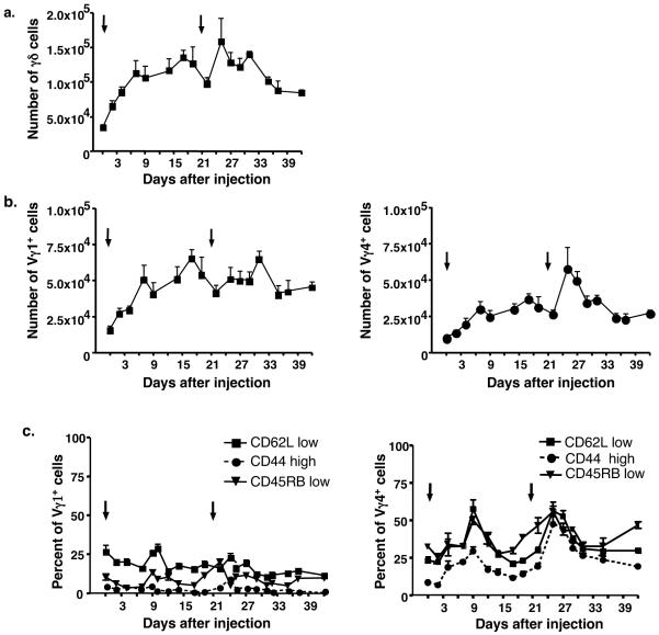
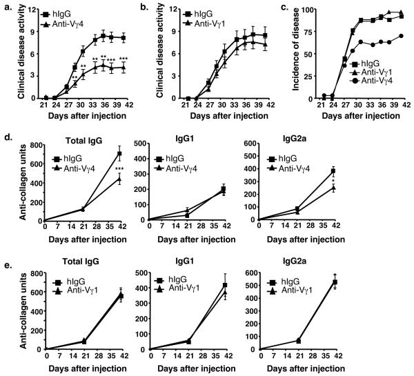
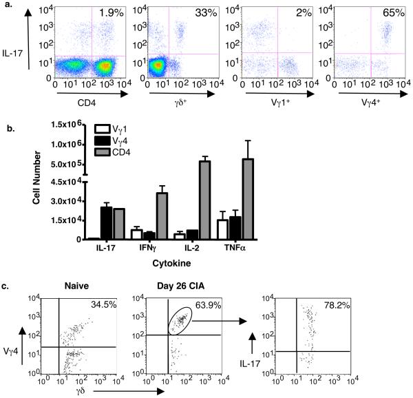
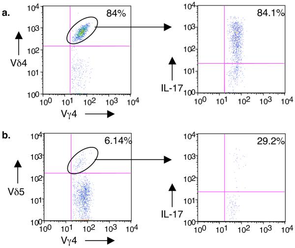
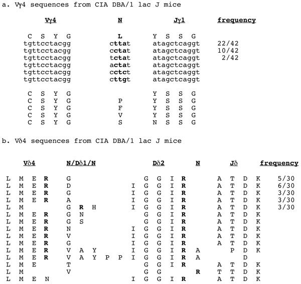
Murine gammadelta T cell subsets, defined by their Vgamma chain usage, have been shown in various disease models to have distinct functional roles. In this study, we examined the responses of the two main peripheral gammadelta T cell subsets, Vgamma1(+) and Vgamma4(+) cells, during collagen-induced arthritis (CIA), a mouse model that shares many hallmarks with human rheumatoid arthritis. We found that whereas both subsets increased in number, only the Vgamma4(+) cells became activated. Surprisingly, these Vgamma4(+) cells appeared to be Ag selected, based on preferential Vgamma4/Vdelta4 pairing and very limited TCR junctions. Furthermore, in both the draining lymph node and the joints, the vast majority of the Vgamma4/Vdelta4(+) cells produced IL-17, a cytokine that appears to be key in the development of CIA. In fact, the number of IL-17-producing Vgamma4(+) gammadelta T cells in the draining lymph nodes was found to be equivalent to the number of CD4(+)alphabeta(+) Th-17 cells. When mice were depleted of Vgamma4(+) cells, clinical disease scores were significantly reduced and the incidence of disease was lowered. A decrease in total IgG and IgG2a anti-collagen Abs was also seen. These results suggest that Vgamma4/Vdelta4(+) gammadelta T cells exacerbate CIA through their production of IL-17.


Figures

Figure 1
Figure 1
The total numbers of γδ T cells (a), Vγ1+ cells, and Vγ4+ cells (b) obtained from the lymph nodes of mice that had received collagen/CFA injections on days 0 and 21 (black arrows). On the indicated days following the initial injection, the draining lymph nodes (inguinal, brachial and popliteal) were removed and cells were stained for γδ T cell subsets. Using FACs analysis, the total number of γδ cells and individual subsets were calculated. Each time point represents the average + SEM for at least 8 different mice. (c) On designated days after collagen/CFA injections (black arrows), γδ T cells were isolated and stained for Vγ1 and Vγ4 expression and for levels of CD62L, CD44, or CD45RB. The mean percentage + SEM of cells having an “activated” phenotype (CD62L low, CD44 high, CD45RB low) is shown.
Figure 2
Figure 2
(a) Clinical disease activity in mice with collagen-induced arthritis. Mice were immunized with collagen/CFA on days 0 and 21. On day 17, mice were given either anti-Vγ4 antibody (black triangle, n=30) or hamster IgG (hIgG) (black square, n=26) intravenously. (b) In a separate experiment, mice were treated with anti-Vγ1 antibody (black triangle, n=30) or hamster IgG (black square, n=25). Clinical disease was assessed three times a week, starting on day 21. Values represent the mean ± SEM of 2 separate experiments (maximum score = 16). **p < 0.01, ***p<.001. Statistical significance was determined using the Mann-Whitney test. (c) Incidence of disease for each of the groups as described in (a & b). The incidence of disease for each hIgG treated group in 2 experiments was pooled and is shown as one group. (d) Anti-collagen antibody levels in mice with CIA that were depleted with an anti-Vγ4 antibody, as compared to hIgG-injected control animals. Mean ± SEM is shown for total IgG, IgG1, and IgG2a antibodies. Mice were treated on day 17 with hIgG (black square, n = 26) or a specific anti-Vγ subset antibody (black triangle, n = 30). *p < 0.05, ***p < 0.001. Data represent the mean ± SEM of 2 separate experiments. (e) Same as in (d) except mice were depleted with an anti-Vγ1 antibody and compared to hIgG. Again, mean ± SEM is shown for 2 separate experiments.
Figure 3
Figure 3
Intracellular cytokine staining of T cells from the draining lymph nodes on day 26. (a) The percentages of CD4+, γδ+, Vγ1+, or Vγ4+ T cells that can produce IL-17 are shown, first gating on cells that stained with CD3. The percentage of Vγ1+ and Vγ4+ cells was then visualized by next gating on cells that stained with a pan-γδ reactive mAb. (b) The total number of Vγ1+, Vγ4+, or CD4+ cells stimulated to produce IL-17, IFNγ, IL-2, or TNFα as determined by intracellular cytokine staining. The total number was calculated based on the percentage that stained in (a). (c) The percentage of Vγ4+ cells in the joints of naïve mice versus CIA mice on day 26 is depicted on the left. The percentage of γδ+/Vγ4+cells that can produce IL-17 is shown on the right.
Figure 4
Figure 4
Vδ usage by Vγ4+ cells in CIA animals. Lymph nodes were analyzed by flow cytometry as for Fig. 3. Cells were triple stained for γδ TCR, Vγ4, and either Vδ4 (a), Vδ5 (b), or Vδ6.3 (data not shown) and the percentage of each V delta subset determined. Then, each Vδ-defined subset (circled population) was examined intracellularly for IL-17 production. The majority of the IL-17-producing Vγ4+ cells co-expressed Vδ4. Vγ4/Vδ6.3+ cells represented less than 0.5% of the Vγ4+ population, and did not produce IL-17 (not shown).
Figure 5
Figure 5
Vγ4 and Vδ4 sequences from CIA-elicited γδ T cells. (a) In the CIA-elicited cells, 37/42 (88%) of the Vγ4+ clones encoded a leucine between the V and the J, and four of the six possible codons were used. In addition, when the codon “cta” was used to form leucine, we found different N/P nucleotides also flanking it. (b) The Vδ4 sequences from CIA-elicited cells revealed a striking length conservation (5-6 amino acids between V and J), a single Dδ2 reading frame, and the conservation of the two arginines, one at the end of the Vδ4 gene and one at the end of the Dδ2 gene. Both arginines were encoded by multiple codons as well.

References

    1. Luross JA, Williams NA. The genetic and immunopathological processes underlying collagen-induced arthritis. Immunology. 2001;103:407–16. - PMC - PubMed
    1. Gregersen PK, Silver J, Winchester RJ. The shared epitope hypothesis. An approach to understanding the molecular genetics of susceptibility to rheumatoid arthritis. Arthritis Rheum. 1987;30:1205–13. - PubMed
    1. Wooley PH, Whalen JD, Chapdelaine JM. Collagen-induced arthritis in mice. VI. Synovial cells from collagen arthritic mice activate autologous lymphocytes in vitro. Cell Immunol. 1989;124:227–38. - PubMed
    1. Andersson EC, et al. Definition of MHC and T cell receptor contacts in the HLA-DR4 restricted immunodominant epitope in type II collagen and characterization of collagen-induced arthritis in HLA-DR4 and human CD4 transgenic mice. Proc. Natl. Acad. Sci. U. S. A. 1998;95:7574–9. - PMC - PubMed
    1. Watson WC, Townes AS. Genetic susceptibility to murine collagen II autoimmune arthritis. Proposed relationship to the IgG2 autoantibody subclass response, complement C5, major histocompatibility complex (MHC) and non-MHC loci. J. Exp. Med. 1985;162:1878–91. - PMC - PubMed

Publication types

MeSH terms