. 2007 Oct 3;2(10):e984.
doi: 10.1371/journal.pone.0000984.

Design and pre-clinical evaluation of a universal HIV-1 vaccine

Affiliations

Design and pre-clinical evaluation of a universal HIV-1 vaccine

Sven Létourneau et al. PLoS One. .

Erratum in

  • PLoS One. 2011;6(3). doi: 10.1371/annotation/fca26a4f-42c1-4772-a19e-aa9d96c4eeb2

Abstract

Background: One of the big roadblocks in development of HIV-1/AIDS vaccines is the enormous diversity of HIV-1, which could limit the value of any HIV-1 vaccine candidate currently under test.

Methodology and findings: To address the HIV-1 variation, we designed a novel T cell immunogen, designated HIV(CONSV), by assembling the 14 most conserved regions of the HIV-1 proteome into one chimaeric protein. Each segment is a consensus sequence from one of the four major HIV-1 clades A, B, C and D, which alternate to ensure equal clade coverage. The gene coding for the HIV(CONSV) protein was inserted into the three most studied vaccine vectors, plasmid DNA, human adenovirus serotype 5 and modified vaccine virus Ankara (MVA), and induced HIV-1-specific T cell responses in mice. We also demonstrated that these conserved regions prime CD8(+) and CD4(+) T cell to highly conserved epitopes in humans and that these epitopes, although usually subdominant, generate memory T cells in patients during natural HIV-1 infection.

Significance: Therefore, this vaccine approach provides an attractive and testable alternative for overcoming the HIV-1 variability, while focusing T cell responses on regions of the virus that are less likely to mutate and escape. Furthermore, this approach has merit in the simplicity of design and delivery, requiring only a single immunogen to provide extensive coverage of global HIV-1 population diversity.


Conflict of interest statement

Competing Interests: The authors have declared that no competing interests exist.

Figures

Figure 1
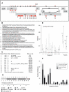
Figure 1. The HIVCONSV immunogen.
(A) Localization of the 14 most highly conserved regions of the HIV-1 proteome. The numbers written vertically under each fragment boundary indicate the first and last aa positions using the HXB2 reference strain numbering (http://www.hiv.lanl.gov/content/hiv-db/LOCATE/locate.html). (B) Predicted aa sequence of the the HIVCONSV immunogen with indicated fragment numbers. (C) Summary of the fragments including: the fragment number; the protein in which it was embedded; the clade of the consensus sequence selected for inclusion in the immunogen, alternating between clades A–D; additional clades that have identical HIVCONSV; and position numbers in the chimeric vaccine. The number of additional clades with identical consensus sequences to selected clade reflects the high level of conservation in these regions, and is encouraging in terms of the global potential of the vaccine. The consensus sequences compared were to the M group consensus, clades A–K, and three very common recombinant circulating forms CRF01 (common in Asia and Africa), CRF02 (common Africa), CRF08 (common in China) retrieved from the Los Alamos database 2004 consensus alignment (http://www.hiv.lanl.gov/content/hiv-db/CONSENSUS/M_GROUP/Consensus.html). (D) Schematic representation of the HIVCONSV immunogen (not drawn to scale) indicating clade anternation (above), overlapping peptide pool derivation and protein origin by colour coding. (E) Hamming distances between the HIVCONSV antigen fragments and the global circulating viral sequences. The full M group alignment, including recombinant sequences, was used for the comparison. The Los Alamos database alignment contains only one sequence person, and contains sequences from between 600 and 1000 individuals in these proteins. The Hamming distance range for 95% of the sequences relative to the vaccine immunogen is given by the vertical lines. The distances between the full length natural proteins were then calculated relative to HXB2 reference strain Env, Vif, Gag and Pol sequences for comparison. Distance measures are minimal estimates, as gaps inserted in regions where insertions and deletions occur were not counted. (F) Numbers of known CD8+ T cell epitopes (defined to within 12 aa or less in the Los Alamos HIV-1 database) in each of the 14 conserved protein fragments included in the HIVCONSV immunogen are shown. When more than one HLA class I presenting molecules can present the same HIV-1 epitope, then each is counted as a distinct epitope; if more than one sequence variant has been described as an epitope presented by the same class I molecule, then these are counted as a distinct epitopes; however, if an HLA serotype and genotype that are potentially the same are each described as presenting the same epitope (like A2 and A*0201) they are scored as a single epitope.
Figure 2
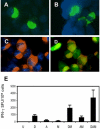
Figure 2. HIVCONSV protein expression in human cells and basic immunogenicity.
A histochemical and DAPI staining of 293T cells transiently transfected with pTH.HIVCONSV DNA (A), or infected with MVA.HIVCONSV (B) or AdHu5.HIVCONSV (C and D). HIVCONSV protein expression was detected using mAb tag Pk at the C-terminus of the immunogen and a primary anti-Pk mAb followed by secondary FITC- (A and B) or AlexaFluor584- (C and D) conjugated detection antibodies. The AdHu5.HIVCONSV vaccine also expressed GFP, which co-localized with the HIVCONSV expression (D). (E) BALB/c mice were immunized using the regimen indicated below, and the HIVCONSV-induced T cell responses were assessed in an ELISPOT assay using the H epitope. Results are shown as a mean±SD (n = 4). U–unimmunized; D–pTH.HIVCONSV DNA; A–AdHu5.HIVCONSV; and M–MVA.HIVCONSV. For doses and timing, see Methods.
Figure 3
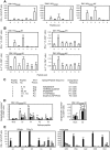
Figure 3. Breadth of HIVCONSV-induced T cell responses in BALB/c mice.
Mice were immunized using the regimen and immunogen indicated above (A, B and C) or below (D) the graphs and the HIVCONSV-specific responses were determined in ex vivo ELISPOT (A and E) or ICS (B and D) assays detecting the indicated cytokines and using for restimulation overlapping peptide pools schematically shown in Fig. 1D (A and B) or individual epitope peptides (D and E). (C) Identified peptides or epitope sequences and their origin, name and T cell reactivity. In (D): white–IFN-γ; black–IL-2; stripy-IFN-γ+IL-2; and grey–TNF-α; *-responses significantly above the no-peptide background (p<0.05). In (E): white–no peptide followed from left to right by epitopes H, G1, G2, P1, P2 and P3. Results are shown as a mean±SD (n = 4). For doses and timing, see Methods.
Figure 4
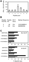
Figure 4. HIVCONSV-induced T cell responses in HLA-A*0201-transgenic mice, strain HHD.
(A) Mice were immunized using the DAM regimen and the vaccine-induced responses were detected in an ex vivo ELISPOT assay. Results are shown as a mean±SD (n = 4). For doses and timing, see Methods. (B) Identified epitope peptides and their origin. (C) Killing of murine EL4 A2-Kd (top) and human JK A2-Kd (bottom) target cells sensitized with the shown peptides in a 51Cr-release assay after a 5-day in vitro peptide re-stimulation. Black, grey and white bars indicated effector to target ratios of 100, 50 and 25 to 1, respectively.
Figure 5
Figure 5. Recognition of HIVCONSV-derived peptides by PBMC from HIV-1-infected patients.
The HIVCONSV-specific memory T cells were assessed in healthy and HIV-1-infected subjects using an IFN-γ ELISPOT assay after a 10-day peptide and cytokine culture. (A) Summed frequencies of HIVCONSV-specific cells detected in healthy (n = 9) and HIV-1-infected (n = 13) subjects. The bars show the group medians of 578 SFU/106 and 8,092 SFU/106 cells for the healthy and infected subjects, respectively. (B) In five subjects indicated below, cultured PBMC were left undepleted (grey) or depleted of CD8+ cells (black) prior to the ELISPOT assay. The difference between undepleted (median = 8,092 SFU/106 cells) and CD8-depleted samples (median = 550 SFU/106 cells) was statistically significant (p = 0.0313). (C) Responses to individual HIVCONSV-derived peptide pools as shown in Fig. 1D determined for the HIV-1-infected (grey) and healthy (black) subjects shown as medians. (D) Responses to individual peptides pools for each HIV-1-infected patient indicated below. Bars show a mean±SD of three assay wells and ‘*’ indicates a positive response according to criteria set in Methods. Due to sample shortage, subject 021 was not tested.

References

    1. Burton DR, Desrosiers RC, Doms RW, Koff WC, Kwong PD, et al. HIV vaccine design and the neutralizing antibody problem. Nat Immunol. 2004;5:233–236. - PubMed
    1. McMichael AJ. HIV vaccines. Annu Rev Immunol. 2006;24:227–255. - PubMed
    1. Duerr A, Wasserheit JN, Corey L. HIV vaccines: new frontiers in vaccine development. Clin Infect Dis. 2006;43:500–511. - PubMed
    1. Hanke T, McMichael AJ, Dorrell L. Clinical experience with plasmid DNA- and modified vaccinia vaccine Ankara (MVA)-vectored HIV-1 clade A vaccine inducing T cells. J Gen Virol. 2007;88:1–12. - PubMed
    1. Gaschen B, Taylor J, Yusim K, Foley B, Gao F, et al. Diversity considerations in HIV-1 vaccine selection. Science. 2002;296:2354–2360. - PubMed

Publication types