Deletion of PKBalpha/Akt1 affects thymic development
- PMID: 17912369
- PMCID: PMC1991598
- DOI: 10.1371/journal.pone.0000992
Deletion of PKBalpha/Akt1 affects thymic development
Abstract
Background: The thymus constitutes the primary lymphoid organ for the majority of T cells. The phosphatidyl-inositol 3 kinase (PI3K) signaling pathway is involved in lymphoid development. Defects in single components of this pathway prevent thymocytes from progressing beyond early T cell developmental stages. Protein kinase B (PKB) is the main effector of the PI3K pathway.
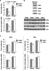
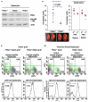
Methodology/principal findings: To determine whether PKB mediates PI3K signaling in the thymus, we characterized PKB knockout thymi. Our results reveal a significant thymic hypocellularity in PKBalpha(-/-) neonates and an accumulation of early thymocyte subsets in PKBalpha(-/-) adult mice. Using thymic grafting and fetal liver cell transfer experiments, the latter finding was specifically attributed to the lack of PKBalpha within the lymphoid component of the thymus. Microarray analyses show that the absence of PKBalpha in early thymocyte subsets modifies the expression of genes known to be involved in pre-TCR signaling, in T cell activation, and in the transduction of interferon-mediated signals.
Conclusions/significance: This report highlights the specific requirements of PKBalpha for thymic development and opens up new prospects as to the mechanism downstream of PKBalpha in early thymocytes.
Conflict of interest statement
Figures


Similar articles
-
Con A activates an Akt/PKB dependent survival mechanism to modulate TCR induced cell death in double positive thymocytes.Mol Immunol. 2003 Jun;39(16):1013-23. doi: 10.1016/s0161-5890(03)00044-0. Mol Immunol. 2003. PMID: 12749908
-
Unequal contribution of Akt isoforms in the double-negative to double-positive thymocyte transition.J Immunol. 2007 May 1;178(9):5443-53. doi: 10.4049/jimmunol.178.9.5443. J Immunol. 2007. PMID: 17442925
-
Constitutively active protein kinase B enhances Lck and Erk activities and influences thymocyte selection and activation.J Immunol. 2003 Aug 1;171(3):1285-96. doi: 10.4049/jimmunol.171.3.1285. J Immunol. 2003. PMID: 12874217
-
Phosphatidylinositol 3-kinase signaling in thymocytes: the need for stringent control.Sci Signal. 2010 Aug 17;3(135):re5. doi: 10.1126/scisignal.3135re5. Sci Signal. 2010. PMID: 20716765 Review.
-
Thymic function in the regulation of T cells, and molecular mechanisms underlying the modulation of cytokines and stress signaling (Review).Mol Med Rep. 2017 Nov;16(5):7175-7184. doi: 10.3892/mmr.2017.7525. Epub 2017 Sep 19. Mol Med Rep. 2017. PMID: 28944829 Free PMC article. Review.
Cited by
-
Thymic development beyond beta-selection requires phosphatidylinositol 3-kinase activation by CXCR4.J Exp Med. 2010 Jan 18;207(1):247-61. doi: 10.1084/jem.20091430. Epub 2009 Dec 28. J Exp Med. 2010. PMID: 20038597 Free PMC article.
-
PI3Ks in lymphocyte signaling and development.Curr Top Microbiol Immunol. 2010;346:57-85. doi: 10.1007/82_2010_45. Curr Top Microbiol Immunol. 2010. PMID: 20563708 Free PMC article. Review.
-
Protein kinase D regulates positive selection of CD4+ thymocytes through phosphorylation of SHP-1.Nat Commun. 2016 Sep 27;7:12756. doi: 10.1038/ncomms12756. Nat Commun. 2016. PMID: 27670070 Free PMC article.
-
AKT1 and AKT2 maintain hematopoietic stem cell function by regulating reactive oxygen species.Blood. 2010 May 20;115(20):4030-8. doi: 10.1182/blood-2009-09-241000. Epub 2010 Mar 30. Blood. 2010. PMID: 20354168 Free PMC article.
-
Phosphoinositide-dependent kinase 1 controls migration and malignant transformation but not cell growth and proliferation in PTEN-null lymphocytes.J Exp Med. 2009 Oct 26;206(11):2441-54. doi: 10.1084/jem.20090219. Epub 2009 Oct 5. J Exp Med. 2009. PMID: 19808258 Free PMC article.
References
-
- Gill J, Malin M, Sutherland J, Gray D, Hollander G, et al. Thymic generation and regeneration. Immunol Rev. 2003;195:28–50. - PubMed
-
- Petrie HT, Zuniga-Pflucker JC. Zoned out: functional mapping of stromal signaling microenvironments in the thymus. Annu Rev Immunol. 2007;25:649–679. - PubMed
-
- Rothenberg EV, Taghon T. Molecular genetics of T cell development. Annu Rev Immunol. 2005;23:601–649. - PubMed
-
- Wilson A, MacDonald HR. Expression of genes encoding the pre-TCR and CD3 complex during thymus development. Int Immunol. 1995;7:1659–1664. - PubMed
-
- Wagner DH., Jr Re-shaping the T cell repertoire: TCR editing and TCR revision for good and for bad. Clin Immunol. 2007;123:1–6. - PubMed
Publication types
MeSH terms
Substances
Grants and funding
LinkOut - more resources
Full Text Sources
Molecular Biology Databases
Miscellaneous

