. 2007 Oct 3;2(10):e993.
doi: 10.1371/journal.pone.0000993.

Structure and reaction mechanism of basil eugenol synthase

Affiliations

Structure and reaction mechanism of basil eugenol synthase

Gordon V Louie et al. PLoS One. .

Abstract

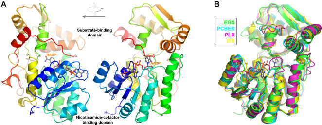
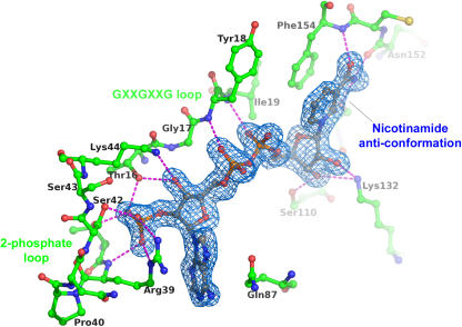
Phenylpropenes, a large group of plant volatile compounds that serve in multiple roles in defense and pollinator attraction, contain a propenyl side chain. Eugenol synthase (EGS) catalyzes the reductive displacement of acetate from the propenyl side chain of the substrate coniferyl acetate to produce the allyl-phenylpropene eugenol. We report here the structure determination of EGS from basil (Ocimum basilicum) by protein x-ray crystallography. EGS is structurally related to the short-chain dehydrogenase/reductases (SDRs), and in particular, enzymes in the isoflavone-reductase-like subfamily. The structure of a ternary complex of EGS bound to the cofactor NADP(H) and a mixed competitive inhibitor EMDF ((7S,8S)-ethyl (7,8-methylene)-dihydroferulate) provides a detailed view of the binding interactions within the EGS active site and a starting point for mutagenic examination of the unusual reductive mechanism of EGS. The key interactions between EMDF and the EGS-holoenzyme include stacking of the phenyl ring of EMDF against the cofactor's nicotinamide ring and a water-mediated hydrogen-bonding interaction between the EMDF 4-hydroxy group and the side-chain amino moiety of a conserved lysine residue, Lys132. The C4 carbon of nicotinamide resides immediately adjacent to the site of hydride addition, the C7 carbon of cinnamyl acetate substrates. The inhibitor-bound EGS structure suggests a two-step reaction mechanism involving the formation of a quinone-methide prior to reduction. The formation of this intermediate is promoted by a hydrogen-bonding network that favors deprotonation of the substrate's 4-hydroxyl group and disfavors binding of the acetate moiety, akin to a push-pull catalytic mechanism. Notably, the catalytic involvement in EGS of the conserved Lys132 in preparing the phenolic substrate for quinone methide formation through the proton-relay network appears to be an adaptation of the analogous role in hydrogen bonding played by the equivalent lysine residue in other enzymes of the SDR family.


Conflict of interest statement

Competing Interests: The authors have declared that no competing interests exist.

Figures

Figure 1
Figure 1. Structure and biosynthesis of the phenylpropenes.
(A) Phenylpropene diversity. The basic skeleton of an allyl phenylpropene is shown, along with the numbering of the carbon atoms used in this manuscript. The iso phenylpropenes differ in the position of the double bond (C7 = C8) in the propene side group. For eugenol (an allyl phenylpropene), the phenyl-ring substituents are 2 = H, 3 = OCH3, 4 = OH, and 5 = H. The possible methylenedioxy-bridge modifications are also shown. (B) Simplified reaction scheme for the biosynthesis of eugenol from L-phenylalanine and the monolignol alcohol, coniferyl alcohol (CFAT: coniferyl alcohol acetyl transferase; EGS: eugenol synthase).
Figure 2
Figure 2. Structure of EGS and comparison with other PIP-family enzymes.
(A) Orthogonal views of EGS/NADP+ monomer. The polypeptide chain of EGS is represented as a ribbon, with coloring varying from blue for the N-terminus to red for the C-terminus. The atoms of the NADP+ cofactor are drawn as balls and sticks, and are colored coded according to element (carbon: gray; nitrogen: blue; oxygen: red; phosphorus: orange). (B) Superposition of the polypeptide-chain backbones of EGS and other PIP-family enzymes (color coding as shown in inset). The NADP+ cofactor of EGS is also shown (the structures of the other PIP-family enzymes were determined in the absence of cofactor).
Figure 3
Figure 3. Interactions between EGS and the NADP+ cofactor.
Only the EGS polypeptide-chain segments that form direct interactions with the NADP+ cofactor are shown. Hydrogen-bond interactions formed by the cofactor are represented as magenta dashed lines. Atom coloring is the same as in Figure 2A, except that the carbon atoms of the polypeptide-chain segments are green. The blue-colored contours envelope regions greater than 3σ in the NADP-omit electron-density map.
Figure 4
Figure 4. Structural comparison of EGS and UDP-galactose epimerase.
(A) Superposition of polypeptide-chain backbones of EGS and UDP-galactose epimerase (color coding as shown in inset). For clarity, only the NADP+ cofactor of EGS is shown. (B) Comparison of NAD(P)-cofactor conformation and substrate-analog binding in EGS and UDP-galactose epimerase. The binding of the EGS competitive inhibitor (7S,8S)-ethyl (7,8-methylene)-dihydroferulate (EMDF) is described in detail in the text and Figure 5. The coloring of the polypeptide-chain segments is the same as in (A). The inset shows the coloring used for the carbon atoms of the nicotinamide cofactors, EMDF bound to EGS, and UDP-glucose bound to UDP-galactose-4-epimerase.
Figure 5
Figure 5. Binding of the competitive inhibitor EMDF by EGS.
(A) Orthogonal views of the 7S,8S- stereoisomer of the competitive inhibitor EMDF bound to EGS. Hydrogen-bond interactions formed by the EMDF molecule (cyan colored carbons) are represented as magenta dashed lines. The blue-colored contours envelope regions greater than 2.5σ in the initial Fobs-Fcalc electron-density map. The direction of view used in the right panel (approximately perpendicular to the plane of the nicotinamide ring) is maintained roughly in figures 5B–D and 6B. The chemical structure of EMDF is shown in the inset. (B) Modeled binding of coniferyl acetate to EGS. The atom coloring is the same as in (A), with magenta carbon atoms for the coniferyl acetate. The chemical structures of coniferyl acetate and EMDF are compared in the inset. The close interaction between the EMDF C7-atom and the hydride donor of the nicotinamide (C4) is shown as a yellow dashed line. (C) Binding of EMDF to the Lys132Gln variant of EGS. Hydrogen-bond interactions formed by the EMDF molecule (cyan colored carbons) are represented as magenta dashed lines. Hydrogen bonds involving the side chain of Gln132 are shown as orange dashed lines. The blue-colored contours envelope regions greater than 2.0σ in the initial Fobs-Fcalc electron-density map. (D) Binding of EMDF to the Lys132Arg variant of EGS (stereo representation). The blue-colored contours envelope regions greater than 2σ in the initial Fobs-Fcalc electron-density map for the EGS-Arg132/EMDF complex (green). The altered positioning of the Arg132 side-chain and neighboring residues (most notably Phe85, Ile88, and Ile129) and the disordering of the C-terminal tail (residues 310–314) are apparent with respect to the holo-EGS-Arg132 structure (magenta). For comparison, the position of the wild-type Lys132 side chain and the key bridging water molecule shown in Figure 5A are also shown (yellow).
Figure 6
Figure 6. Hydrogen-bonding interactions in the EGS active-site and proposed reaction mechanism of EGS.
(A) Hydrogen-bonding network involving the Lys132 side-chain amino group, the 4-hydroxyl group of EMDF, and the bridging water molecule. Inferred hydrogen-atom positions are shown in blue. Hydrogen-bond interactions are represented as magenta dashed lines. (B) Comparison of the hydrogen-bond interactions made by the catalytic lysine residue in EGS (Lys132) and the SDR UDP-galactose-4-epimerase (Lys153). The inset shows the coloring used for the carbon atoms of EMDF bound to EGS, and UDP-glucose bound to UDP-galactose-4-epimerase. Hydrogen bonds are drawn as thin dashed lines. Water molecules are drawn as red spheres. Those outlined in the pale red form part of a postulated proton-relay network in UDP-galactose-4-epimerase, whereas the presumed catalytic water molecule in EGS is outlined in green and marked with an asterisk. (C) Proposed reaction mechanism of EGS (and IGS) involving a quinone-methide intermediate. The catalytic base (B:) that promotes deprotonation of the 4-hydroxyl group of the substrate is the hydroxide ion that is activated by the side-chain amino group of Lys132. The loss of acetate generates the quinone-methide intermediate. The attack at C7 of this intermediate by the NADPH-derived hydride yields the allyl phenylpropene eugenol, whereas hydride attack at C9 presumably yields the iso-phenylpropene isoeugenol.
Figure 7
Figure 7. Chemoenzymatic synthesis of coniferyl acetate.
Coniferyl acetate was obtained as a colorless oil with a yield of nearly 100%.
Figure 8
Figure 8. Chemical synthesis of (7S,8S)-ethyl (7,8-methylene)-dihydroferulate (EMDF).
EMDF was obtained with an overall yield of 10%.
Figure 9
Figure 9. Double reciprocal plot for EGS activity in the presence and absence of EMDF.
The plots illustrate the mixed nature of the EMDF EGS inhibitor. Ki was estimated from nonlinear fitting to a modified Michaelis-Menten equation.

References

    1. Gang DR, Wang J, Dudareva N, Nam KH, Simon JE, et al. An investigation of the storage and biosynthesis of phenylpropenes in sweet basil. Plant Physiol. 2001;125:539–555. - PMC - PubMed
    1. Pichersky E, Noel JP, Dudareva N. Biosynthesis of plant volatiles: nature's diversity and ingenuity. Science. 2006;311:808–811. - PMC - PubMed
    1. Dexter R, Qualley A, Kish CM, Ma CJ, Koeduka T, et al. Characterization of a petunia acetyltransferase involved in the biosynthesis of the floral volatile isoeugenol. Plant J. 2007;49:265–275. - PubMed
    1. Koeduka T, Fridman E, Gang DR, Vassao DG, Jackson BL, et al. Eugenol and isoeugenol, characteristic aromatic constituents of spices, are biosynthesized via reduction of a coniferyl alcohol ester. Proc Natl Acad Sci. 2006;103:10128–10133. - PMC - PubMed
    1. Vassao DG, Gang DR, Koeduka T, Jackson B, Pichersky E, et al. Chavicol formation in sweet basil (Ocimum basilicum): cleavage of an esterified C9 hydroxyl group with NAD(P)H-dependent reduction. Org Biomol Chem. 2006;4:2733–2744. - PubMed

Publication types

LinkOut - more resources