. 2008 Aug;28(5):769-79.
doi: 10.1007/s10571-007-9219-0. Epub 2007 Oct 3.

The neuroprotective potential of phase II enzyme inducer on motor neuron survival in traumatic spinal cord injury in vitro

Affiliations

The neuroprotective potential of phase II enzyme inducer on motor neuron survival in traumatic spinal cord injury in vitro

Xiao-Yun Liu et al. Cell Mol Neurobiol. 2008 Aug.

Abstract

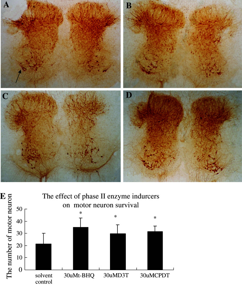
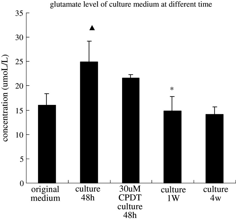
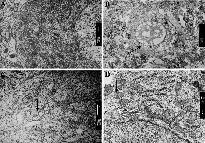
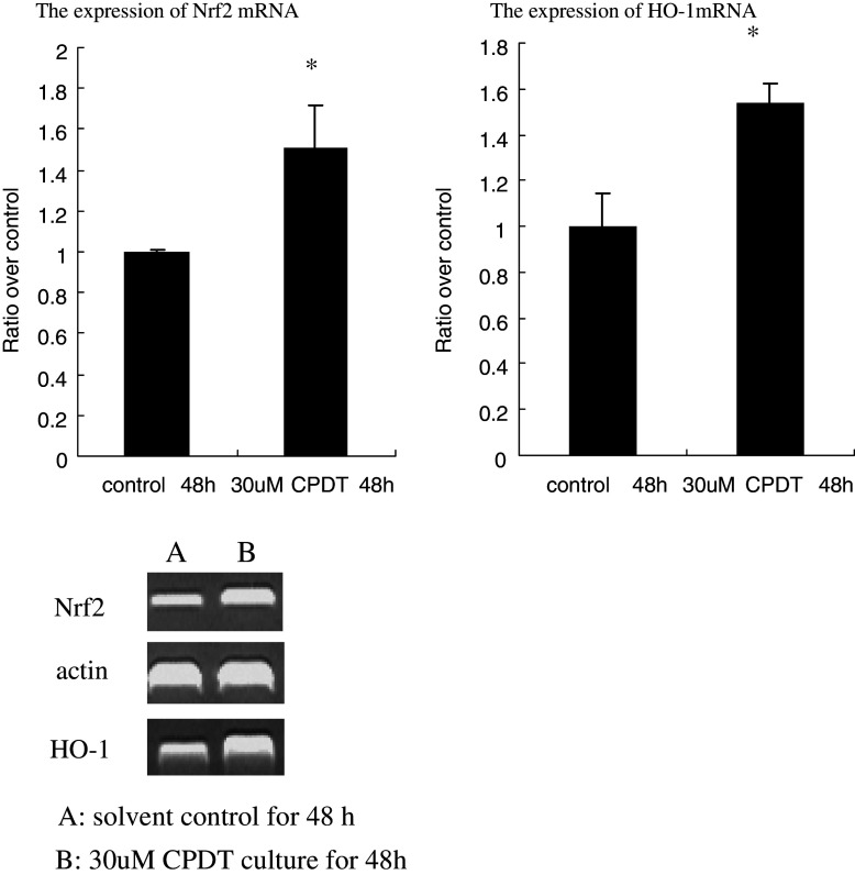
(1) Phase II enzyme inducer is a kind of compound which can promote the expression of antioxidative enzymes through nuclear factor erythroid 2-related factor 2 (Nrf2) activation. Recently, it has been reported that these compounds show neuroprotective effect via combating oxidative stress. The purpose of this study is to determine whether phase II enzyme inducers have neuroprotective effects on traumatic spinal cord injury. (2) An organotypic spinal cord culture system was used, Phase II enzyme inducers were added to culture medium for 1 week, motor neurons were counted by SMI-32 staining, glutamate, Nrf2, and Heme oxygenase-1(HO-1) mRNA were tested. (3) This study showed motor neuron loss within 1 week in culture. After 1 week in culture, the system was stable. Moreover, Glutamate was increased when in culture 48 h and decreased after 1 week in culture. There was no significant change between 1 and 4 weeks in culture. Necrotic motor neuron and damaged mitochondrial were observed in culture 48 h. Furthermore, phase II enzyme inducers: tert-butyhydroquinone (t-BHQ), 3H-1,2-dithiole-3-thione (D3T), and 5,6-dihydrocyclopenta-1,2-dithiole-3-thione (CPDT) were shown to promote motor neuron survival after dissection, it was due to increasing Nrf2 and HO-1 mRNA expression and protecting mitochondrial not due to decreasing glutamate level. (4) The loss of motor neuron due to dissection can mimic severe traumatic spinal cord injury. These results demonstrate that glutamate excitotoxicity and the damage of mitochondrial is possibly involve in motor neuron death after traumatic spinal cord injury and phase II enzyme inducers show neuroprotective potential on motor neuron survival in traumatic spinal cord injury in vitro.


Figures

Fig. 1
Fig. 1
Motor neuron death after dissection in an organotypic spinal cord slice culture. Lumber spinal cord explants (350-μm sections) were prepared from 7-day old rats, harvested at culture for 48 h, 1 week, 4 weeks. Motor neurons were immnostained with SMI-32 (an anti-neurofilament monoclonal antibody). (A) an image of SMI-32-stained slices when culture for 48 h. (B) the enlargement of ventral horn of A, arrow points to one of the motor neurons. (C) an image of SMI-32-stained slices when culture for 1 week, arrow points to one of the motor neurons. (D) an image of SMI-32-stained slices when culture for 4 weeks. (E) average number of motor neurons per slice (mean ± SD, n = 10–15) in each group. Significantly different marked by an asterisk (compared with culture 48 h group < 0.01)
Fig. 2
Fig. 2
Neuroprotective effect on motor neurons by CPDT, D3T, t-BHQ against traumatic spinal cord injury. Lumber spinal cord explants (350-μm sections) were prepared from 7-day old rats and were treated with CPDT, D3T, t-BHQ at 30μM as well as solvent control for 1 week. The media and the test compounds were changed biweekly. At the end of the treatment, the explants were harvested and motor neurons were immnostained with SMI-32. (A) solvent control (B) add 30 μM CPDT to culture medium for one week (C) add 30 μM D3T to culture medium for one week (D) add 30 μM t-BHQ to culture medium for one week, (E) average number of motor neurons per explant (mean ± SD, n = 10–15) in each treatment group. Significant different is marked by an asterisk (compared with solvent control < 0.05)
Fig. 3
Fig. 3
The effect of CPDT on extracellular glutamate levels. The media were changed biweekly along with the test compounds. The media, include culture for 48 h, add 30 μM CPDT to medium for 48 h, culture for 1 W, and culture for 4 W, were harvested 48 h after last medium change. Glutamate concentrations in the media were measured using a biochemical assay kit, based on the glutamic dehydrogenase-catalyzed conversion of glutamate to α-ketoglutarate with concomitant conversion of NAD+ to NADH. Each value is a mean ± SD (n = 6). Glutamate concentration in the original medium was 16 μM. (▴ Compared with original medium, < 0.01. *Compared with culture 48 h, < 0.01)
Fig. 4
Fig. 4
Ultrastructural observation of motor neuron. (A) normal nuclei and cytoplasma organella of the ventral motor neuron in 7-day-old rat (B) a necrotic motor neuron in 48 h culture (arrow). (C) mitochondrial damage (arrow) was observed in 48 h culture. (D) mitochondrial protection (arrow) of CPDT in 48 h culture
Fig. 5
Fig. 5
Activation of Nrf2 and Nrf2 target genes by CPDT in rat spinal cord explants. Lumber spinal cord explants (350-μm sections) were prepared from 7-day old rats and were treated with CPDT at 30 μM as well as solvent control for 48 h. RNA were extracted from the explants (a pool of 30 slices for each sample) and examined for levels of Nrf2, HO-1 mRNA by RT-PCR. β-actin was used as a control. The results are representative of triplicate experiments. Significant different is marked by an asterisk (compared with solvent control, < 0.05)

References

    1. Baptiste DC, Fehlings MG (2006) Pharmacological approaches to repair the injured spinal cord. J Neurotrauma 23(3–4):318–334 - PubMed
    1. Banik NL, Shields DC, Ray S, Davis B, Matzelle D, Wilford G, Hogan EL (1998) Role of calpain in spinal cord injury: effects of calpain and free radical inhibitors. Ann N Y Acad Sci 844:131–137 - PubMed
    1. Cao Z, Hallur S, Qiu HZ, Peng X, Li Y (2004) Induction of endogenous glutathione by the chemoprotective agent, 3H-1, 2-dithiole-3-thione, in human neuroblastoma SH-SY5Y cells affords protection against peroxynitrite-induced cytotoxicity. Biochem Biophys Res Commun 316(4):1043–1049 - PubMed
    1. Dykens JA, Stern A, Trenkner E (1987) Mechanism of kainate toxicity to cerebellar neurons in vitro is analogous to reperfusion tissue injury. J Neurochem 49(4):1222–1228 - PubMed
    1. Fahey JW, Talalay P (1999) Antioxidant functions of sulforaphane:a potent inducer of phase II detoxication enzymes. Food Chem Toxicol 37:973–979 - PubMed

Publication types

MeSH terms