. 2007 Dec 1;505(4):352-62.
doi: 10.1002/cne.21485.

Chemical and morphological alterations of spines within the hippocampus and entorhinal cortex precede the onset of Alzheimer's disease pathology in double knock-in mice

Affiliations

Chemical and morphological alterations of spines within the hippocampus and entorhinal cortex precede the onset of Alzheimer's disease pathology in double knock-in mice

Chiye Aoki et al. J Comp Neurol. .

Abstract

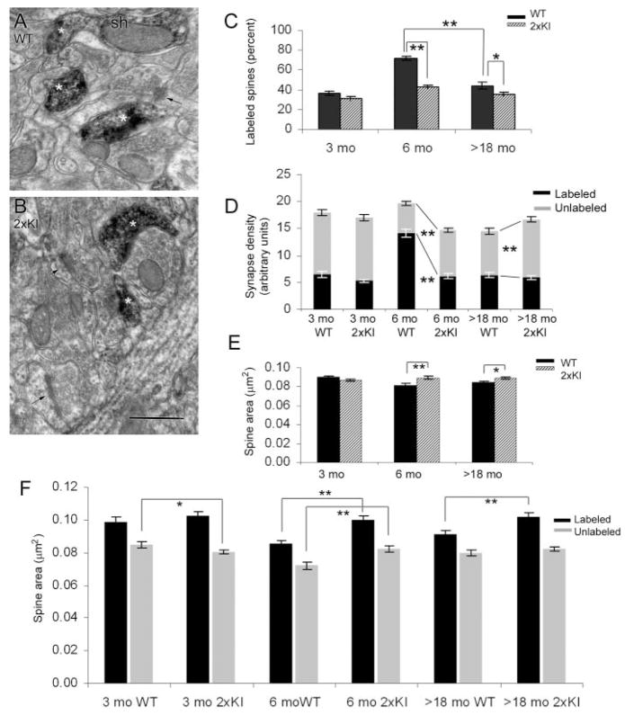
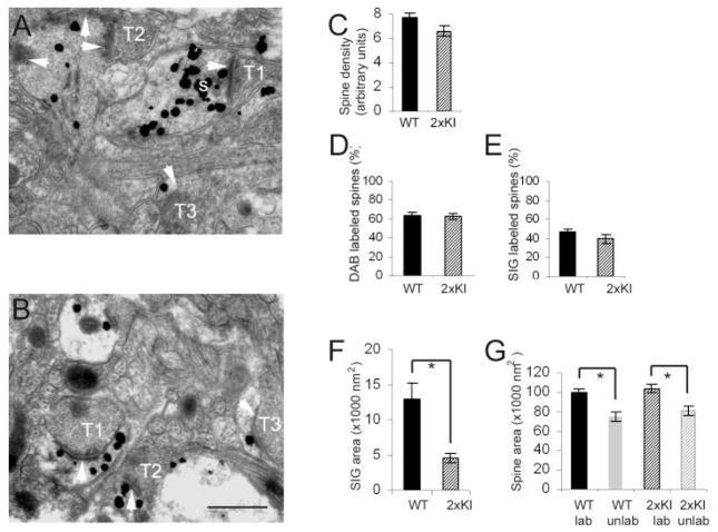
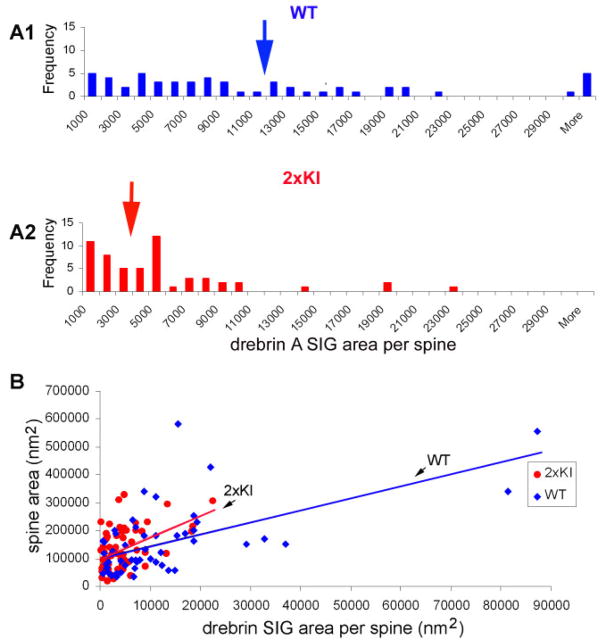
Mice with knock-in of two mutations that affect beta amyloid processing and levels (2xKI) exhibit impaired spatial memory by 9-12 months of age, together with synaptic plasticity dysfunction in the hippocampus. The goal of this study was to identify changes in the molecular and structural characteristics of synapses that precede and thus could exert constraints upon cellular mechanisms underlying synaptic plasticity. Drebrin A is one protein reported to modulate spine sizes and trafficking of proteins to and from excitatory synapses. Thus, we examined levels of drebrin A within postsynaptic spines in the hippocampus and entorhinal cortex. Our electron microscopic immunocytochemical analyses reveal that, by 6 months, the proportion of hippocampal spines containing drebrin A is reduced and this change is accompanied by an increase in the mean size of spines and decreased density of spines. In the entorhinal cortex of 2xKI brains, we detected no decrement in the proportion of spines labeled for drebrin A and no significant change in spine density at 6 months, but rather a highly significant reduction in the level of drebrin A immunoreactivity within each spine. These changes are unlike those observed for the somatosensory cortex of 2xKI mice, in which synapse density and drebrin A immunoreactivity levels remain unchanged at 6 months and older. These results indicate that brains of 2xKI mice, like those of humans, exhibit regional differences of vulnerability, with the hippocampus exhibiting the first signatures of structural changes that, in turn, may underlie the emergent inability to update spatial memory in later months.


Figures

Fig. 1
Fig. 1
Dendritic spines of the hippocampus immunolabeled for drebrin A. A,B: Examples of digitally captured electron micrographs from the stratum radiatum of the CA1 of a WT and a 2xKI mouse, respectively, at the age of 6 months. The tissues were processed to immunolabel for drebrin A, by using HRP-DAB as the electron-dense tag. Asterisks indicate examples of spines that are immunolabeled for drebrin A, and arrows point to unlabeled spines. Sh, example of a dendritic shaft that is contiguous with the labeled spine and is also immunoreactive. C: The proportion of synapses labeled for drebrin A is markedly different at 6 months. The synapses encountered from each age-genotype were divided into multiple groups (at least 60), each consisting of 25 synapses. For each group, the proportion of synapses immunolabeled for drebrin was determined. The graph shows the mean and SEM values of the percent labeled. **, P < 0.0001; *, P < 0.001 (two-way ANOVA). D: Spine density difference across genotypes is most prevalent at 6 months. The same sets of synapses analyzed in C were subjected to synapse density measurements. The number of labeled and unlabeled synapses appearing in each micrograph equal to 29 μm2 (arbitrary unit) was determined. The graph represents the mean and SEM values obtained per age-genotype (minimum of 60 micrographs). The dark bars represent the density of labeled synapses per unit area, and the light bars represent the unlabeled synapses per unit area. **, P < 0.0001. E: The areas of spines differ the most at 6 months. The same sets of synapses analyzed for data shown in C and D were subjected to spine profile area comparisons across genotypes. The solid bars represent the mean values of spines in the WT brains, and the striped bars represent the mean values of spine sizes in the 2xKI brains. **, P < 0.0001; *, P < 0.05. F: Both the labeled and unlabeled spines are larger in the hippocampus of 2xKI mice at 6 months. The same sets of synapses analyzed in C–E were first categorized as labeled or unlabeled spines and then subjected to area comparisons across genotypes. Both the labeled and unlabeled spines exhibit size differences at 6 months. (P < 0.0001). At >18 months, only the drebrin A-labeled spines are larger in the hippocampus of 2xKI mice (P < 0.0001). At 3 months, the unlabeled spines of 2xKI hippocampi are slightly smaller (P < 0.05). The numbers of observations were 1,078 and 2,042 for 3-month WT and 2xKI mice, respectively; 1,177 and 879 for 6-month WT and 2xKI; and 1,306 and 1,507 for >18-month WT and 2xKI mice. For abbreviations, see list. Scale bar = 500 nm in B (applies to A,B).
Fig. 2
Fig. 2
At 6 months, spines of the WT entorhinal cortex contain greater amounts of drebrin A immunoreactivity compared with 2xKI spines. A,B: Examples of spine labeling in layer 1 of the entorhinal cortex of a WT and a 2xKI animal, respectively. The large maximally electron-dense particles, such as the one tagged with a white letter s in A, represent silver-intensified gold (SIG) immunolabeling for drebrin A. White arrows point to the postsynaptic densities (PSD). T1, T2, and T3 are presynaptic axon terminals. C: Mean and SEM values of the number of postsynaptic spines per unit area (spine density). The samples were taken from Vibratome sections that were semi-adjacent to the ones shown in A and B. These tissues were not immunolabeled for drebrin A and were postfixed by using the osmium procedure. Altogether, 514 spines from WT tissue and 484 spines from 2xKI tissue were encountered within fields of equal total area (30 micrographs × 29 μm2 per micrograph shot at a magnification of 25,000× = 870 μm2) for each animal. We detected no difference across the genotypes (t = 1.09, P >0.05). D: Mean and SEM values of the proportion of spines encountered from tissue that underwent drebrin A immunolabeling by the HRP-DAB procedure. For each animal, a minimum of 30 micrographs at a magnification of 25,000× were captured to sample 15–17 groups of 25 spines (375 spines per animal), for assessing the proportion of spines immunolabeled for drebrin A. We detected no difference across the genotypes (t = 0.21, P >0.05). E–G: Mean and SEM values of three types of measurements made from Vibratome sections that underwent the SIG immunolabeling procedure to detect drebrin A. E: Proportion of postsynaptic spines with drebrin A immunoreactivity. Sixteen groups of 10 synapses were sampled from the WT tissue, and 17 groups of 10 synapses were sampled from the 2xKI tissue. F: Mean and SEM values of area occupied by SIG particles within each labeled spine encountered. G: Cytoplasmic area captured within individual spine profiles within single 2D images that were labeled and unlabeled. Asterisks indicate statistical significance (P < 0.001) by Student’s t-test in F and two-way ANOVA in G. For abbreviations, see list. Scale bar = 500 nm in B (applies to A,B).
Fig. 3
Fig. 3
Spines with low levels of drebrin A immunoreactivity are more numerous in the entorhinal cortex of 2xKI brains. SIG immunolabeling for drebrin A indicates that the levels of drebrin A can vary by more than 40-fold. A1,A2: Outcome of a frequency analysis performed to compare the relative number of spines containing varying levels of drebrin A immunoreactivity. Levels of drebrin A within spines was quantified by measuring the area occupied by SIG particles, using Image J software. This analysis indicates that, in the 2xKI entorhinal cortex, spines containing low levels of drebrin A immunoreactivity are more numerous than those containing high levels of drebrin A immunoreactivity. In contrast, the entorhinal cortices of WT brains contain a more even distribution of spines across the range of drebrin A immunoreactivity. B: Drebrin A immunoreactivity correlates weakly with dendritic spine size (r = 0.524887 for WT, 0.407118 for 2xKI, P < 0.05). [Color figure can be viewed in the online issue, which is available at www.interscience.wiley.com.]

Similar articles

Cited by

References

    1. Aoki C, Fujisawa S, Mahadomrongkul V, Shah PJ, Nader K, Erisir A. NMDA receptor blockade in intact adult cortex increases trafficking of NR2A subunits into spines, postsynaptic densities, and axon terminals. Brain Res. 2003;963:139–149. - PubMed
    1. Aoki C, Sekino Y, Hanamura K, Fujisawa S, Mahadomrongkul V, Ren Y, Shirao T. Drebrin A is a postsynaptic protein that localizes in vivo to the submembranous surface of dendritic sites forming excitatory synapses. J Comp Neurol. 2005;483:383–402. - PubMed
    1. Braak H, Braak E. Neuropathological stageing of Alzheimer-related changes. Acta Neuropathol (Berl) 1991;82:239–259. - PubMed
    1. Calabrese B, Shaked GM, Tabarean IV, Braga J, Koo EH, Halpain S. Rapid, concurrent alterations in pre- and postsynaptic structure induced by naturally-secreted amyloid-beta protein. Mol Cell Neurosci. 2007;35:183–193. - PMC - PubMed
    1. Calon F, Lim GP, Yang F, Morihara T, Teter B, Ubeda O, Rostaing P, Triller A, Salem N, Jr, Ashe KH, Frautschy SA, Cole GM. Docosahexaenoic acid protects from dendritic pathology in an Alzheimer’s disease mouse model. Neuron. 2004;43:633–645. - PMC - PubMed

Publication types