

Femtosecond laser and microkeratome corneal flaps: comparison of stromal wound healing and inflammation
- PMID: 17912936
- PMCID: PMC2698458
- DOI: 10.3928/1081-597X-20070901-05
Femtosecond laser and microkeratome corneal flaps: comparison of stromal wound healing and inflammation
Abstract
Purpose: To examine early postoperative wound healing in rabbit corneas that had LASIK flaps formed with three different models (15 KHz, 30 KhZ, and 60 KHz) of a femtosecond laser compared with flaps formed with a microkeratome.
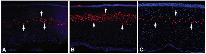
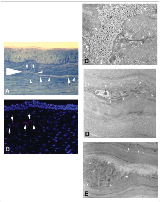
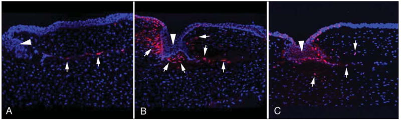
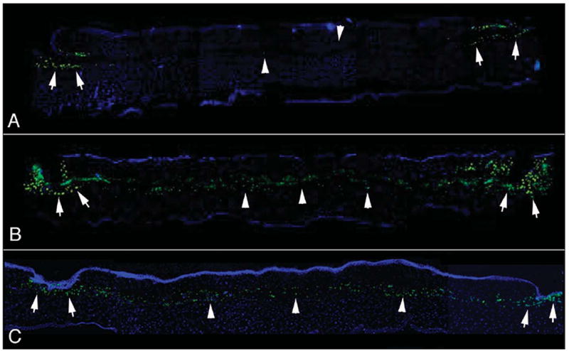
Methods: Thirty-nine rabbit eyes were randomized to receive either no surgery or corneal flaps formed with one of the lasers or the microkeratome. Sixteen eyes also had lamellar cuts with no side cuts with the 30 KHz laser. Animals were sacrificed and corneas processed as frozen sections or fixed for transmission electron microscopy. Frozen sections were evaluated with the TUNEL assay to detect apoptosis, immunocytochemistry for Ki67 to detect cell mitosis, and immunocytochemistry for CD11b to detect mononuclear cells.
Results: Rabbit corneas that had flaps formed with the 15 KHz laser had significantly more stromal cell death, greater stromal cell proliferation, and greater monocyte influx in the central and peripheral comea at 24 hours after surgery than corneas that had flaps formed with the 30 KHz or 60 KHz laser or the microkeratome. Results of the 60 KHz laser and microkeratome were not significantly different for any of the parameters at 24 hours, except for mitotic stromal cells at the flap margin. Transmission electron microscopy revealed that the primary mode of stromal cell death at 24 hours after laser ablation was necrosis.
Conclusions: Stromal cell necrosis associated with femtosecond laser flap formation likely contributes to greater inflammation after LASIK performed with the femtosecond laser, especially with higher energy levels that result in greater keratocyte cell death.
Figures
Similar articles
-
Effect of femtosecond laser energy level on corneal stromal cell death and inflammation.J Refract Surg. 2009 Oct;25(10):869-74. doi: 10.3928/1081597X-20090917-08. Epub 2009 Oct 12. J Refract Surg. 2009. PMID: 19835327 Free PMC article.
-
Wound Healing, Inflammation, and Corneal Ultrastructure After SMILE and Femtosecond Laser-Assisted LASIK: A Human Ex Vivo Study.J Refract Surg. 2018 Jun 1;34(6):393-399. doi: 10.3928/1081597X-20180425-02. J Refract Surg. 2018. PMID: 29889292
-
Conventional Versus Inverted Side-cut Flaps for Femtosecond Laser-Assisted LASIK: Laboratory and Clinical Evaluation.J Refract Surg. 2017 Feb 1;33(2):96-103. doi: 10.3928/1081597X-20161102-02. J Refract Surg. 2017. PMID: 28192588 Clinical Trial.
-
Cellular effects after laser in situ keratomileusis flap formation with femtosecond lasers: a review.Cornea. 2012 Feb;31(2):198-205. doi: 10.1097/ICO.0b013e3182068c42. Cornea. 2012. PMID: 22157568 Review.
-
Microkeratome versus femtosecond flaps: accuracy and complications.Curr Opin Ophthalmol. 2014 Jul;25(4):270-4. doi: 10.1097/ICU.0000000000000070. Curr Opin Ophthalmol. 2014. PMID: 24837579 Review.
Cited by
-
Effect of Myopic Femtosecond Laser-Assisted LASIK on Anterior Chamber Inflammation (Flare Values) and Corneal Endothelium: A Prospective before and after Study.J Ophthalmol. 2021 Nov 26;2021:2395028. doi: 10.1155/2021/2395028. eCollection 2021. J Ophthalmol. 2021. PMID: 34868671 Free PMC article.
-
Surface quality of human corneal lenticules after femtosecond laser surgery for myopia comparing different laser parameters.Graefes Arch Clin Exp Ophthalmol. 2011 Sep;249(9):1417-24. doi: 10.1007/s00417-010-1578-4. Epub 2011 Jan 15. Graefes Arch Clin Exp Ophthalmol. 2011. PMID: 21240524
-
Effect of age on visual and refractive results after LASIK: mechanical microkeratome versus femtosecond laser.Int J Ophthalmol. 2019 Mar 18;12(3):488-495. doi: 10.18240/ijo.2019.03.21. eCollection 2019. Int J Ophthalmol. 2019. PMID: 30918820 Free PMC article.
-
[Alternatives to femtosecond laser technology: subnanosecond UV pulse and ring foci for creation of LASIK flaps].Ophthalmologe. 2014 Jun;111(6):531-8. doi: 10.1007/s00347-013-2994-8. Ophthalmologe. 2014. PMID: 24942119 German.
-
Corneal molecular and cellular biology update for the refractive surgeon.J Refract Surg. 2009 May;25(5):459-66. doi: 10.3928/1081597X-20090422-09. J Refract Surg. 2009. PMID: 19507799 Free PMC article. Review.
References
-
- Nordan LT, Slade SG, Baker RN, Suarez C, Juhasz T, Kurtz R. Femtosecond laser flap creation for laser in situ keratomileusis: six-month follow-up of initial U.S. clinical series. J Refract Surg. 2003;19:8–14. - PubMed
-
- Binder PS. Flap dimensions created with the IntraLase FS laser. J Cataract Refract Surg. 2004;30:26–32. - PubMed
-
- Kim JY, Kim MJ, Kim TI, Choi HJ, Pak JH, Tchah H. A femtosecond laser creates a stronger flap than a mechanical microkeratome. Invest Ophthalmol Vis Sci. 2006;47:599–604. - PubMed
-
- Holzer MP, Rabsilber TM, Auffarth GU. Femtosecond laser-assisted corneal flap cuts: morphology, accuracy, and histopathology. Invest Ophthalmol Vis Sci. 2006;47:2828–2831. - PubMed
-
- Mohan RR, Hutcheon AEK, Choi R, Hong J-W, Lee J-S, Mohan RR, Ambrósio R, Jr, Zieske JD, Wilson SE. Apoptosis, necrosis, proliferation, and myofibroblast generation in the stroma following LASIK and PRK. Exp Eye Res. 2003;76:71–87. - PubMed
Publication types
MeSH terms
Substances
Grants and funding
LinkOut - more resources
Full Text Sources
Other Literature Sources
Research Materials
