Review
. 2007 Dec;17(6):706-15.
doi: 10.1016/j.sbi.2007.08.019. Epub 2007 Oct 29.

Sensor complexes regulating two-component signal transduction

Affiliations
Review

Sensor complexes regulating two-component signal transduction

Hendrik Szurmant et al. Curr Opin Struct Biol. 2007 Dec.

Abstract

Two-component signal transduction systems consisting of a sensor histidine kinase and a response regulator/transcription factor interpret a multitude of environmental and cellular signals and coordinate the expression of a wide array of genes in bacteria. Signal recognition by sensor histidine kinases is the province of a sensor complex consisting of several protein domains that together serve to augment or attenuate the activity of the histidine kinase and thereby of gene expression. Recent investigations have shown the diverse strategies bacteria use to assemble protein domains into the sensor complexes to accomplish signaling. Structural studies of such domains are leading to an understanding of the mechanisms by which sensor complexes recognize signals and regulate kinase activity.


Figures
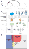
Fig. 1
Fig. 1
Two-component system concept and histidine kinase modular architecture (A) Two component signal transduction systems comprise a sensor histidine kinase that upon stimulus perception achieved by a sensor complex of domains autophosphorylates on a conserved histidine residue. The phosphoryl group is subject to transfer to an aspartate residue on the response regulator, which initiates the cellular response usually through regulation of the expression of target genes. (B) The sensor histidine kinase is a large modular protein consisting of the conserved catalytic core at the C-terminus and several optional N-terminal elements involved in signal sensing or transmission of a conformational change to the catalytic core. Any of these optional domains can occur as tandem repeats in histidine kinase proteins and their multiplicity comprises the sensor complex. Example structures for periplasmic domains are from left to right CitA, DcuS, PhoQ and LuxQ sensing domains; the example structure for the HAMP domain is from Archaeoglobus fulgidus protein AF1503; example structures for the cytoplasmic sensing domains from left to right are FixL, photoactive yellow protein (PYP), and bacteriophytochrome (BphP) sensing domains; the example structure for the catalytic core is of SK853 comprising HisKA and ATP-binding domains. (C) Venn diagram displaying the occurrence of the common cytoplasmic elements HAMP (Red), GAF (Yellow), and PAS (Blue) in combination with the histidine kinase HisKA (Gray) domain as recognized by the Smart Database. The majority (62%) of all HisKA domains are paired with at least one of these three elements.
Fig. 2
Fig. 2
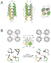
The HAMP domain structure implies a rotational signal transduction mechanism. (A) The HAMP domain of Archaeoglobus fulgidus protein Af1503 displays a parallel four helical coiled-coil with each monomer contributing two helices. Interface residues are in red and blue. (B) Interface residues are arranged as knobs to knobs packing (left) which could convert to knobs into holes packing via a 26° rotation of all helices. The first of four layers of interacting residues is shown. Reprinted with permission from [6].
Fig. 3
Fig. 3
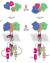
The LuxPQ structure implies a signal transduction mechanism involving asymmetric dimer formation. (A) LuxP and the sensing domain of LuxQ form a stable complex in apo- (left) and in halo-form (right). Upon ligand A2 binding the LuxPQ complex dimerizes along an asymmetric interface. (B) Model for histidine kinase inhibition via asymmetric dimerization. Reprinted with permission from [10].
Fig. 4
Fig. 4
The PhoQ charge-repulsion sensing mechanism. The PhoQ periplasmic sensing domains form a dimer with calcium ions bound on one surface of the dimer (A). The N and C termini of the subunits are both attached to transmembrane helices that are proposed to protrude into the membrane from the calcium bound dimer surface. This surface is highly electro-negative (B) and would be repulsed from the similarly charged membrane surface were it not for the divalent cations bridging the two surfaces (C). The structure shown in (C) is proposed to be the “off” state of the kinase. Disruption of the cation bridging either through cation depletion, in vitro, or through intercalation in the membrane by host defense peptides, in vivo, is proposed to cause repulsion of the dimer/transmembrane surfaces with consequent conformational alteration of the PhoQ sensing domains and conversion of the kinase to the “on” state.
Fig. 5
Fig. 5
Diverse structures and mechanisms of auxiliary proteins. (A) Periplasmic binding proteins are soluble proteins, which regulate an associated kinase by direct interaction in response to ligand binding shown here utilizing the LuxPQ example of Vibrio harveii. Individual structures of Apo- and AI2 ligand-complexed LuxP are displayed (B) YycH and YycI are membrane tethered periplasmic proteins that negatively regulate the kinase YycG through direct interaction and might respond to unidentified ligands X and Y. The structures of YycH and YycI display a common fold over two domains. (C) The pXO2-61 protein of Bacillus anthracis display a myoglobin-like fold highly homologous in sequence and likely in structure to the sensing domain of the sporulation histidine kinase BA2291 and a second protein pXO1-118. Plasmid encoded proteins pXO2-61 and pXO1-118 are believed to compete with the associated kinase for unknown activation signals X and Y.

References

    1. Hoch JA. Two-component and phosphorelay signal transduction. Curr Opin Microbiol. 2000;3:165–170. - PubMed
    1. Galperin MY. A census of membrane-bound and intracellular signal transduction proteins in bacteria: bacterial IQ, extroverts and introverts. BMC Microbiol. 2005;5:35. - PMC - PubMed
    1. Marina A, Waldburger CD, Hendrickson WA. Structure of the entire cytoplasmic portion of a sensor histidine-kinase protein. Embo J. 2005;24:4247–4259. The first crystal structure of a full-length histidine kinase catalytic core. - PMC - PubMed
    1. Mascher T, Helmann JD, Unden G. Stimulus perception in bacterial signal-transducing histidine kinases. Microbiol Mol Biol Rev. 2006;70:910–938. - PMC - PubMed
    1. Galperin MY, Nikolskaya AN, Koonin EV. Novel domains of the prokaryotic two-component signal transduction systems. FEMS Microbiol Lett. 2001;203:11–21. - PubMed

Publication types

LinkOut - more resources