. 2007;35(19):6648-62.
doi: 10.1093/nar/gkm739. Epub 2007 Oct 2.

The ePHD protein SPBP interacts with TopBP1 and together they co-operate to stimulate Ets1-mediated transcription

Affiliations

The ePHD protein SPBP interacts with TopBP1 and together they co-operate to stimulate Ets1-mediated transcription

Eva Sjøttem et al. Nucleic Acids Res. 2007.

Abstract

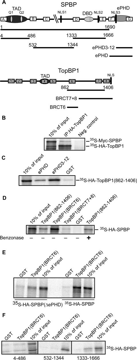
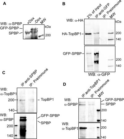
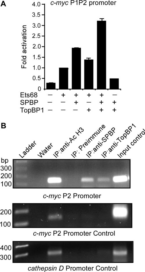
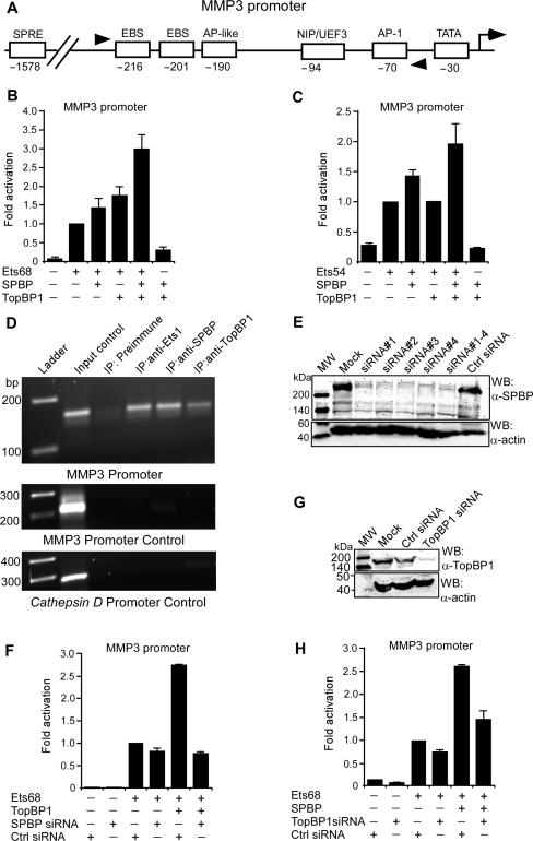
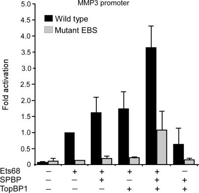
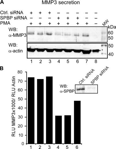
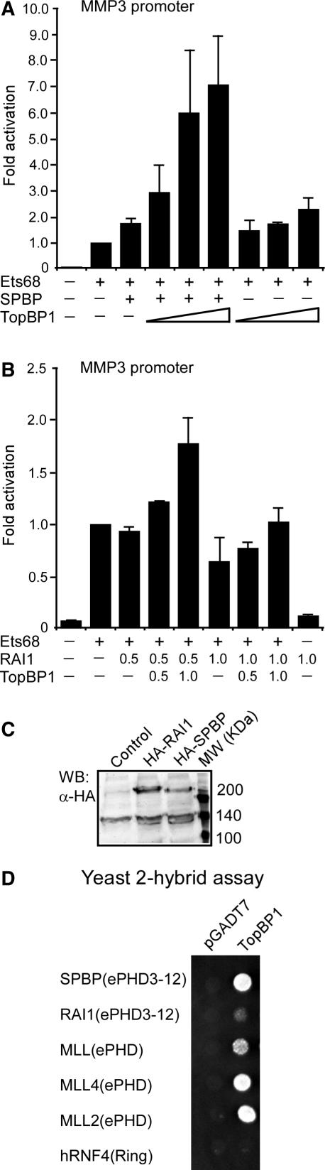
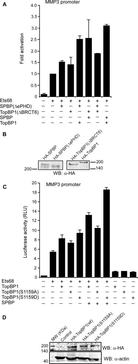
SPBP (Stromelysin-1 PDGF responsive element binding protein) is a ubiquitously expressed 220 kDa nuclear protein shown to enhance or repress the transcriptional activity of various transcription factors. A yeast two-hybrid screen, with the extended plant homeodomain (ePHD) of SPBP as bait, identified TopBP1 (topoisomerase II beta-binding protein 1) as a candidate interaction partner of SPBP. TopBP1 has eight BRCA1 carboxy-terminal (BRCT) domains and is involved in DNA replication, DNA damage responses and in the regulation of gene expression. The interaction between SPBP and TopBP1 was confirmed in vitro and in vivo, and was found to be mediated by the ePHD domain of SPBP and the BRCT6 domain of TopBP1. Both SPBP and TopBP1 enhanced the transcriptional activity of Ets1 on the c-myc P1P2- and matrix metalloproteinase-3 (MMP3) promoters. Together they displayed a more than additive effect. Both proteins were associated with these promoters. The involvement of TopBP1 was dependent on the serine 1159 phosphorylation site, known to be important for transcriptional activation. Depletion of endogenous SPBP by siRNA treatment reduced MMP3 secretion by 50% in phorbol ester-stimulated human fibroblasts. Taken together, our results show that TopBP1 and SPBP interact physically and functionally to co-operate as co-activators of Ets1.


Figures

Figure 1.
Figure 1.
SPBP and TopBP1 interact in vitro via the ePHD and BRCT6 domains. (A) Schematic representation of the domain structure of human SPBP (1960 amino acids) and human TopBP1 (1522 amino acids). Deletion mutants of SPBP and TopBP1 used in this study and the TopBP1 region isolated in the yeast two-hybrid screen are indicated. The eight BRCT domains of TopBP1 are represented by numbered boxes. The open boxes designated A-G of SPBP represent regions with strong homology to human RAI1. TAD: trans-activation domain, DBD: DNA-binding domain, NLS: Nuclear Localization Signal, ePHD: extended PHD domain, Q1/Q2: Glutamine-rich stretches. (B) In vitro co-immunoprecipitation showing interaction between SPBP and TopBP1 full-length proteins. Full-length HA-TopBP1 was in vitro co-translated together with Myc-SPBP in the presence of [35S]methionine. Immunoprecipitations were performed using an anti-HA antibody. Precipitated proteins as well as 10% input of in vitro translated proteins were resolved by SDS–PAGE (5%). Negative control is the HA-tag co-translated together with Myc-tagged full-length SPBP and immunoprecipitated with an anti-HA antibody. (C) GST pull-down assays demonstrating interaction both between the complete ePHD and the ePHD(3–12) domain of SPBP, and TopBP1(862–1406). HA-tagged TopBP1(862–1406) was in vitro translated in the presence of [35S]methionine and incubated with equal amounts of either GST, GST-SPBP(ePHD) or GST-SPBP(ePHD3-12). The pulled down proteins, together with in vitro translated HA-TopBP1(862–1406) corresponding to 10% of the input were separated by SDS–PAGE (10%). (D) BRCT6 domain of TopBP1 binds strongly to SPBP, while BRCT7 + 8 shows weak affinity for SPBP. Radiolabelled in vitro translated full-length HA-SPBP was allowed to bind to equal amounts of the different GST-fused fragments of TopBP1. The interaction between TopBP1(862–1406) and full-length HA-SPBP was tested with, or without the nuclease benzonase. Samples and 10% of the input were resolved by SDS–PAGE (5%). (E and F) In vitro GST pull-down assays demonstrating that the SPBP(1333–1666) region interacts weakly with TopBP1. HA-tagged SPBP, SPBP(ΔePHD), SPBP(4–486), SPBP(532–1344) and SPBP(1333–1666) were in vitro translated in the presence of [35S]methionine and incubated with equal amounts of either GST or GST-TopBP1(BRCT6). The pulled-down proteins, together with 10% of the input were separated by SDS–PAGE.
Figure 2.
Figure 2.
SPBP and TopBP1 interact in vivo. (A) Expression of GFP-SPBP stably integrated into the genome of the U2OS-TA cell line. Growing the cells in the presence of doxycycline represses the expression of the integrated GFP-SPBP (left lane). Removal of doxycycline induces expression of GFP-SPBP at a similar level as the endogenous protein (middle lane). Total cellular extracts from U2OS-TA cells containing the GFP-SPBP minigene grown in the presence or absence of doxycycline were separated by SDS–PAGE (5%) and immunoblotted using an anti-SPBP antibody. (B) GFP-SPBP and HA-TopBP1 interact in U2OS-GFP-SPBP cells. U2OS-GFP-SPBP cells were transfected with a HA-TopBP1 expressing plasmid. One day after transfection immunoprecipitations were performed using an anti-GFP antibody. The co-precipitated proteins were detected by western blotting using an anti-TopBP1 antibody (upper panel). Immunoprecipitated SPBP was visualized using the anti-GFP antibody (lower panel). (C) SPBP co-precipitate endogenous TopBP1. Immunoprecipitations from U2OS-GFP-SPBP cells were performed using an anti-SPBP antibody. The co-precipitated proteins were detected by western blotting using an anti-TopBP1 antibody (upper panel). Immunoprecipitated SPBP was visualized using an anti-SPBP antibody (lower panel). The two SPBP bands correspond to GFP-SPBP (upper band) and endogenous SPBP (lower band). (D) Endogenous TopBP1 co-precipitate SPBP. Immunoprecipitations were performed using an anti-TopBP1 antibody. The co-precipitated proteins were detected by western blotting using an anti-SPBP antibody (upper panel). Immunoprecipitated TopBP1 was visualized using the anti-TopBP1 antibody (lower panel). The two SPBP bands correspond to GFP-SPBP (upper band) and endogenous SPBP (lower band).
Figure 3.
Figure 3.
SPBP and TopBP1 associate with the c-myc promoter and co-operate to enhance Ets1-mediated transcriptional activation of this promoter. (A) When SPBP and TopBP1 are co-expressed with Ets68, a more than additive activation of Ets68-mediated transcription of the c-myc P1P2 promoter is observed. Transient transfection assays were carried out in HeLa cells using 0.5 μg of the reporter p1p2mycluc, together with expression vectors for Ets68 (0.1 μg), HA-SPBP (1.0 μg) and/or HA-TopBP1 (1.0 μg) as indicated. The data represent the mean of three independent experiments, each performed in triplicate. (B) Chromatin immunoprecipitation assays show that both SPBP and TopBP1 associate with the c-myc promoter. Extracts from U2OS-GFP-SPBP cells (1.5 × 10⁷ cells per antibody) were immunoprecipitated with preimmune serum (IgG), polyclonal anti-SPBP antibody, polyclonal anti-TopBP1 antibody and anti-acetylated histone H3 antibody (positive control). Input control (1:50) was included (lane 7). PCR analyses on the immunoprecipitated chromatin were carried out using primers flanking the c-myc P2 promoter (position −28 and +200 respectively), primers aligning to positions −4107 and −3949 of the c-myc P2 promoter (c-myc P2 Promoter Control), and primers aligning to positions −3351 and −3069 of the cathepsin D promoter (cathepsin D Promoter Control). The 1 kb DNA ladder is shown to the left.
Figure 4.
Figure 4.
SPBP and TopBP1 co-operate to enhance Ets1-mediated transcriptional activation of the MMP3 promoter. (A) Schematic representation of the human MMP3 promoter. Arrowheads indicate the position of forward and reverse primers (−231 and −65, respectively) used for ChIP. EBS: Ets1-binding sites, NIP/UEF3: nuclear inhibitory protein/urokinase-type plasminogen activator enhancer factor 3, SPRE: Stromelysin-1 PDGF-responsive element. (B) SPBP and TopBP1 co-operate to enhance Ets68-mediated activation of the MMP3 promoter. Transient transfections were carried out in HeLa cells as described in Figure 3A except that the reporter plasmid contains 1448 bp of the human MMP3 promoter in front of the luciferase gene. The data represent the mean of four independent experiments, each performed in triplicate. (C) SPBP and TopBP1 co-operate to enhance Ets54-mediated transcriptional activation of the MMP3 promoter. Transient transfections were carried out in HeLa cells as described in (B) except that an expression vector for murine Ets54 (27) was used instead of the Ets68 expression vector. The data represent the mean of two independent experiments, each performed in triplicate. (D) Chromatin immunoprecipitation assays show that Ets1, SPBP and TopBP1 are associated with the MMP3 promoter. Extracts from U2OS-GFP-SPBP cells (1.5 × 10⁷ cells per antibody) were immunoprecipitated with preimmune serum (IgG), polyclonal anti-Ets1 antibody, polyclonal anti-SPBP antibody and polyclonal anti-TopBP1 antibody. Input control (1:50) was included (lane 2). PCR analyses on the immunoprecipitated chromatin were carried out using primers aligning at position −231 and −65 of the MMP3 promoter. Control PCR analysis was carried out using primers aligning to positions −3537 and −3326 of the MMP3 promoter, and positions −3351 and −3069 of the cathepsin D promoter. The 1 kb DNA ladder is shown to the left. (E) Western blot showing knockdown of SPBP in HeLa cells using four different siRNA oligos. Transient transfections were carried out using siRNA oligos 1–4 alone and mixed (50 nM) as indicated. Cell lysates were separated by SDS–PAGE (6%), and immunoblotted using the anti-SPBP antibody (upper panel). The blots were re-probed with an anti-β-actin antibody in order to determine the amount of loaded protein (lower panel). (F) Down-regulation of SPBP using siRNA decreased the transcriptional activation of MMP3 promoter mediated by either Ets1 alone or Ets1 and TopBP1 together. Transient transfections were carried out in HeLa cells using the pGL3-MMP3 reporter plasmid (30 ng), together with the expression plasmids for Ets68 (10 ng) and HA-TopBP1 (50 ng), and a SPBP-specific siRNA (50 nM) or scrambled siRNA as indicated. Cells were harvested 40 h post-transfection. The data represent the mean of three independent experiments performed in triplicate. (G) Western blot showing siRNA-mediated knockdown of TopBP1 in HeLa cells. Transient transfections were carried out using 50 nM TopBP1 siRNA or 50 nM Control siRNA. Cell lysates were separated by SDS–PAGE (6%), and immunoblotted using an anti-TopBP1 antibody (upper panel). β-actin was used as loading control (lower panel). (H) Down-regulation of TopBP1 using siRNA decreased the transcriptional activation of the MMP3 promoter mediated by either Ets1 alone or Ets1 and SPBP together. Transient transfections were carried out as described in (F) except that an expression plasmid for SPBP was used instead of the TopBP1 expression plasmid, and TopBP1 specific siRNA was used instead of the SPBP siRNA. The data represent the mean of two independent experiments performed in triplicate.
Figure 5.
Figure 5.
The stimulation of Ets1 activation of the MMP3 promoter by SPBP and TopBP1 is dependent on the tandem Ets1-binding sites. Transient transfections were carried out in HeLa cells as described in Figure 3A except that two different luciferase reporter gene plasmids were used, one containing the wild-type human MMP3 promoter (1448 bp upstream sequence), and one containing the human MMP3 promoter with the tandem Ets1-binding sites mutated. The data represent the mean of two independent experiments, each performed in triplicate.
Figure 6.
Figure 6.
The level of endogenous SPBP affects the level of phorbol ester-induced MMP3 secretion. (A) A western blot showing the effect of down-regulation of endogenous SPBP on the level of secreted MMP3 induced by PMA in IMR90 fibroblasts. Cells were starved in MEM with 0.1% FBS for 24 h before being transfected two following days with SPBP siRNA (50 nM) or scrambled siRNA (50 nM). The cells were starved in MEM without serum for 24 h post-transfection before stimulation with MEM containing 1 ng/ml PMA for 20 h. Medium samples were analysed by SDS–PAGE (7.5%) and immunoblotting with an MMP3 antibody. Medium from three parallel wells per siRNA are shown (lanes 1–3 and 4–6, respectively) in addition to untreated cells showing the induction of MMP3 in the presence of PMA (compare lanes 7 and 8). The levels of β-actin in the cells from which the medium were harvested are shown in the lower panel. The cells in each well were harvested in 30 μl 2× SDS loading buffer and the extract separated by SDS–PAGE (10%). (B) Knockdown of endogenous SPBP reduces the MMP3 secretion in PMA-stimulated IMR90 cells by 50%. The amount of secreted MMP3 was normalized to the level of β-actin expression in each well. Quantification was performed using the LumiAnalyst software (Roche). A western blot of SPBP expression in total cellular extract from IMR90 cells transfected with scrambled siRNA and SPBP siRNA are shown in the inserted panel.
Figure 7.
Figure 7.
The SPBP homologue RAI1 does not stimulate Ets1-mediated transcriptional activation of the MMP3 promoter or co-operate with TopBP1. (A) TopBP1 potentiates the SPBP-enhanced Ets1 transactivation in a dose-dependent manner. Transient transfections were carried out in HeLa cells as described in Figure 4 except that 0.5 μg of the SPBP expression plasmid and increasing amounts of TopBP1 expression plasmids (0.5, 1.0 and 1.5 μg, respectively) were used. The data represent the mean of two individual experiments performed in triplicate. (B) RAI1 does not enhance Ets1 transactivation or act together with TopBP1 to enhance Ets1-mediated transcription. Transient transfections were carried out in HeLa cells as described in (A) except that 0.5 μg or 1.0 μg of RAI1 expression plasmid was used instead of the SPBP expressing plasmid. The data represent the mean of two individual experiments performed in triplicate. (C) Western blot documenting similar expression levels of HA-SPBP and HA-RAI1 in HeLa cells. Total cellular extracts from HeLa cells transfected with expression plasmids for HA-RAI1 or HA-SPBP as indicated were analysed by 6% SDS–PAGE and immunoblotted using an anti-HA antibody. (D) Yeast two-hybrid assay showing that the ePHD domain of RAI1 interacts very weakly with TopBP1 compared to the SPBP ePHD domain, and the ePHD domains of MML protein family. One colony containing AD or DBD plasmids was picked and re-suspended 200 μl dH2O. Ten microlitres of the cell suspension was spotted on QDO medium and allowed to grow at 30°C for 3 days.
Figure 8.
Figure 8.
The co-operation between SPBP and TopBP1 is dependent on the interaction between the ePHD- and BRCT6 domains and on TopBP1 oligomerization. (A) Deletion of the SPBP ePHD domain or the BRCT6 domain of TopBP1 abolishes their ability to co-operate to enhance Ets1 transactivation. Transient transfections were carried out in HeLa cells using the pGL3-MMP3 reporter (0.5 μg), together with expression vectors for Ets68 (100 ng), HA-SPBP (1 μg), HA-SPBP(ΔePHD) (1 μg), HA-TopBP1 (1 μg) and/or HA-TopBP1(ΔBRCT6) (1 μg) as indicated. Cells were harvested 20 h post-transfection. The data represent the mean of four independent experiments, each performed in triplicate. (B) Western blot showing similar expression levels of the full-length- and deletion constructs of SPBP and TopBP1. Total cellular extracts from HeLa cells transfected with expression plasmids for HA-SPBP, HA-SPBP(ΔePHD), HA-TopBP1 or HA-TopBP1(ΔBRCT6) as indicated were analysed by 6% SDS–PAGE and immunoblotted using an anti-HA antibody. (C) TopBP1-mediated enhancement of Ets1 transcriptional activity and the co-operation with SPBP is dependent on phosphorylation and oligomerization of TopBP1. HeLa cells were transfected with the pGL3-MMP3 reporter (0.5 μg), expression vectors for Ets68 (100 ng), HA-TopBP1(S1159A) (1 μg), HA-TopBP1(S1159D) (1 μg), HA-TopBP1(1 μg) and/or HA-SPBP(1 μg) as indicated. Cells were harvested 20 h post-transfection. The mean luciferase activities obtained in a representative experiment performed in triplicate is shown. (D) Western blot showing similar expression levels of wild-type and mutated HA-TopBP1. Anti-HA immunoblot performed as in (B).

Similar articles

Cited by

References

    1. Rekdal C, Sjøttem E, Johansen T. The nuclear factor SPBP contains different functional domains and stimulates the activity of various transcriptional activators. J. Biol. Chem. 2000;275:40288–40300. - PubMed
    1. Sanz L, Moscat J, Diaz-Meco MT. Molecular characterization of a novel transcription factor that controls stromelysin expression. Mol. Cell. Biol. 1995;15:3164–3170. - PMC - PubMed
    1. Lyngsø C, Bouteiller G, Damgaard CK, Ryom D, Sanchez-Munoz S, Norby PL, Bonven BJ, Jørgensen P. Interaction between the transcription factor SPBP and the positive cofactor RNF4. An interplay between protein binding zinc fingers. J. Biol. Chem. 2000;275:26144–26149. - PubMed
    1. Kirstein M, Sanz L, Quinones S, Moscat J, Diaz-Meco MT, Saus J. Cross-talk between different enhancer elements during mitogenic induction of the human stromelysin-1 gene. J. Biol. Chem. 1996;271:18231–18236. - PubMed
    1. Gburcik V, Bot N, Maggiolini M, Picard D. SPBP is a phosphoserine-specific repressor of estrogen receptor alpha. Mol. Cell. Biol. 2005;25:3421–3430. - PMC - PubMed

Publication types

MeSH terms