Review
. 2007;35(22):7429-55.
doi: 10.1093/nar/gkm711. Epub 2007 Oct 2.

Human telomere, oncogenic promoter and 5'-UTR G-quadruplexes: diverse higher order DNA and RNA targets for cancer therapeutics

Affiliations
Review

Human telomere, oncogenic promoter and 5'-UTR G-quadruplexes: diverse higher order DNA and RNA targets for cancer therapeutics

Dinshaw J Patel et al. Nucleic Acids Res. 2007.

Abstract

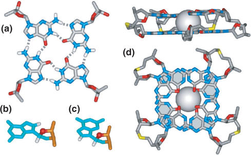
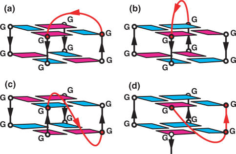
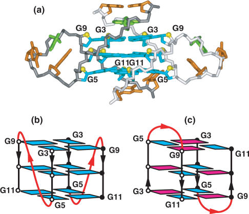
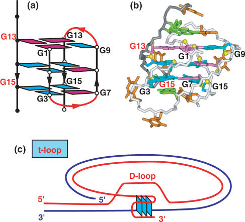
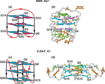
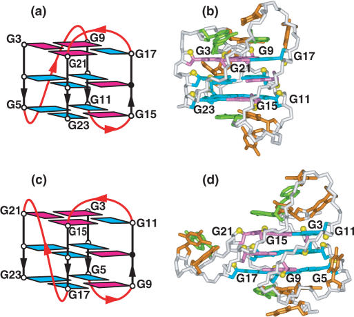
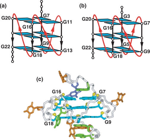
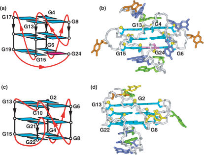
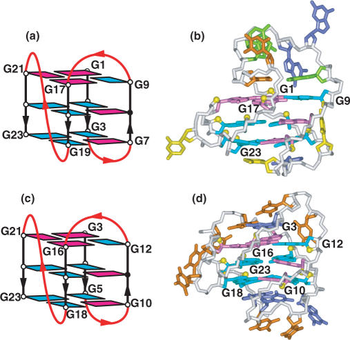
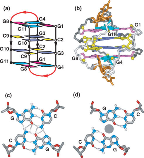
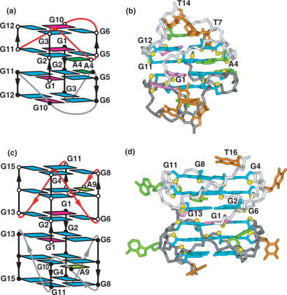
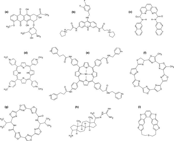
Guanine-rich DNA sequences can form G-quadruplexes stabilized by stacked G-G-G-G tetrads in monovalent cation-containing solution. The length and number of individual G-tracts and the length and sequence context of linker residues define the diverse topologies adopted by G-quadruplexes. The review highlights recent solution NMR-based G-quadruplex structures formed by the four-repeat human telomere in K(+) solution and the guanine-rich strands of c-myc, c-kit and variant bcl-2 oncogenic promoters, as well as a bimolecular G-quadruplex that targets HIV-1 integrase. Such structure determinations have helped to identify unanticipated scaffolds such as interlocked G-quadruplexes, as well as novel topologies represented by double-chain-reversal and V-shaped loops, triads, mixed tetrads, adenine-mediated pentads and hexads and snap-back G-tetrad alignments. The review also highlights the recent identification of guanine-rich sequences positioned adjacent to translation start sites in 5'-untranslated regions (5'-UTRs) of RNA oncogenic sequences. The activity of the enzyme telomerase, which maintains telomere length, can be negatively regulated through G-quadruplex formation at telomeric ends. The review evaluates progress related to ongoing efforts to identify small molecule drugs that bind and stabilize distinct G-quadruplex scaffolds associated with telomeric and oncogenic sequences, and outlines progress towards identifying recognition principles based on several X-ray-based structures of ligand-G-quadruplex complexes.


Figures

Figure 1.
Figure 1.
(a) Schematic alignment of four guanines in a plane to form the G–G–G–G tetrad (G-tetrad). Each guanine uses its Watson–Crick and major groove edges to form a pair of hydrogen bonds. This leaves the minor groove edge available for further recognition. Schematic illustrating (b) anti and (c) syn guanine glycosidic torsion angle alignments. The H8 and sugar H1′ protons are closer to each other (2.5 Å) in the syn alignment compared to a longer distance (3.7 Å) in the anti alignment, a feature that can be readily monitored by NMR (256). (d) Two views of K+ cation-coordination between adjacent G-tetrad planes. Each tetrad provides four inwardly pointing guanine carbonyls, such that the dehydrated K+ cation participates in a directly coordinated tetragonal bipyramidal arrangement (10).
Figure 2.
Figure 2.
Schematic illustrating (a) edge-wise, (b) diagonal, (c) double-chain-reversal or propeller and (d) V-shaped loops. The loops connect individual strands or columns bridging two G-tetrad planes. Color-coding for schematics is as follows: anti guanines in blue and syn guanines in magenta. G-rich columns in black and connecting loops in red.
Figure 3.
Figure 3.
(a) X-ray structure of the two-repeat human telomere bimolecular G-quadruplex formed by the d(TAGGGTTAGGGT) sequence for crystals grown from K+ solution (coordinates deposition: 1K8P) (17). The bases are color coded as follows: guanine (blue), adenine (green) and thymine (orange). All strands are parallel in this bimolecular quadruplex and the loops are of the double-chain-reversal or propeller type. NMR-based folding topologies of interconverting (b) all parallel-stranded and (c) anti-parallel-stranded conformations of the two-repeat human telomere bimolecular G-quadruplex formed by the d(TAGGGTTAGGGT) sequence in K+ solution (94).
Figure 4.
Figure 4.
NMR-based (a) (3 + 1) folding topology and (b) solution structure of the three-repeat human telomere bimolecular G-quadruplex formed by the d[G3(T2AG3)2T] sequence in Na+ solution (coordinates deposition: 2AQY) (96). All three G-tracts from one strand and the 3′-terminal G-tract from the partner strand are used to form the bimolecular G-quadruplex. (c) Schematic of hypothetical G-quadruplex formation when the 3′-end overhang (in red) invades the adjacent double-stranded segment of the telomere to form the so-called t-loop (97).
Figure 5.
Figure 5.
NMR-based (a) folding topology and (b) solution structure of the four-repeat human telomere unimolecular G-quadruplex formed by the d[AG3(T2AG3)3] sequence in Na+ solution (coordinates deposition: 143D) (9). The loop types starting from the 5′-end are edge-wise, diagonal and edge-wise. Individual strands have both a parallel and anti-parallel neighbor, with the G-tetrads adopting syn–syn–anti–anti alignments. X-ray-based (c) folding topology and (d) crystal structure of the four-repeat human telomere unimolecular G-quadruplex formed by the d[AG3(T2AG3)3] sequence for crystals grown from K+ solution (coordinates deposition: 1KF1) (17). All three loops are of the double-chain-reversal or propeller type, all strands are parallel and all guanines adopt anti alignments.
Figure 6.
Figure 6.
NMR-based (a) folding topology and (b) solution structure of the four-repeat human telomere unimolecular G-quadruplex formed by the d[TAG3(T2AG3)3] sequence (form-1) in K+ solution (coordinates deposition: 2JSM, 2JSK) (101). The loop types starting from the 5′-end are double-chain-reversal, edge-wise and edge-wise. Two other groups have independently investigated the same system and come to similar conclusions (102–105). NMR-based (c) folding topology and (d) solution structure of the four-repeat human telomere unimolecular G-quadruplex formed by the d[TAG3(T2AG3)3TT] sequence (form-2) in K+ solution (coordinates deposition: 2JSL, 2JSQ) (101). The loop types starting from the 5′-end are edge-wise, edge-wise and double-chain-reversal. One other group has independently investigated the same system and come to similar conclusions (106).
Figure 7.
Figure 7.
NMR-based folding topology of unimolecular G-quadruplexes formed by (a) myc-2345 and (b) thymine for guanine-containing variant myc-1245 sequences in K+ solution (126). (c) NMR-based solution structure of the thymine for guanine-containing variant myc-2345 promoter unimolecular G-quadruplex in K+ solution (coordinates deposition: 1XAV) (128).
Figure 8.
Figure 8.
NMR-based (a) folding topology and (b) solution structure of the unimolecular G-quadruplex formed by the myc-23456 sequence in K+ solution (coordinates deposition: 2A5P) (129). NMR-based (c) folding topology and (d) solution structure of the unimolecular G-quadruplex formed by the c-kit1 sequence in K+ solution (coordinates deposition: 2O3M) (135).
Figure 9.
Figure 9.
NMR-based (a) (3 + 1) G-quadruplex folding topology and (b) solution structure of the thymine for guanine-containing variant bcl-2 2345 promoter unimolecular G-quadruplex in K+ solution (coordinates deposition: 2F8U) (139). NMR-based (c) (3 + 1) G-quadruplex topology and (d) solution structure adopted by the four-repeat Tetrahymena telomere unimolecular G-quadruplex formed by the d[(T2G4)4] sequence in Na+ solution (coordinates deposition: 186D) (7).
Figure 10.
Figure 10.
NMR-based (a) folding topology and (b) solution structure of the bimolecular G-quadruplex formed by the d(GCGGT3GCGG) sequence in Na+ solution (coordinates deposition: 1A6M) (164). Schematics illustrating mixed tetrad pairing alignments observed for (c) direct and (d) slipped G–C–G–C tetrads, associated with pairing along the major groove edges. A monovalent cation (gray ball) most likely bridges the acceptor atoms along the major groove edges of opposing guanines in the slipped G–C–G–C tetrad.
Figure 11.
Figure 11.
Schematics illustrating base triad pairing alignments for (a) A–(T-A) (183), (b) G–(C-A) (20) triads. Schematics illustrating (c) A–(G–G–G–G) pentad (18,171) and (d) A–(G–G–G–G)–A hexad (16) alignments. NMR-based (e) folding topology and (f) solution structure of end-to-end stacked tetramolecular G-quadruplexes formed by four d(GGAGGAG) strands in 150 mM Na+ solution (coordinates deposition: 1EEG) (16).
Figure 12.
Figure 12.
NMR-based (a) folding topology and (b) solution structure of the V-shaped interlocked bimolecular G-quadruplex formed by the d(G3AG2T3G3AT) sequence in Na+ solution (coordinates deposition: 1JJP) (18). NMR-based (c) folding topology and (d) solution structure of interlocked bimolecular G-quadruplex formed by the d(G4TG3AG2AG3T) 93del sequence in K+ solution (coordinates deposition: 1Y8D) (171).
Figure 13.
Figure 13.
Chemical formulas of (a) daunomycin (222), (b) 9-benzylamino-substituted acridine (227), (c) bisquinolinium-substituted phenanthroline (X=NCH3 +, Y=CH; X=CH, Y= NCH3+) (229), (d) 5,10,15,20-tetrakis-(N-methyl-4-pyridyl)porphyrin (TMPyP4), (e) Mn(III) porphyrin with flexible cationic arms (234), (f) telomestatin (235), (g) oxazole-containing 24-membered macrocycle (HXDV) (240), (h) steroid diamine funtumine substituted by a guanylhydrazone moiety (244) and (i) left-handed chiral cyclic-helicene with a short linker (207).
Figure 14.
Figure 14.
(a) X-ray structure of the complex of three molecules of daunomycin (in red) bound to the tetramolecular d(TGGGGT) G-quadruplex (coordinates deposition: 1O0K) (222). (b) Overlap of daunomycin molecules over the terminal G-tetrad. (c) X-ray structure of the complex of disubstituted aminoalkylamido acridine (in red) bound to the bimolecular d(G4T4G4) G-quadruplex (coordinates deposition: 1L1H) (221). (d) Overlap of the substituted acridine over the terminal G-tetrad. (e) X-ray structure of the porphyrin TMPyP4 (in red) bound to the d(TAG3T2AG3) bimolecular G-quadruplex (coordinates deposition: 2HRI) (233). (f) Overlap of the porphyrin ring of TMPyP4 with a terminal base pair.

References

    1. Gellert M, Lipsett MN, Davies DR. Helix formation by guanylic acid. Proc. Natl Acad. Sci. USA. 1962;48:2013–2018. - PMC - PubMed
    1. Zimmerman SB, Cohen GH, Davies DR. X-ray fiber diffraction and model-building study of polyguanylic acid and polyinosinic acid. J. Mol. Biol. 1975;92:181–192. - PubMed
    1. Sen D, Gilbert W. Formation of parallel four-stranded complexes by guanine-rich motifs in DNA and its implications for meiosis. Nature. 1988;334:364–366. - PubMed
    1. Wang Y, Patel DJ. Guanine residues in d(T2AG3) and d(T2G4) form parallel-stranded potassium cation stabilized G-quadruplexes with anti glycosidic torsion angles in solution. Biochemistry. 1992;31:8112–8119. - PubMed
    1. Laughlan G, Murchie AI, Norman DG, Moore MH, Moody PC, Lilley DM, Luisi B. The high-resolution crystal structure of a parallel-stranded guanine tetraplex. Science. 1994;265:520–524. - PubMed

Publication types