

Cortical granule exocytosis in C. elegans is regulated by cell cycle components including separase
- PMID: 17913784
- PMCID: PMC5507579
- DOI: 10.1242/dev.011361
Cortical granule exocytosis in C. elegans is regulated by cell cycle components including separase
Abstract
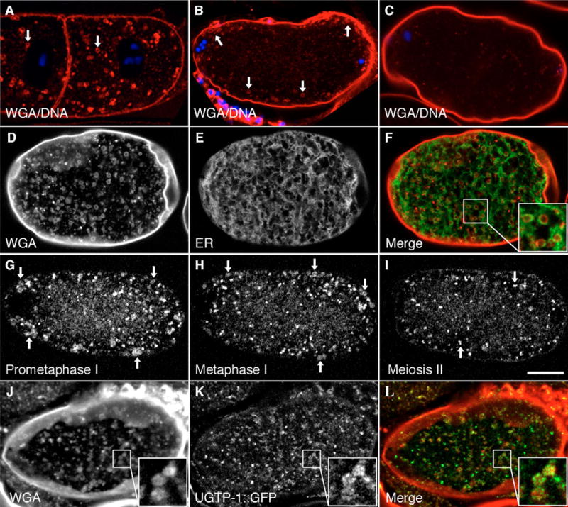
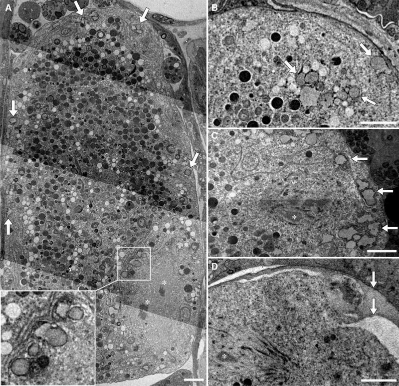
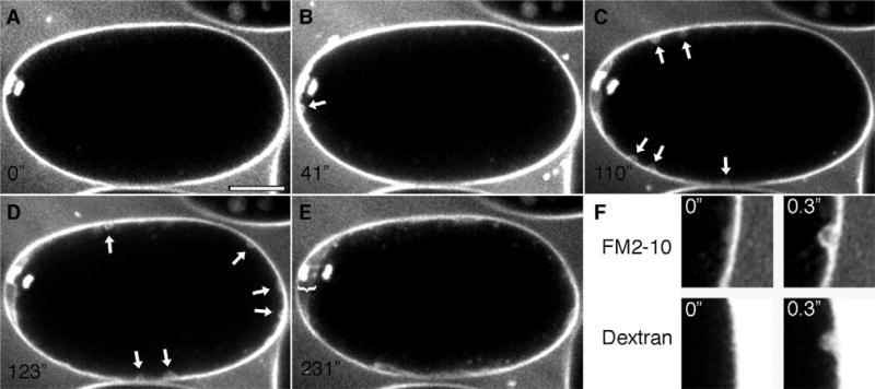
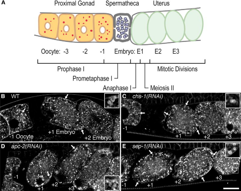
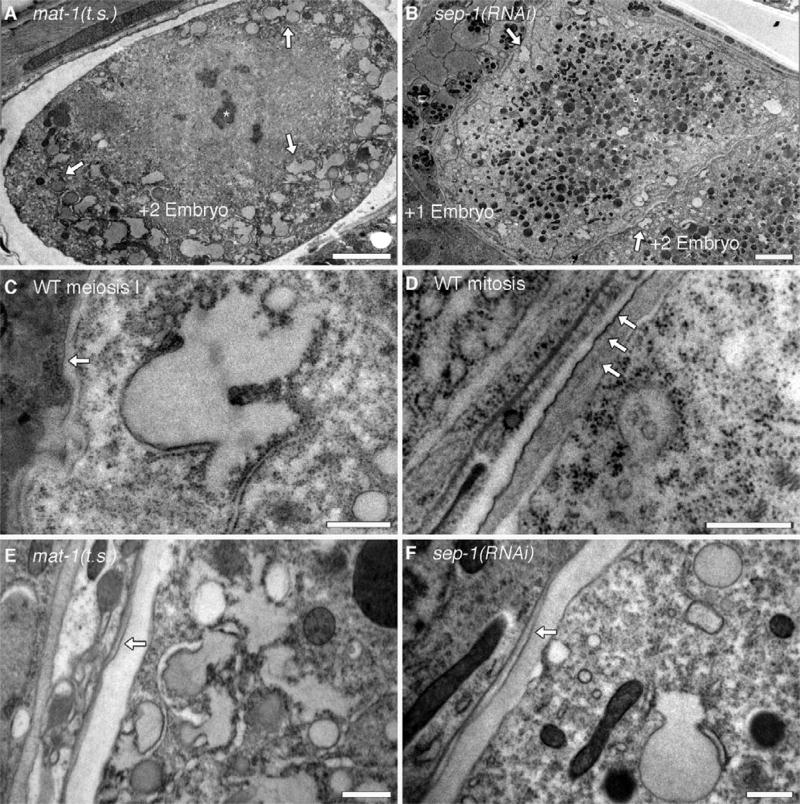
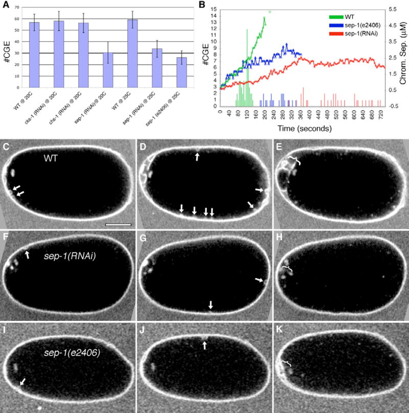
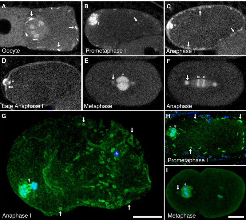
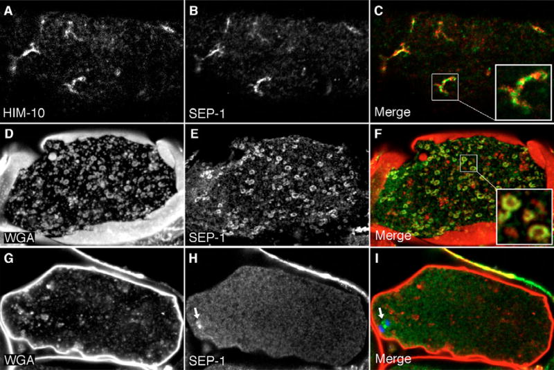
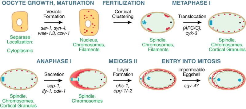
In many organisms, cortical granules undergo exocytosis following fertilization, releasing cargo proteins that modify the extracellular covering of the zygote. We identified cortical granules in Caenorhabditis elegans and have found that degranulation occurs in a wave that initiates in the vicinity of the meiotic spindle during anaphase I. Previous studies identified genes that confer an embryonic osmotic sensitivity phenotype, thought to result from abnormal eggshell formation. Many of these genes are components of the cell cycle machinery. When we suppressed expression of several of these genes by RNAi, we observed that cortical granule trafficking was disrupted and the eggshell did not form properly. We conclude that osmotic sensitivity phenotypes occur because of defects in trafficking of cortical granules and the subsequent formation of an impermeable eggshell. We identified separase as a key cell cycle component that is required for degranulation. Separase localized to cortically located filamentous structures in prometaphase I upon oocyte maturation. After fertilization, separase disappeared from these structures and appeared on cortical granules by anaphase I. RNAi of sep-1 inhibited degranulation in addition to causing extensive chromosomal segregation failures. Although the temperature-sensitive sep-1(e2406) allele exhibited similar inhibition of degranulation, it had minimal effects on chromosome segregation. These observations lead us to speculate that SEP-1 has two separable yet coordinated functions: to regulate cortical granule exocytosis and to mediate chromosome separation.
Figures
Similar articles
-
Rab6 is required for the exocytosis of cortical granules and the recruitment of separase to the granules during the oocyte-to-embryo transition in Caenorhabditis elegans.J Cell Sci. 2012 Dec 1;125(Pt 23):5897-905. doi: 10.1242/jcs.116400. Epub 2012 Sep 19. J Cell Sci. 2012. PMID: 22992455
-
Protein phosphatase 5 is a negative regulator of separase function during cortical granule exocytosis in C. elegans.J Cell Sci. 2011 Sep 1;124(Pt 17):2903-13. doi: 10.1242/jcs.073379. J Cell Sci. 2011. PMID: 21878498 Free PMC article.
-
Protease dead separase inhibits chromosome segregation and RAB-11 vesicle trafficking.Cell Cycle. 2017 Oct 18;16(20):1902-1917. doi: 10.1080/15384101.2017.1363936. Epub 2017 Aug 18. Cell Cycle. 2017. PMID: 28820333 Free PMC article.
-
The P Granules of C. elegans: A Genetic Model for the Study of RNA-Protein Condensates.J Mol Biol. 2018 Nov 2;430(23):4702-4710. doi: 10.1016/j.jmb.2018.08.007. Epub 2018 Aug 8. J Mol Biol. 2018. PMID: 30096346 Free PMC article. Review.
-
The eggshell in the C. elegans oocyte-to-embryo transition.Genesis. 2012 Apr;50(4):333-49. doi: 10.1002/dvg.20823. Epub 2011 Dec 27. Genesis. 2012. PMID: 22083685 Review.
Cited by
-
Maternal MEMI Promotes Female Meiosis II in Response to Fertilization in Caenorhabditis elegans.Genetics. 2016 Dec;204(4):1461-1477. doi: 10.1534/genetics.116.192997. Epub 2016 Oct 11. Genetics. 2016. PMID: 27729423 Free PMC article.
-
Separase Cleaves the N-Tail of the CENP-A Related Protein CPAR-1 at the Meiosis I Metaphase-Anaphase Transition in C. elegans.PLoS One. 2015 Apr 28;10(4):e0125382. doi: 10.1371/journal.pone.0125382. eCollection 2015. PLoS One. 2015. PMID: 25919583 Free PMC article.
-
A Flexible Network of Lipid Droplet Associated Proteins Support Embryonic Integrity of C. elegans.Front Cell Dev Biol. 2022 Apr 4;10:856474. doi: 10.3389/fcell.2022.856474. eCollection 2022. Front Cell Dev Biol. 2022. PMID: 35445028 Free PMC article.
-
Dynamic zinc fluxes regulate meiotic progression in Caenorhabditis elegans†.Biol Reprod. 2022 Aug 9;107(2):406-418. doi: 10.1093/biolre/ioac064. Biol Reprod. 2022. PMID: 35466369 Free PMC article.
-
Conservation of the separase regulatory domain.Biol Direct. 2018 Apr 27;13(1):7. doi: 10.1186/s13062-018-0210-0. Biol Direct. 2018. PMID: 29703221 Free PMC article.
References
-
- Abramoff MD, Magelhaes PJ, Ram SJ. Image Processing with ImageJ. Biophoton. Int. 2004;11:36–42.
-
- Albertson R, Riggs B, Sullivan W. Membrane traffic: a driving force in cytokinesis. Trends Cell Biol. 2005;15:92–101. - PubMed
-
- Bard F, Casano L, Mallabiabarrena A, Wallace E, Saito K, Kitayama H, Guizzunti G, Hu Y, Wendler F, Dasgupta R, et al. Functional genomics reveals genes involved in protein secretion and Golgi organization. Nature. 2006;439:604–607. - PubMed
Publication types
MeSH terms
Substances
Grants and funding
LinkOut - more resources
Full Text Sources
Other Literature Sources
Molecular Biology Databases
