. 2007 Dec;81(24):13723-34.
doi: 10.1128/JVI.01079-07. Epub 2007 Oct 3.

Human seminal plasma abrogates the capture and transmission of human immunodeficiency virus type 1 to CD4+ T cells mediated by DC-SIGN

Affiliations

Human seminal plasma abrogates the capture and transmission of human immunodeficiency virus type 1 to CD4+ T cells mediated by DC-SIGN

Juan Sabatté et al. J Virol. 2007 Dec.

Abstract

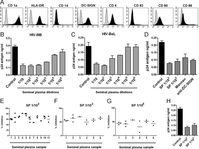
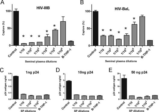
Dendritic cell-specific intercellular adhesion molecule 3-grabbing nonintegrin (DC-SIGN) is expressed by dendritic cells (DCs) at mucosal surfaces and appears to play an important role in the dissemination of human immunodeficiency virus type 1 (HIV-1) infection. DC-SIGN binds HIV-1 gp120 and efficiently transmits the virus to T CD4(+) cells, which become the center of viral replication. Semen represents the main vector for HIV-1 dissemination worldwide. In the present study we show that human seminal plasma (SP), even when used at very high dilutions (1:10(4) to 1:10(5)), markedly inhibits the capture and transmission of HIV-1 to T CD4(+) cells mediated by both DCs and B-THP-1-DC-SIGN cells. In contrast, SP does not inhibit the capture of HIV-1 by DC-SIGN-negative target cells, such as the T-cell line SupT-1, monocytes, and activated peripheral blood mononuclear cells. The SP inhibitor has a high molecular mass (>100 kDa) and directly interacts with DC-SIGN-positive target cells but not with HIV-1. Moreover, the inhibitor binds to concanavalin A, suggesting that it contains high-mannose N-linked carbohydrates. Of note, using biotin-labeled SP we found that the binding of SP components to DCs was abrogated by mannan, while their interaction with B-THP-1 cells was almost completely dependent on the expression of DC-SIGN. Since epithelium integrity is often compromised after vaginal or anal intercourse, as well as in the presence of ulcerative-sexually transmitted diseases, our results support the notion that components of the SP might be able to access to the subepithelium, inhibiting the recognition of HIV-1 gp120 by DC-SIGN-positive DCs.


Figures

FIG. 1.
FIG. 1.
Capture of HIV-1 by DCs is inhibited by SP. (A) Representative histograms of the phenotype of immature DCs. (B and C) DCs were incubated for 30 min at 37°C with different SP dilutions and then cultured with HIV-1IIIB (CXCR4) or HIV-1BaL (CCR5) (5 ng of p24 for 90 min at 37°C). Cells were then washed thoroughly, lysed, and assayed for p24 antigen by ELISA. The results from a representative experiment (n = 9) performed in triplicate are shown. (D) DCs were incubated for 30 min at 37°C in the absence or presence of different SP dilutions, mannan (1 mg/ml), or a blocking MAb directed to DC-SIGN used at a concentration three- to fivefold higher than those needed to saturate all binding sites, as determined by FACS analysis. DCs were then cultured with HIV-1BaL (5 ng of p24) for 90 min at 37°C. After this period, cells were washed thoroughly, lysed, and assayed for p24 antigen by ELISA. A representative experiments (n = 5) made by triplicate is shown. (E to G) SP and DCs from different donors were studied in capture assays. Each point represents a distinct DC donor. Experiments were carried out as described above, using SP dilutions of 1:102 (E), 1:103 (F), and 1:104 (G) and HIV-1BaL (5 ng of p24). The results are expressed as the percent inhibition of HIV-1 capture for each DC donor. (H) A 100-μl portion of an SP dilution of 1:5 was mixed with 100 μl of two HIV-1BAL stocks previously adjusted to 1 or 10 μg of p24/ml. Both mixtures were incubated for 15 min at 37°C. After this period, 10 μl of the first mixture and 1 μl of the second mixture were added to 106 DCs suspended in 90 and 99 μl of culture medium, respectively, to yield DC suspensions exposed to SP dilutions of 1:102 and 1:103 in the presence of 5 ng of p24. After incubation for 90 min at 37°C, the DCs were washed thoroughly, pelleted, lysed, and assayed for HIV p24 by ELISA. The results of representative experiments (n = 3) performed in triplicate are shown.
FIG. 2.
FIG. 2.
SP inhibits the capture of HIV-1 mediated by DC-SIGN. (A and B) B-THP-1-DC-SIGN cells were incubated for 30 min at 37°C without (controls) or with different SP dilutions and then were cultured with HIV-1IIIB or HIV-1BaL (5 ng of p24) for 90 min at 37°C. The cells were then washed thoroughly, lysed, and assayed for p24 antigen by ELISA. The capture mediated by B-THP-1 cells, which do not express DC-SIGN, is also shown. The results are expressed as the percentage of HIV-1 capture compared to control cells. The data represent the arithmetic means ± the SEM of five to six experiments carried out in triplicate. Asterisk represents statistical significance (P < 0.05) versus controls. (C to E) Capture assays by B-THP-1-DC-SIGN cells was performed as described above, using different amounts of HIV-1BaL as indicated for each panel. The results of representative experiments are shown (n = 3 to 4).
FIG. 3.
FIG. 3.
SP does not inhibit the capture of HIV-1 mediated by DC-SIGN negative target cells. Activated PBMC (10 U of IL-2/ml plus 10 μg of PHA/ml for 48 h), monocytes, and the cell line SupT-1 were incubated for 30 min at 37°C without (controls) or with a SP dilution of 1:102 and then were exposed to HIV-1IIIB (CXCR4) (5 ng of p24) for 90 min at 37°C. The cells were then washed thoroughly, lysed, and evaluated for p24 antigen by ELISA. The data represent the arithmetic means ± the SEM of three experiments carried out in triplicate.
FIG. 4.
FIG. 4.
Infection of DCs by HIV-1 is inhibited by SP. (A) Infection of DCs was performed by incubating cells for 30 min at 37°C without (controls) or with SP dilutions of 1:102 or 1:103, mannan (1 mg/ml), or saturating concentrations of a blocking MAb directed to DC-SIGN. Cells were then exposed to HIV-1BaL (5 ng of p24) for 90 min at 37°C, washed thoroughly, and cultured for several days. Supernatants harvested at days 3, 6, and 9 were assayed for HIV p24 antigen by ELISA. The results of a representative experiment (n = 4) performed in triplicate are shown. (B) Transient exposure to SP does not result in a loss of DC viability. DCs were incubated in the absence or presence of a SP dilution of 1:102 for 2 h at 37°C. Cells were then washed and cultured for different periods at 37°C (0 h to 9 days). Cell viability was then analyzed using the MTT assay as described in Materials and Methods. Optical densities were measured at 570 nm. Values for control cells (i.e., cells not pretreated with SP) were considered, at each time point, as 100% viability. The results of a representative experiment (n = 3) performed in triplicate are shown. (C) Infection of GHOST cells is not inhibited by SP. GHOST cells (DC-SIGN negative, CD4+, CXCR4+, and CCR5+) were exposed to HIV-1BaL (5 ng of p24) for 90 min at 37°C, washed thoroughly, and cultured for 48 h at 37°C. Infection of GHOST cells was analyzed by flow cytometry as described in Materials and Methods. The results of a representative experiment (n = 5) are shown.
FIG. 5.
FIG. 5.
SP inhibits trans-infection of T CD4+ cells. DCs (A) or B-THP-1-DC-SIGN cells (B and C) were incubated for 30 min at 37°C without (controls) or with different SP dilutions, mannan (1 mg/ml), or saturating concentrations of a blocking MAb directed to DC-SIGN. Cells were then exposed to HIV-1BaL (5 ng of p24) for 90 min at 37°C. After this period, cells were thoroughly washed, and trans-infection of activated PBMC (A), the T-cell line SupT-1 (B), and GHOST cells (DC-SIGN negative, CD4+, CXCR4+, and CCR5+) (C) was assessed as described in Materials and Methods. The results of representative experiments (n = 3 to 6) are shown. Trans-infection mediated by B-THP-1 cells, which do not express DC-SIGN, is also shown in panels B and C.
FIG. 6.
FIG. 6.
Analysis of the inhibitory activity mediated by SP. (A) B-THP-1-DC-SIGN cells were incubated without (controls) or with different SP dilutions, for 30 min at 37°C. Cells were then washed four times and cultured with HIV-1BaL (5 ng of p24) for 90 min at 37°C. Cells were then washed thoroughly, lysed, and assayed for p24 antigen by ELISA. (B) Viral stocks (HIV-1BaL) were incubated without or with different SP dilutions for 30 min at 37°C. HIV-1 stocks were then washed three times by ultracentrifugation. B-THP-1-DC-SIGN cells were exposed to untreated (controls) or SP-pretreated HIV-1 (5 ng of p24) for 90 min at 37°C. Cells were then washed thoroughly, lysed, and assayed for p24 antigen by ELISA. (C and D) DCs were cultured for different periods in the absence or presence of SP (dilution 1:102) or mannan (1 mg/ml). After the cells were washed, the expression of DC-SIGN (C) and CD4 (D) was analyzed by flow cytometry. Dotted histograms represent the expression of DC-SIGN (C) or CD4 (D) by control DCs. The results of a representative experiment (n = 3) are shown. (E) Aliquots of SP (dilution 1:10) were subjected to different treatments. They were heated for 10 min at 95°C, treated with trypsin (3 h at 37°C and 10 min at 95°C), treated with an antibody directed to LeX, or filtered in centrifugal filter devices with 100-kDa cutoffs and then diluted with culture medium to its original volume. The ability of each SP sample to inhibit the capture of HIV-1BaL (5 ng of p24) by B-THP-1-DC-SIGN cells was then assessed by using a final dilution of 1:103. (F) ConA-Sepharose chromatography of SP (1 ml of SP diluted 1:10 with culture medium) was performed as described in Materials and Methods. The ConA-interacting fraction was eluted using 5% α-methyl-d-mannoside, thoroughly dialyzed, and used at final dilutions of 1:103, 1:104, or 1:105. The ability to inhibit the capture of HIV-1 by B-THP-1-DC-SIGN cells was then assessed. We also studied, as a control, the ability of the same SP sample, not subjected to chromatography, to inhibit HIV-1 capture. In panels A, B, E, and F the data represent the arithmetic means ± the SEM of three to five experiments carried out in triplicate. Asterisk represents statistical significance (P < 0.05) versus controls.
FIG. 7.
FIG. 7.
Analysis of the role of DC-SIGN in the binding of biotin-labeled SP to DCs and B-THP-1-DC-SIGN cells. Biotinylation of SP was performed as described under Materials and Methods. (A) DCs (1.5 × 10⁶ cells/ml) were incubated for 30 min at 4°C with different dilutions of biotin-labeled SP. Cells were then washed, and the binding of biotin-labeled SP to DCs was evaluated by flow cytometry using FITC-avidin. The results of a representative experiment (n = 4) are shown. (B) DCs (1.5 × 10⁶ cells/ml) were incubated for 30 min at room temperature with or without mannan (1 mg/ml). The cells were then incubated with dilutions of biotin-labeled SP of 1:102 or 1:103 for 30 min at 4°C. The cells were then washed, and the binding of biotin-labeled SP to DCs was evaluated by flow cytometry using FITC-avidin. The results of a representative experiment (n = 3) are shown. (C and D) The expression of DC-SIGN (C) and the binding of biotin-labeled SP (1:102) (D) were measured by flow cytometry in B-THP-1 cells, B-THP-1-DC-SIGN cells, and mixed cell suspensions containing equal numbers of each cell type. The results of a representative experiment (n = 4) are shown.

Similar articles

Cited by

References

    1. Allen, R. D., and T. K. Roberts. 1986. The relationship between the immunosuppressive and cytotoxic effect of human seminal plasma. Am. J. Reprod. Immunol. Microbiol. 11:59-64. - PubMed
    1. Appelmelk, B. J., I. van Die, S. J. van Vliet, C. M. Vandenbroucke-Grauls, T. B. Geijtenbeek, and Y. van Kooyk. 2003. Carbohydrate profiling identifies new pathogens that interact with dendritic cell-specific ICAM-3-grabbing nonintegrin on dendritic cells. J. Immunol. 170:1635-1639. - PubMed
    1. Berlier, W., M. Cremel, H. Hamzeh, R. Lévy, F. Lucht, T. Bourlet, B. Pozzetto, and O. Delézayi. 2006. Seminal plasma promotes the attraction of Langerhans cells via the secretion of CCL20 by vaginal epithelial cells: involvement in the sexual transmission of HIV. Hum. Reprod. 21:1135-1142. - PubMed
    1. Bouhlal, H., N. Chomont, N. Haeffner-Cavaillon, M. D. Kazatchkine, L. Belec, and H. Hocini. 2002. Opsonization of HIV-1 by semen complement enhances infection of human epithelial cells. J. Immunol. 169:3301-3306. - PubMed
    1. Bouvet, J. P., G. Gresenguet, and L. Belec. 1997. Vaginal pH neutralization by semen as a cofactor of HIV transmission. Clin. Microbiol. Infect. 3:19. - PubMed

Publication types

MeSH terms

Substances