. 2007 Dec;81(24):13354-64.
doi: 10.1128/JVI.01196-07. Epub 2007 Oct 3.

Chaperones activate hepadnavirus reverse transcriptase by transiently exposing a C-proximal region in the terminal protein domain that contributes to epsilon RNA binding

Affiliations

Chaperones activate hepadnavirus reverse transcriptase by transiently exposing a C-proximal region in the terminal protein domain that contributes to epsilon RNA binding

Michael Stahl et al. J Virol. 2007 Dec.

Abstract

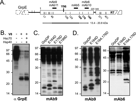
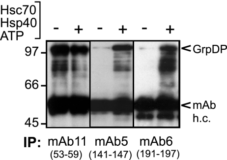
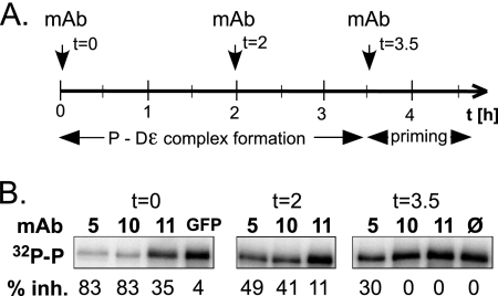
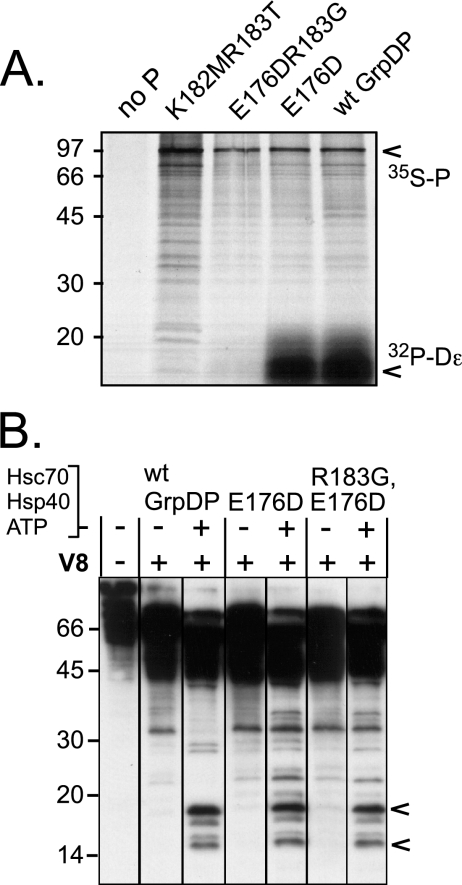
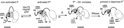
All hepatitis B viruses replicate by protein-primed reverse transcription, employing a specialized reverse transcriptase, P protein, that carries a unique terminal protein (TP) domain. To initiate reverse transcription, P protein must bind to a stem-loop, epsilon, on the pregenomic RNA template. TP then provides a Y residue for covalent attachment of the first nucleotide of an epsilon-templated DNA oligonucleotide (priming reaction) that serves to initiate full-length minus-strand DNA synthesis. epsilon binding requires the chaperone-dependent conversion of inactive P protein into an activated, metastable form designated P*. However, how P* differs structurally from P protein is not known. Here we used an in vitro reconstitution system for active duck hepatitis B virus P combined with limited proteolysis, site-specific antibodies, and defined P mutants to structurally compare nonactivated versus chaperone-activated versus primed P protein. The data show that Hsp70 action, under conditions identical to those required for functional activation, transiently exposes the C proximal TP region which is, probably directly, involved in epsilon RNA binding. Notably, after priming and epsilon RNA removal, a very similar new conformation appears stable without further chaperone activity; hence, the activation of P protein is triggered by energy-consuming chaperone action but may be completed by template RNA binding.


Figures

FIG. 1.
FIG. 1.
P-protein domain structure and activation. (A) Wild-type DHBV P protein (wt P) and the recombinant P proteins (rec P) used in the present study. Like all hepadnaviral P proteins, DHBV P protein consists of conserved RT and RH domains plus an N-terminal TP domain and a functionally dispensable spacer. Y96 in TP serves as attachment site for the 5′ end of the DNA primer oligonucleotide. Numbers represent amino acid positions. In the recombinant proteins the spacer is replaced by a short SPG linker and the C-terminal amino acids past position 761 are deleted (6); the heterologous domains (GrpE or NusA) enhance solubility. (B) Schematic representation of primed P protein. Bound ɛ RNA undergoes a conformational alteration, which opens up the upper ɛ stem (3) and appropriately positions the Y residue in TP and the template region in ɛ so as to allow the RT domain to copy the DNA primer (thick curved line connected to TP). DNA synthesis ceases until ɛ is replaced as a template by the DR1* element close to the opposite end of the pgRNA. (C) Stepwise formation of primed P protein. Inactive P is transiently activated into P* by the ATP-consuming activity of Hsc70 and Hsp40; Hsp90 plus Hop, or Bag-1S, can further stimulate P* formation. P* is metastable, and its steady-state concentration is determined by the rates of P* production and decay. Only P* can bind ɛ RNA, forming a stable initiation-competent complex. If provided with deoxynucleoside triphosphates, the complex carries out synthesis of the primer oligonucleotide.
FIG. 2.
FIG. 2.
Hsc70 and Hsp40 plus ATP induce a specific conformational change involving the TP domain. (A) V8 fragments (14 to 18 kDa) from GrpDP are only formed in the presence of Hsc70 plus Hsp40 plus ATP. GrpDP was incubated with the indicated factors and then subjected to limited V8 proteolysis. Products were analyzed by SDS-PAGE and immunoblotting, with MAb9 recognizing an epitope involving TP residues 53 to 59. Numbers on the left show the positions of marker proteins of the indicated sizes in kilodaltons. (B) Chaperone-induced 14- to 18-kDa fragments occur independently of the heterologous fusion partner and of the spacer region. NusDP, containing NusA instead of GrpE as a fusion partner, and GrpDP including the authentic P-protein spacer region were incubated with Hsc70, Hsp40 plus ATP, or not and then subjected to V8 digestion as in panel A. (C) Limited proteolysis with Asp-N protease also detects a chaperone-dependent conformational alteration involving TP. GrpDP was incubated with Hsc70, Hsp40 plus ATP, or not and then subjected to proteolysis with Asp-N protease. Reactions were analyzed as in panel A. (D) Hsp90/Hop and Bag-1S do not induce detectable additional conformational changes in TP. GrpDP was incubated with Hsc70 and Hsp40 plus ATP as in panel A, but in addition with Hsp90/Hop or Bag-1S. Reactions were analyzed as in panel A.
FIG. 3.
FIG. 3.
The chaperone-induced conformation of P protein is metastable. GrpDP was incubated in the presence of Hsc70, Hsp40, and ATP as in Fig. 2, and then the reaction mixture was split. One-half was treated with hexokinase and glucose for ATP depletion (panel −ATP); the other half was not (panel +ATP). The reactions were further incubated at 30°C for 3 h, and aliquots were taken at the indicated time points, subjected to V8 protease cleavage, and analyzed by immunoblotting. Note that the chaperone-induced 14- to 18-kDa fragment (arrowheads) remained present at essentially at the same concentration in the presence of ATP and yet disappeared upon ATP depletion; most of the other proteolysis products remained unaffected.
FIG. 4.
FIG. 4.
Mapping the borders of the chaperone-induced 14- to 18-kDa V8 fragments. (A) Potential V8 cleavage sites in and around the TP part of GrpDP. TP is shown as a bar, flanked by the GrpE and the RT domain. Each vertical line represents an E residue. The positions of some of the E residues, of Y96, and of the MAb epitopes are indicated. The candidate region producing the 14- to 18-kDa fragments is represented by the straight line with the minimal (solid part) and maximal (dashed extensions) segments plus their calculated molecular masses. (B) Cleavage between GrpE and TP is chaperone independent. GrpDP was incubated with the indicated chaperones and then subjected to V8 proteolysis. Products were monitored by immunoblotting with an anti-GrpE antibody. GrpE was efficiently clipped off with or without chaperones. (C) E164 is involved in the C-terminal V8 cleavage. GrpDP and the indicated mutants were incubated with Hsc70, Hsp40, and ATP and then subjected to V8 proteolysis. Products were separated by SDS-PAGE in a Tris-Tricine gel system and analyzed by immunoblotting with MAb9. Note the mobility shift for the 14- to 18-kDa fragments from mutant E164D. (D) E176 and E199 are also exposed by chaperone action. GrpDP and the indicated mutants were treated as described above, and then the products were detected by using MAb9 (left panel) or MAb6 (right panel). Note the upward shift of the fragments in question with the double mutant and the exclusive recognition of these upshifted fragments by MAb6.
FIG. 5.
FIG. 5.
Efficient immunoprecipitation of GrpDP by MAbs recognizing the C-proximal TP part requires chaperone activity. GrpDP was incubated with or without chaperones and immunoprecipitated with MAb5, MAb6, or MAb11. Bound proteins were analyzed by immunoblotting with MAb9. Bands corresponding to GrpDP and to the heavy chains of the immunoprecipitation (IP) MAbs (MAb h.c.) are indicated on the right.
FIG. 6.
FIG. 6.
A conformation similar to that transiently induced by chaperones is stabilized in primed, RNA-deprived P protein. (A) Primed wild-type GrpDP protein. GrpDP was incubated with Hsc70, Hsp40, ATP, and Dɛ RNA and then subjected to priming conditions in the presence of [α-32P]dATP. Prior to V8 proteolysis, some samples were treated with increasing amounts of RNase A (0.1, 1.0, or 10 μg per reaction), as indicated by “+,” “++,” or “+++.” Proteolysis products were detected by SDS-PAGE, followed by phosphorimaging. (B) V8 fragments (14 to 20 kDa) from primed RNA-deprived P protein are stable without ATP-consuming chaperone activity. Reconstituted primed P-Dɛ complexes were subjected to ATP depletion or not; both samples were then treated with RNase and further incubated. Over a time course of 2 h aliquots were removed and probed by V8 proteolysis. Products were analyzed by SDS-PAGE, followed by phosphorimaging. The signal intensities of the 14- to 20-kDa fragments after 2 h were still ca. 70% as high as those immediately after RNase treatment, irrespective of ATP depletion. (C) Mutants E164D and E164,176D are functionally active. The mutant proteins were analyzed in parallel to wild-type GrpPD as in panel A. No significant differences in priming signal intensities and formation of stable 45-kDa V8 fragments were detectable in the absence of RNase treatment. (D) E164 and E176 are involved in generating the chaperone-specific small V8 fragments from primed P protein. In vitro-primed P proteins were treated with 10 μg of RNase A per reaction and subjected to V8 proteolysis as in panel A, followed by immunoprecipitation with either MAb9 or MAb6. Precipitated proteins were analyzed by SDS-PAGE using the Tris-Tricine system. Control lanes on the left show primed untreated P protein, RNA-deprived V8-treated P protein without immunoprecipitation, and primed untreated P protein incubated with an anti-GFP MAb. wt, wild type.
FIG. 7.
FIG. 7.
Selective inhibition of P-Dɛ complex formation and priming by MAbs recognizing the C proximal TP region. (A) Experimental setup. GrpDP was incubated with Hsc70, Hsp40, ATP, and Dɛ RNA for 3.5 h and then subjected to priming conditions in the presence of [α-32P]dATP. MAbs directed against different epitopes in TP and against a GFP antibody or no antibody as controls were added at the indicated time points. (B) Effects of MAbs on the formation of primed P protein. Samples from the priming reactions were analyzed by SDS-PAGE and autoradiography. Signals for 32P-labeled P protein were quantified by phosphorimaging. Relative inhibition (percent inhibition values) was derived by normalizing the signal intensities to that of the reaction without added antibody (0% inhibition).
FIG. 8.
FIG. 8.
RNA-binding deficiency of P mutant R183G is not caused by a gross structural alteration. (A) The single R183G mutation ablates Dɛ RNA binding as efficiently as a K182MR183T double mutation. The indicated proteins were in vitro translated in RL in the presence of [35S]methionine and 32P-labeled Dɛ RNA and immobilized via their His tags to Ni-NTA agarose. Protein and potentially bound RNA were analyzed by SDS-PAGE and autoradiography. The positions of 35S-labeled P protein and of 32P-labeled RNA are indicated by arrowheads. (B) The RNA-binding-deficient R183G mutant undergoes the same chaperone-induced conformational change as RNA-binding-competent P protein. The indicated proteins were incubated with Hsc70, Hsp40, and ATP, or not and subjected to limited V8 proteolysis, followed by immunoblotting with MAb9. All samples were run on the same gel, except that the lanes have been reordered for ease of comparison between chaperone-treated and nontreated samples from one protein. The R183G mutant produced essentially the same chaperone-induced fragments as wild-type GrpDP and the parental, RNA-binding-competent variant E176D. wt, wild type.
FIG. 9.
FIG. 9.
Structural model for hepadnaviral P protein activation. Initially, P protein is in a stable nonactivated state, in which the C-proximal TP region including E164, E176, and E199 and the epitopes for MAb5, MAb6, and MAb10 is occluded, whereas the N-proximal E residues in TP and the epitopes for MAb9 and MAb11 are, and remain, accessible. Occlusion might involve the RH domain, as suggested by the partial chaperone independence of P proteins with large C-terminal truncations (2, 43). ATP-consuming Hsc70 plus Hsp40 action transiently exposes the C-proximal sites, including R183, which is involved in RNA binding. In this metastable activated state P*, the corresponding MAbs, and ɛ can compete for binding to the C-proximal TP region. ɛ binding generates a stable priming-competent P*-Dɛ complex in which the RNA blocks the access of V8 protease and the respective MAbs. RNase A treatment creates a new state P′ in which the same sites are exposed as in chaperone-activated P*; however, different from P*, P′ remains stable without ATP-consuming chaperone activity. P′, here artificially created by RNA digestion, may resemble P protein in transition from ɛ-dependent initiation to elongation mode, which likely accompanies the replacement of ɛ by DR1* as the RNA template.

Similar articles

Cited by

References

    1. Beck, J., and M. Nassal. 1996. A sensitive procedure for mapping the boundaries of RNA elements binding in vitro translated proteins defines a minimal hepatitis B virus encapsidation signal. Nucleic Acids Res. 24:4364-4366. - PMC - PubMed
    1. Beck, J., and M. Nassal. 2003. Efficient Hsp90-independent in vitro activation by Hsc70 and Hsp40 of duck hepatitis B virus reverse transcriptase, an assumed Hsp90 client protein. J. Biol. Chem. 278:36128-36138. - PubMed
    1. Beck, J., and M. Nassal. 1998. Formation of a functional hepatitis B virus replication initiation complex involves a major structural alteration in the RNA template. Mol. Cell. Biol. 18:6265-6272. - PMC - PubMed
    1. Beck, J., and M. Nassal. 2007. Hepatitis B virus replication. World J. Gastroenterol. 13:48-64. - PMC - PubMed
    1. Beck, J., and M. Nassal. 2004. In vitro reconstitution of epsilon-dependent duck hepatitis B virus replication initiation. Methods Mol. Med. 95:315-325. - PubMed

Publication types

MeSH terms

LinkOut - more resources