. 2007 Dec;81(24):13335-45.
doi: 10.1128/JVI.01238-07. Epub 2007 Oct 3.

NSm protein of Rift Valley fever virus suppresses virus-induced apoptosis

Affiliations

NSm protein of Rift Valley fever virus suppresses virus-induced apoptosis

Sungyong Won et al. J Virol. 2007 Dec.

Abstract

Rift Valley fever virus (RVFV) is a member of the genus Phlebovirus within the family Bunyaviridae. It can cause severe epidemics among ruminants and fever, myalgia, a hemorrhagic syndrome, and/or encephalitis in humans. The RVFV M segment encodes the NSm and 78-kDa proteins and two major envelope proteins, Gn and Gc. The biological functions of the NSm and 78-kDa proteins are unknown; both proteins are dispensable for viral replication in cell cultures. To determine the biological functions of the NSm and 78-kDa proteins, we generated the mutant virus arMP-12-del21/384, carrying a large deletion in the pre-Gn region of the M segment. Neither NSm nor the 78-kDa protein was synthesized in arMP-12-del21/384-infected cells. Although arMP-12-del21/384 and its parental virus, arMP-12, showed similar growth kinetics and viral RNA and protein accumulation in infected cells, arMP-12-del21/384-infected cells induced extensive cell death and produced larger plaques than did arMP-12-infected cells. arMP-12-del21/384 replication triggered apoptosis, including the cleavage of caspase-3, the cleavage of its downstream substrate, poly(ADP-ribose) polymerase, and activation of the initiator caspases, caspase-8 and -9, earlier in infection than arMP-12. NSm expression in arMP-12-del21/384-infected cells suppressed the severity of caspase-3 activation. Further, NSm protein expression inhibited the staurosporine-induced activation of caspase-8 and -9, demonstrating that other viral proteins were dispensable for NSm's function in inhibiting apoptosis. RVFV NSm protein is the first identified Phlebovirus protein that has an antiapoptotic function.


Figures
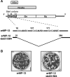
FIG. 1.
FIG. 1.
(A) Schematic diagram of anti-genomic-sense M segments of arMP-12 and arMP-12-del21/384. Vertical lines in the pre-Gn region represent five in-frame translation initiation codons. Shaded boxes at the top represent the coding regions of the NSm and 78-kDa proteins. The sequences around the first, second, and third AUG codons and the deleted region of pre-Gn in arMP-12-del21/384 are shown at the bottom. (B) Plaque morphologies of arMP-12 and arMP-12-del21/384. Vero E6 cells were infected with arMP-12 or arMP-12-del21/384, and plaques were fixed and stained with 0.75% crystal violet, 10% formaldehyde, and 5% ethanol at 4 days p.i.
FIG. 2.
FIG. 2.
Replication of arMP-12 and arMP-12-del21/384 in VeroE6 cells and J774.1 cells. (A) Vero E6 cells were either mock infected or independently infected with arMP-12 or arMP-12-del21/384 at an MOI of 1. Intracellular RNAs were extracted at 8 h p.i., and the accumulation of viral RNA was examined by Northern blot analysis with viral-sense-specific RNA probes (23). L, M, and S segments are indicated with arrowheads. The M segment of arMP-12-del21/384 migrated faster than that of arMP-12 due to a large deletion in the pre-Gn region. (B) Vero E6 cells were either mock infected or independently infected with the indicated viruses at an MOI of 3, and total proteins were collected at 8 h p.i. Western blot analysis using an anti-RVFV antibody was performed to demonstrate the accumulation of Gn/Gc, NSs, and N proteins. An anti-L antibody and an anti-actin antibody were used to detect L protein and actin protein, respectively, while an anti-NSm antibody was used to detect both the NSm and 78-kDa proteins (51). (C) Vero E6 cells were either mock infected or independently infected with the indicated viruses at an MOI of 3. Cells were radiolabeled with 100 μCi/ml at 8 h p.i. for 30 min. Cytoplasmic extracts were incubated with an anti-NSm antibody or with preimmune serum (Pre), and radioimmunoprecipitation (IP) was performed as described previously (51). (D) Vero E6 and J774.1 cells were independently infected with arMP-12 or arMP-12-del21/384 at the indicated MOI, and the culture supernatants were collected from triplicates at the indicated time p.i. Titers were determined by a plaque assay in Vero E6 cells.
FIG. 3.
FIG. 3.
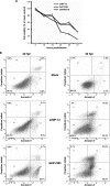
Cell viability assay (A) and annexin V/PI staining and flow cytometric analysis (B) of infected cells. (A) Vero E6 cells were independently infected with arMP-12, arMP-12-delNSm/78, or arMP-12-del21/384 at an MOI of 10. At the indicated time p.i., infected cells were treated with 0.5 mg of MTT/ml and incubated for 4 h. The metabolic reaction was terminated, and MTT formazan crystals in living cells were dissolved by addition of an MTT solubilization solution. Absorbance was measured at a wavelength of 570 nm, and the background absorbance at 690 nm was subtracted. Cell viability was calculated as a percentage relative to the absorbance of mock-infected control cultures at the corresponding time p.i. Data are the results of triplicate experiments. (B) Vero E6 cells were either mock infected or independently infected with arMP-12 or arMP-12-del21/384 at an MOI of 3. Cells, including floating cells, were collected at 20 and 40 h p.i. and were stained with annexin V and PI for 15 min. A total of 1 × 10⁴ stained cells were analyzed by flow cytometry. Dot plots are divided into quadrants of cell populations: Q1 (staining PI positive and annexin V negative), Q2 (staining positive with both), Q3 (staining negative with both), and Q4 (staining annexin V positive and PI negative). Q4 represents the early-apoptotic cell population. The percentage of cells in each quadrant is also presented.
FIG. 4.
FIG. 4.
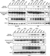
Activation of caspase-3 and cleavage of PARP in virus-infected cells. Vero E6, 293, and J774.1 cells were either mock infected or infected with arMP-12 or arMP-12-del21/384 at an MOI of 3. Total-cell lysates were collected at the indicated time p.i. and subjected to Western blot analysis. Anti-cleaved caspase-3 (Asp175), an anti-PARP antibody, and an anti-actin antibody were used to demonstrate cleaved caspase-3 (p17), both uncleaved and cleaved PARP, and actin, respectively. RVFV N and NSs proteins were detected by an anti-RVFV antibody. Lanes STP and DMSO, 293 cells treated with 3 μM STP in DMSO and with DMSO only, respectively. The specific band signal in each immunoblot was quantified by densitometric scanning and normalized against an actin control for cleaved caspase-3 or against total PARP (uncleaved and cleaved PARP) for cleaved PARP. Increases (n-fold) over the signal from mock-infected cells are given below the lanes.
FIG. 5.
FIG. 5.
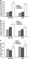
Enzymatic activities of caspases in virus-infected cells. Vero E6 and 293 cells were either mock infected or independently infected with arMP-12 or arMP-12-del21/384 at an MOI of 3. Cells were collected at 16 h p.i., and 100 μg of proteins from each sample was incubated with a colorimetric peptide substrate for caspase-3/7 (A), caspase-8 (B), or caspase-9 (C) for 2 h. Color changes were measured at 405 nm, and the background reading from cell lysates and buffers was subtracted from the sample reading. Data are representative of at least three independent experiments, and statistical significance was determined by a paired Student t test. Asterisks represent P values for the differences between arMP-12 and arMP-12-del21/384 infections (P < 0.001, P < 0.001, and P < 0.05 for panels A, B, and C, respectively).
FIG. 6.
FIG. 6.
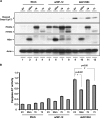
Effects of expression of NSm and the 78-kDa and 73-kDa proteins on arMP-12-del21/384 replication-induced apoptosis. (A) 293 cells in 12-well plates were independently transfected with 1 μg of either pCAGGS (EV), pCAGGS-NSm (NSm), pCAGGS-78 (78), or pCAGGS-73 (73). At 48 h posttransfection, cells were either mock infected or independently infected with arMP-12 or arMP-12-del21/384 (del21/384) at an MOI of 3. Total-cell lysates were collected at 16 h p.i. and analyzed by Western blot analysis with an anti-cleaved caspase-3 antibody detecting cleaved caspase 3 (p17), an anti-NSm antibody detecting NSm and the 73-kDa and 78-kDa proteins, and an anti-actin antibody. Experiments were performed three times, and similar results were obtained. A set of representative data is shown. (B) 293 cells in six-well plates were transfected with 2.5 μg of plasmids as described for panel A and then either mock infected or independently infected with arMP-12 or arMP-12-del21/384 at 48 h posttransfection. At 16 h p.i., cell lysates were collected and the caspase-3/7 enzymatic activities of lysates were analyzed as described in the legend to Fig. 5. All samples were prepared in triplicate, and statistical significance was determined by a paired Student t test (*, P < 0.001; #, P < 0.05).
FIG. 7.
FIG. 7.
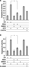
Inhibition of STP-induced apoptosis by NSm expression. 293 cells were independently transfected with pCAGGS (EV) or pCAGGS-NSm, followed by treatment with 3 μM STP or DMSO at 48 h posttransfection. The enzymatic activities of caspase-8 (A) and caspase-9 (B) were measured with 50 μg of lysates at 3 h after treatment with STP or DMSO as described in the legend to Fig. 5. As a control, cells were treated with 20 μM synthetic caspase-8 inhibitor (Z-IETD) or caspase-9 inhibitor (Z-LEHD) 1 h prior to STP treatment. Asterisks represent P values of <0.001 for panel A and <0.01 for panel B. Data are results of triplicate experiments.

Similar articles

Cited by

References

    1. Aggarwal, B. B., W. J. Kohr, P. E. Hass, B. Moffat, S. A. Spencer, W. J. Henzel, T. S. Bringman, G. E. Nedwin, D. V. Goeddel, and R. N. Harkins. 1985. Human tumor necrosis factor. Production, purification, and characterization. J. Biol. Chem. 260:2345-2354. - PubMed
    1. Balachandran, S., C. N. Kim, W. C. Yeh, T. W. Mak, K. Bhalla, and G. N. Barber. 1998. Activation of the dsRNA-dependent protein kinase, PKR, induces apoptosis through FADD-mediated death signaling. EMBO J. 17:6888-6902. - PMC - PubMed
    1. Balkhy, H. H., and Z. A. Memish. 2003. Rift Valley fever: an uninvited zoonosis in the Arabian peninsula. Int. J. Antimicrob. Agents 21:153-157. - PubMed
    1. Banerjee, S., K. Narayanan, T. Mizutani, and S. Makino. 2002. Murine coronavirus replication-induced p38 mitogen-activated protein kinase activation promotes interleukin-6 production and virus replication in cultured cells. J. Virol. 76:5937-5948. - PMC - PubMed
    1. Billecocq, A., M. Spiegel, P. Vialat, A. Kohl, F. Weber, M. Bouloy, and O. Haller. 2004. NSs protein of Rift Valley fever virus blocks interferon production by inhibiting host gene transcription. J. Virol. 78:9798-9806. - PMC - PubMed

Publication types

MeSH terms

LinkOut - more resources