. 2007 Dec;81(24):13608-21.
doi: 10.1128/JVI.01509-07. Epub 2007 Oct 3.

Identification of regions and residues in feline junctional adhesion molecule required for feline calicivirus binding and infection

Affiliations

Identification of regions and residues in feline junctional adhesion molecule required for feline calicivirus binding and infection

Robert J Ossiboff et al. J Virol. 2007 Dec.

Abstract

The feline junctional adhesion molecule A (fJAM-A) is a functional receptor for feline calicivirus (FCV). fJAM-A is a member of the immunoglobulin superfamily (IgSF) and consists of two Ig-like extracellular domains (D1 and D2), a membrane-spanning domain, and a short cytoplasmic tail. To identify regions of fJAM-A that interact with FCV, we purified recombinant fJAM-A ectodomain and D1 and D2 domains. We found that preincubation of FCV with the ectodomain or D1 was sufficient to inhibit FCV infection in plaque reduction assays. In enzyme-linked immunosorbent assays, FCV binding to fJAM-A ectodomain was concentration dependent and saturable; however, FCV bound D1 alone weakly and was unable to bind D2. To characterize FCV binding to surface-expressed fJAM-A, we transfected truncated and chimeric forms of fJAM-A into a nonpermissive cell line and assayed binding by flow cytometry. Only D1 was necessary for FCV binding to cells; all other domains could be replaced. Using a structure-guided mutational approach, we identified three mutants of fJAM-A within D1 (D42N, K43N, and S97A) that exhibited significantly decreased capacities to bind FCV. In contrast to our finding that D1 mediated FCV binding, we found that all domains of fJAM-A were necessary to confer susceptibility to FCV infection. Furthermore, surface expression of fJAM-A was not sufficient to permit FCV infection by all of the isolates we investigated. This indicates that (i) other cellular factors are required to permit productive FCV infection and (ii) individual FCV isolates differ in the factors they require.


Figures

FIG. 1.
FIG. 1.
Characterization of recombinant fJAM-A ectodomain and fJAM-A-specific rabbit antisera. (A) GST-Ecto, GST-D1, and GST-D2 were expressed in E. coli (BL21). Soluble recombinant proteins were purified by affinity chromatography. (B) The fJAM-A regions of the recombinant proteins were released by 3C protease cleavage, and the predicted sizes of the fusion and cleaved proteins were verified by SDS-polyacrylamide gel electrophoresis. (C) A rabbit polyclonal antiserum against soluble fJAM-A ectodomain was prepared, and its capacity to specifically recognize the purified fusion and cleavage proteins was confirmed by immunoblotting; a higher concentration of antibody was used to detect the cleaved D2 (indicated by an asterisk). Membranes were also probed with anti-GST. (D) Surface expression of fJAM-A in CHO cells by fluorescence microscopy. CHO-K1 cells were transfected with pCI-fJAM-A to express full-length fJAM-A or the empty vector, pCI-Neo. The cells were fixed at 24 h p.t. and immunostained with fJAM-A rabbit antiserum, followed by Alexa 594-conjugated goat anti-rabbit IgG. Nuclei were stained with DAPI. (E) The fJAM-A rabbit antiserum recognizes JAM-A on feline CRFK but not human HeLa cells. CRFK or HeLa cells were immunostained with either anti-fJAM-A or anti-hJAM-A (MAb BV16), followed by Alexa 594-conjugated goat anti-rabbit or Alexa 594-conjugated goat anti-mouse IgG; nuclei were stained with DAPI.
FIG. 2.
FIG. 2.
Inhibition of FCV infection of CRFK cells by fJAM-A antiserum. Serial dilutions of anti-fJAM-A or a 1:10 dilution of rabbit preimmune serum was preincubated with monolayers of CRFK cells for 1 h on ice. The cells were then inoculated with ∼20 PFU of FCV-5 and incubated for an additional 1 h at room temperature. The cells were then overlaid with EMEM-5% fetal bovine serum and 1% Bacto Agar and cultured at 37°C for 48 h. The plaques in each well were counted, and the results were expressed as the percentage of plaque reduction from infected monolayers that were untreated. The results shown are the mean plaque reductions plus standard deviations of six replicate wells of a single representative experiment.
FIG. 3.
FIG. 3.
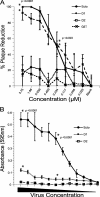
Binding of fJAM-A ectodomain and the D1 and D2 Ig-like domains to FCV and their effects on infectivity. (A) FCV-5 was incubated with purified fJAM-A ectodomain (Ecto), D1, D2, or GST for 1 h on ice and then adsorbed to a monolayer of CRFK cells for 1 h at room temperature. The cells were then overlaid with EMEM, 5% fetal bovine serum, and 1% Bacto Agar and incubated at 37°C for 48 h. The plaques in each well were counted, and the data were expressed as the percentage of plaque reduction relative to monolayers infected with untreated virus. The data shown are the means of six replicates ± standard deviations from one representative experiment. ANOVA was performed on three concentrations (3.75, -0.234, and 0.029 μM) to determine statistical differences; significant concentrations are indicated by asterisks. (B) ELISA plates were coated with 5 μM solutions of soluble fJAM-A ectodomain, D1, or D2. Serial dilutions of FCV-5 were incubated with the immobilized proteins for 1 h, and then the plates were washed extensively. Bound FCV-5 was detected with rabbit anti-FCV serum, followed by HRP-conjugated goat anti-rabbit IgG. Colorimetric HRP substrate was added, and the amount of bound FCV-5 was quantified by absorbance at 595 nm. The means and standard deviations are shown; n = 3. As for panel A, ANOVA was performed on three concentrations; significant concentrations are indicated by asterisks.
FIG. 4.
FIG. 4.
FCV binding to CHO cells expressing fJAM-A deletion and chimeric mutants. (A) A panel of fJAM-A deletion and chimeric mutants was created to investigate FCV binding. Chimeric receptors were generated by exchanging single Ig-like loops from the IgSF proteins fJAM-A (red), hJAM-A (yellow), and hCAR (blue). Deletion constructs lacking single Ig-like domains, as well as a construct in which the transmembrane and cytoplasmic domains were replaced with a GPI anchor, were generated. (B) CHO-S cells were transfected with each construct. At 24 h p.t., FCV was adsorbed to the cells on ice for 30 min. After being washed with cold PBS, the bound virus and cell surface fJAM-A were detected with mouse anti-FCV MAb and rabbit anti-fJAM-A antibodies, followed by Alexa 488-conjugated goat anti-mouse IgG and Alexa 647-conjugated goat anti-rabbit IgG. Virus binding and receptor expression were analyzed by flow cytometry. (C) Virus binding was measured by determining the percentage of receptor-positive cells that were positive for virus. The means (n ≥ 3) (1 × 10⁴ cells) and standard deviations are shown.
FIG. 5.
FIG. 5.
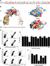
FCV binding to fJAM-A D1 point mutants. (A) The amino acid sequences of the D1 domain of fJAM-A (residues 26 to 125) and hJAM-A (27 to 126) were aligned, and nonidentical residues were identified (highlighted by red boxes). (B) Identified residues were mapped on the hJAM-A crystal structure (Protein Data Bank identification no. 1NBQ; red and blue residues), and 11 surface-exposed residues were selected to mutate to the hJAM-A sequence (blue residues; images were created in PyMOL [Delano Scientific]). A dimerization mutant (E60R/K62E) that reverses the charges on two of the four charged residues in the dimerization motif was also created. (C) CHO-S cells were transfected with each of the constructs. At 24 h p.t., cells were incubated with FCV-5 on ice for 30 min, followed by immunostaining to detect surface expression of receptor and FCV binding, and then analyzed by flow cytometry. (D) After gating was performed for receptor-positive cells, virus binding was measured and expressed as a percentage. The means of at least three replicates (1 × 10⁴ cells each) plus standard deviations are shown. To further investigate the decreased binding observed with the point mutant D42N, additional mutants were made to reverse or eliminate the charge on residue 42, as well as to alter other charged residues in the vicinity of residue 42. (E and F) Virus binding to cells expressing the constructs was measured by flow cytometry, and virus binding was determined as for panel D (F). The means (n ≥ 3; 1 × 10⁴ cells each) plus standard deviations are shown. Constructs indicated by asterisks were bound by significantly lower levels of virus, as determined by ANOVA (P < 0.0001).
FIG. 6.
FIG. 6.
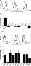
FCV infection of cells expressing chimeric, deletion, or select D1 point mutant constructs. (A) F9 was bound to two sets of cells expressing chimeric or deletion constructs for 30 min on ice. One set of F9-inoculated cells was resuspended in growth medium and placed at 37°C for 24 h; the other set of cells was washed and immediately immunostained for the virus and receptor to determine background binding of virus. After 24 h, the infected cells were washed and immunostained for virus and receptor as described above. Bound (solid lines) and 24-h-incubated (dashed lines) samples were then analyzed by flow cytometry. (B) Infectivity was measured by determining the change in mean log virus fluorescence intensity between bound and incubated samples. The averages of the changes in the geometric means (n ≥ 4) plus standard errors are shown. (C) Cells expressing select D1 point mutant constructs were prepared and analyzed in the same fashion. (D) The change in the mean virus fluorescence intensity was used as a measure for infection, and the averages of the changes in geometric means (n ≥ 4) plus standard errors are shown.
FIG. 7.
FIG. 7.
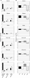
FCV isolate infectivity in nonpermissive cell lines expressing fJAM-A. (A) FCV isolates (MOI = 0.5) were incubated with monolayers of CHO-K1 cells expressing fJAM-A (transiently or stably) for 24 or 48 h. Virus was also incubated for 24 h with empty CHO-K1 cells, CHO-K1 cells transfected with the empty vector (CHO K1 Mock), and CRFK cells. The infectivities of the samples were determined by plaque assay, and the change in titer from virus-bound-only samples was calculated. The mean log10 changes in titers of three replicates ± standard deviations from a representative experiment are shown. (B) Monolayers of four additional nonpermissive cell lines (CHL, Flp-In T-REx 293, Vero, and HeLa) were transiently transfected with fJAM-A and incubated with virus (MOI = 0.5) for 24 (CHL, Vero, and HeLa) or 48 (Flp-In T-REx 293) hours. The infectivities of the samples were determined by plaque assay, and the changes in titers from virus-bound-only samples were calculated. The mean log10 titers of three replicates ± standard deviations are shown.

Similar articles

Cited by

References

    1. Babinska, A., M. H. Kedees, H. Athar, T. Ahmed, O. Batuman, Y. H. Ehrlich, M. M. Hussain, and E. Kornecki. 2002. F11-receptor (F11R/JAM) mediates platelet adhesion to endothelial cells: role in inflammatory thrombosis. Thromb. Haemost. 88:843-850. - PubMed
    1. Babinska, A., M. H. Kedees, H. Athar, T. Sobocki, M. B. Sobocka, T. Ahmed, Y. H. Ehrlich, M. M. Hussain, and E. Kornecki. 2002. Two regions of the human platelet F11-receptor (F11R) are critical for platelet aggregation, potentiation and adhesion. Thromb. Haemost. 87:712-721. - PubMed
    1. Baldwin, S. L., T. D. Powell, K. S. Sellins, S. V. Radecki, J. John Cohen, and M. J. Milhausen. 2004. The biological effects of five feline IFN-alpha subtypes. Vet. Immunol. Immunopathol. 99:153-167. - PubMed
    1. Barton, E. S., J. C. Forrest, J. L. Connolly, J. D. Chappell, Y. Liu, F. J. Schnell, A. Nusrat, C. A. Parkos, and T. S. Dermody. 2001. Junction adhesion molecule is a receptor for reovirus. Cell 104:441-451. - PubMed
    1. Bazzoni, G., O. M. Martinez-Estrada, F. Orsenigo, M. Cordenonsi, S. Citi, and E. Dejana. 2000. Interaction of junctional adhesion molecule with the tight junction components ZO-1, cingulin, and occludin. J. Biol. Chem. 275:20520-20526. - PubMed

Publication types

MeSH terms

Substances

LinkOut - more resources