. 2007 Dec;81(24):13552-65.
doi: 10.1128/JVI.01576-07. Epub 2007 Oct 3.

Analysis of Venezuelan equine encephalitis virus capsid protein function in the inhibition of cellular transcription

Affiliations

Analysis of Venezuelan equine encephalitis virus capsid protein function in the inhibition of cellular transcription

Natalia Garmashova et al. J Virol. 2007 Dec.

Abstract

The encephalitogenic New World alphaviruses, including Venezuelan (VEEV), eastern (EEEV), and western equine encephalitis viruses, constitute a continuing public health threat in the United States. They circulate in Central, South, and North America and have the ability to cause fatal disease in humans and in horses and other domestic animals. We recently demonstrated that these viruses have developed the ability to interfere with cellular transcription and use it as a means of downregulating a cellular antiviral response. The results of the present study suggest that the N-terminal, approximately 35-amino-acid-long peptide of VEEV and EEEV capsid proteins plays the most critical role in the downregulation of cellular transcription and development of a cytopathic effect. The identified VEEV-specific peptide C(VEE)33-68 includes two domains with distinct functions: the alpha-helix domain, helix I, which is critically involved in supporting the balance between the presence of the protein in the cytoplasm and nucleus, and the downstream peptide, which might contain a functional nuclear localization signal(s). The integrity of both domains not only determines the intracellular distribution of the VEEV capsid but is also essential for direct capsid protein functioning in the inhibition of transcription. Our results suggest that the VEEV capsid protein interacts with the nuclear pore complex, and this interaction correlates with the protein's ability to cause transcriptional shutoff and, ultimately, cell death. The replacement of the N-terminal fragment of the VEEV capsid by its Sindbis virus-specific counterpart in the VEEV TC-83 genome does not affect virus replication in vitro but reduces cytopathogenicity and results in attenuation in vivo. These findings can be used in designing a new generation of live, attenuated, recombinant vaccines against the New World alphaviruses.


Figures
FIG. 1.
FIG. 1.
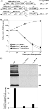
Effects of capsid protein mutant expression on cellular transcription and cell growth. (A and B) Schematic representation of VEEV replicons expressing GFP or mutated capsid proteins. The arrows indicate the positions of the subgenomic promoters. Replicons expressed either CVEE with mutated protease (mutCVEE) (A) or CVEE having no mutations in the protease domain (B). VEErepL/CVEEfrsh/Pac was included in the experiments presented in panels B, D, and F as an additional control. Cells were electroporated by 5 μg of the in vitro-synthesized RNAs. Different dilutions of the electroporated cells were seeded into 100-mm tissue culture dishes. Puromycin selection was performed as described in Materials and Methods. Purr cell colonies were stained with crystal violet on days 4 to 9 posttransfection, depending on their growth rates. The results are presented in CFU per μg of RNA used for transfection. The ranges indicate variations between different experiments. (C and D) Analysis of cell growth after transfection with VEEV replicons expressing GFP and different capsid proteins. Equal numbers of electroporated cells were seeded into six-well Costar plates. Puromycin selection (10 μg/ml) was performed between 6 and 48 h posttransfection. Then, the cells were incubated in puromycin-free media, and viable cells were counted at the indicated times. The data were normalized on the number of viable, adherent cells determined at 6 h posttransfection. The error bars indicate standard deviations. (E and F) Analysis of cellular transcription. RNA labeling in the electroporated cells was performed for 2 h with [3H]uridine at 24 h posttransfection. RNA samples were isolated and analyzed by gel electrophoresis under the conditions described in Materials and Methods. Autoradiographs of the gels are presented. The positions of the ribosomal 28S and 18S and preribosomal 45S RNAs are indicated. For quantitative analysis, aliquots of the RNA samples derived from equal numbers of cells were washed on Whatman 3MM filters with trichloroacetic acid, as described in Materials and Methods, and the radioactivity was measured by liquid scintillation counting. One of two reproducible experiments is presented.
FIG. 2.
FIG. 2.
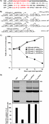
The N-terminal CVEE fragment inhibits cellular transcription and causes CPE development. (A) Schematic representation of VEEV genome-based replicons expressing the amino-terminal fragments of CVEE fused with GFP and analysis of their abilities to establish persistent replication and develop Purr foci. The arrows indicate the positions of the subgenomic promoters. The numbers indicate the last capsid-specific amino acids in fusion proteins. (B) Analysis of cell growth after transfection with VEEV replicons expressing GFP or the indicated fusions. The data were normalized on the number of viable, adherent cells determined at 6 h posttransfection. The error bars indicate standard deviations. (C) Inhibition of transcription in the BHK-21 cells transfected with VEEV replicons expressing the indicated proteins. RNA labeling with [3H]uridine was performed for 2 h at 24 h posttransfection. Electroporation, RNA labeling, and analysis were performed as described in the legend to Fig. 1 and in Materials and Methods. The positions of the ribosomal 28S and 18S and preribosomal 45S RNAs are indicated. Quantitative analysis of residual cellular transcription was performed as indicated in the legend to Fig. 1. The constructs presented were analyzed in the same set of experiments as the expression cassettes presented in Fig. 4. Therefore, the same controls were used.
FIG. 3.
FIG. 3.
Analysis of the effects of the N-terminal VEEV capsid fragment on cellular transcription and cell viability. (A) Sequence alignment of CVEE30-68 peptide with the corresponding capsid fragments of other alphaviruses: VEEV (25), EEEV (45), SINV, and SFV (44). Helix I sequences are indicated in red. Residues identical to those in the VEEV sequence are indicated by dashes. The stars indicate the positions of the deletions introduced for better alignment of the sequences. (B) Schematic representation of VEEV genome-based replicons expressing the amino-terminal fragments of CVEE fused with GFP and analysis of their abilities to establish persistent replication and to develop Purr foci. The arrows indicate the positions of the subgenomic promoters. The numbers indicate the last capsid-specific amino acids in fusion proteins. The results are presented in CFU per μg of RNA used for transfection. The ranges indicate variations between the experiments. (C) Analysis of cell growth after transfection with VEEV replicons expressing GFP and the indicated fusions. The data were normalized on the number of viable, adherent cells determined at 6 h posttransfection. The error bars indicate standard deviations. (D) Analysis of cellular transcription. RNA labeling with [3H]uridine was performed for 2 h at 24 h posttransfection. RNA samples were analyzed by gel electrophoresis under the conditions described in the legend to Fig. 1 and Materials and Methods. The positions of the ribosomal 28S and 18S and preribosomal 45S RNAs are indicated. Quantitative analysis of residual cellular transcription was performed as indicated in the legend to Fig. 1.
FIG. 4.
FIG. 4.
Comparative analysis of the effects of CVEE and a CVEE-GFP fusion on cellular transcription and cell viability. (A) Schematic representation of VEEV genome-based replicons expressing CVEE and a CVEE-GFP fusion and analysis of the replicons' abilities to establish persistent replication and develop Purr foci. The arrows indicate the positions of the subgenomic promoters. (B) Analysis of cell growth after transfection with VEEV replicons expressing either GFP or the indicated capsid variants. The data were normalized on the number of viable, adherent cells determined at 6 h posttransfection. The error bars indicate standard deviations. (C) Analysis of cellular transcription. RNA labeling with [3H]uridine was performed for 2 h at 24 h posttransfection. Electroporation, RNA labeling, and analysis were performed as described in the legend to Fig. 1 and in Materials and Methods. The positions of the ribosomal 28S and 18S and preribosomal 45S RNAs are indicated. Quantitative analysis of residual cellular transcription was performed as indicated in the legend to Fig. 1. (D) Analysis of GFP and CVEE-GFP expression by the indicated replicons. Cell lysates were prepared at 20 h posttransfection and analyzed by Western blotting using GFP-specific antibody and infrared dye 800CW-labeled secondary antibodies. The images were acquired on an Odyssey Infrared Imager (LI-COR).
FIG. 5.
FIG. 5.
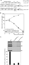
Analysis of the effects of the N-terminal deletions in capsid protein on cellular transcription and cell viability. (A) Schematic representation of VEEV genome-based replicons expressing the deleted forms of capsid protein fused with GFP and analysis of their abilities to establish persistent replication and develop Purr foci. The arrows indicate the positions of the subgenomic promoters. The numbers indicate the first amino acids of CVEE after deletion. In all of the constructs, the initiating capsid protein-specific AUG was present in its natural position. (B) Analysis of the growth of the cells carrying VEEV replicons expressing GFP or the indicated fusions. The data were normalized on the number of viable, adherent cells determined at 6 h posttransfection. The error bars indicate standard deviations.
FIG. 6.
FIG. 6.
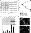
Effects of the CVEE peptides fused with either GFP or GFPNLS on cell viability and cellular transcription. (A) Schematic representation of VEEV genome-based replicons expressing the deleted forms of capsid protein fused with GFP and analysis of their abilities to establish persistent replication and to develop Purr foci. The arrows indicate the positions of the subgenomic promoters. In all of the constructs, the initiating AUG was created upstream of the studied peptide. (B) Analysis of cell growth after transfection of VEEV replicons encoding the indicated fusions. The data were normalized on the number of viable, adherent cells determined at 6 h posttransfection. The error bars indicate standard deviations. (C) Analysis of cellular transcription. RNA labeling with [3H]uridine was performed for 2 h at 24 h posttransfection. Electroporation, RNA labeling, and analysis were performed as described in the legend to Fig. 1 and in Materials and Methods. The positions of the ribosomal 28S and 18S and preribosomal 45S RNAs are indicated. Quantitative analysis of residual cellular transcription was performed as indicated in the legend to Fig. 1. (D) Intracellular distribution of CVEE30-60-GFP and CVEE30-60-GFPNLS fusions. BHK-21 cells were transfected with the corresponding replicons, and the intracellular distributions of the fusions were analyzed at 12 h posttransfection on an inverted fluorescence microscope (Leica).
FIG. 7.
FIG. 7.
Comparative analysis of the effects of CVEE30-68 and CEEE33-71 peptides fused with GFPNLS on cellular transcription and cell viability. (A) Schematic representation of VEEV genome-based replicons expressing GFP or fusion proteins and analysis of the replicons' abilities to establish persistent replication and develop Purr foci. The initiating AUG was created upstream of the studied peptides. (B) Analysis of cell growth after transfection with VEEV replicons expressing the indicated proteins. The data were normalized on the number of viable, adherent cells determined at 6 h posttransfection. The error bars indicate standard deviations. (C) Analysis of cellular transcription. RNA labeling with [3H]uridine was performed for 2 h at 24 h posttransfection. Electroporation, RNA labeling, and analysis were performed as described in the legend to Fig. 1 and in Materials and Methods. The positions of the ribosomal 28S and 18S and preribosomal 45S RNAs are indicated. Quantitative analysis of residual cellular transcription was performed as indicated in the legend to Fig. 1.
FIG. 8.
FIG. 8.
Intracellular distributions of different CVEE-GFP fusions. (A and B) BHK-21 cells were transfected with the replicons expressing CVEE-GFP (a), CVEEΔ35-47-GFP (b), and CVEEfrsh-GFP (c) proteins, and the intracellular distributions of the fusions were analyzed at 12 h posttransfection. The images were acquired on a confocal microscope. Different magnifications are presented in panels A and B. (C) BHK-21 cells were transfected with VEErepL/CVEE-GFP/Pac, and, at 12 h posttransfection, the cells were permeabelized with 0.5% Triton X-100, stained with MAb 414 antibody and AlexaFluor 546-labeled secondary antibodies, and analyzed on a confocal microscope. (a) Distribution of CVEE/GFP on the nuclear membrane. (b) MAb 414 staining of the same cell. (c) Overlay of the images (the enlarged fragment is indicated in a and b). The bars in panel A correspond to 20 μm, and in panels B and C, to 5 μm.
FIG. 9.
FIG. 9.
Replication of VEEV TC-83 with mutated capsid protein. (A) Schematic representation of the viral genomes. In VEEV/CSIN1, the natural N-terminal fragment, located upstream of the protease domain (aa 1 to 110), was replaced by its SINV-specific counterpart (aa 1 to 98), indicated by a black box. In VEEV/Cfrsh, the capsid protein gene contained a frameshift mutation that changed the peptide between aa 57 and 86. (B) BHK-21 cells were electroporated with 5 μg of in vitro-synthesized RNAs. One-fifth of the samples were seeded into 35-mm culture dishes. At the indicated times posttransfection, the media were replaced by fresh media, and virus titers in the culture fluids were determined by a plaque assay on BHK-21 cells. Note that cells transfected with VEEV/CSIN1 and VEEV/Cfrsh RNAs continued to release virus after 24 h posttransfection, when VEEV TC-83 RNA-transfected cells developed a profound CPE. (C) Survival of mice infected with the indicated viruses. Six-day-old NIH Swiss mice were inoculated i.c. with the indicated doses of viruses. The animals were monitored for 2 months. No deaths occurred after day 9 postinfection in any of these experiments.

Similar articles

Cited by

References

    1. Aguilar, P. V., S. C. Weaver, and C. F. Basler. 2007. Capsid protein of eastern equine encephalitis virus inhibits host cell gene expression. J. Virol. 81:3866-3876. - PMC - PubMed
    1. Alevizatos, A. C., R. W. McKinney, and R. D. Feigin. 1967. Live, attenuated Venezuelan equine encephalomyelitis virus vaccine. I. Clinical effects in man. Am. J. Trop. Med. Hyg. 16:762-768. - PubMed
    1. Berge, T. O., I. S. Banks, and W. D. Tigertt. 1961. Attenuation of Venezuelan equine encephalomyelitis virus by in vitro cultivation in guinea pig heart cells. Am. J. Hyg. 73:209-218.
    1. Black, B. L., R. B. Rhodes, M. McKenzie, and D. S. Lyles. 1993. The role of vesicular stomatitis virus matrix protein in inhibition of host-directed gene expression is genetically separable from its function in virus assembly. J. Virol. 67:4814-4821. - PMC - PubMed
    1. Bredenbeek, P. J., I. Frolov, C. M. Rice, and S. Schlesinger. 1993. Sindbis virus expression vectors: packaging of RNA replicons by using defective helper RNAs. J. Virol. 67:6439-6446. - PMC - PubMed

Publication types

MeSH terms