. 2007 Dec;81(24):13533-43.
doi: 10.1128/JVI.01669-07. Epub 2007 Oct 3.

Human papillomavirus type 16 E7 oncoprotein associates with the centrosomal component gamma-tubulin

Affiliations

Human papillomavirus type 16 E7 oncoprotein associates with the centrosomal component gamma-tubulin

Christine L Nguyen et al. J Virol. 2007 Dec.

Abstract

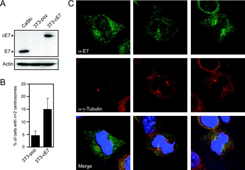
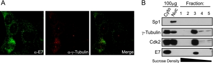
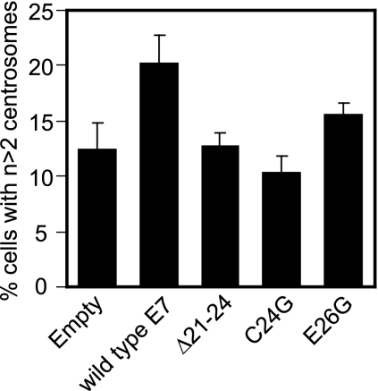
Expression of a high-risk human papillomavirus (HPV) E7 oncoprotein is sufficient to induce aberrant centrosome duplication in primary human cells. The resulting centrosome-associated mitotic abnormalities have been linked to the development of aneuploidy. HPV type 16 (HPV16) E7 induces supernumerary centrosomes through a mechanism that is at least in part independent of the inactivation of the retinoblastoma tumor suppressor pRb and is dependent on cyclin-dependent kinase 2 activity. Here, we show that HPV16 E7 can concentrate around mitotic spindle poles and that a small pool of HPV16 E7 is associated with centrosome fractions isolated by sucrose density gradient centrifugation. The targeting of HPV16 E7 to the centrosome, however, was not sufficient for centrosome overduplication. Nonetheless, we found that HPV16 E7 can associate with the centrosomal regulator gamma-tubulin and that the recruitment of gamma-tubulin to the centrosome is altered in HPV16 E7-expressing cells. Since the association of HPV16 E7 with gamma-tubulin is independent of pRb, p107, and p130, our results suggest that the association with gamma-tubulin contributes to the pRb/p107/p130-independent ability of HPV16 E7 to subvert centrosome homeostasis.


Figures

FIG. 1.
FIG. 1.
HPV16 E7 can concentrate around mitotic spindle poles. (A) Western blot analysis of E7 expression levels in stable NIH 3T3, 3T3-poz (control), and 3T3-cE7 cells compared to HPV-positive CaSki cervical cancer cells. Actin is shown as a loading control. (B) Percentage of 3T3-cE7 cells with more than two centrosomes compared to control 3T3-poz cells. Bar graphs indicate averages of four experiments in which >150 cells were counted per experiment. Error bars indicate the standard errors between experiments. (C) Representative images of mitotic NIH 3T3 cells with stable expression of C-terminally HA- and FLAG-tagged HPV16 E7. Coimmunofluorescence was performed using an E7-specific antibody (α-E7) and a γ-tubulin antibody (α-γ-Tubulin) as a centrosomal marker. Nuclei were visualized with Hoechst 33258 DNA dye.
FIG. 2.
FIG. 2.
HPV16 E7 cofractionates with centrosomal components after sucrose density gradient centrifugation. (A) Immunofluorescence analysis of endogenous E7 and γ-tubulin expression in HPV16-positive CaSki cervical carcinoma cells. (B) Detection of HPV16 E7 in centrosome-containing fractions. Cytoplasmic lysates of the HPV16-positive CaSki cervical cancer cell line were subjected to sucrose density gradient centrifugation. Aliquots (150 μl) of individual fractions (fractions 1 to 5 of 25 fractions) along with 100-μg aliquots of total cytoplasmic (Cyto) and nuclear (Nuc) extracts were analyzed by Western blotting. E7 cofractionates with the centrosomal components γ-tubulin and cdk2 in fraction 3. Sp1 is shown as a nuclear marker, and the black bar indicates the direction of sucrose density. Consistent with its cytoplasmic localization, the majority of HPV16 E7 is contained in the later, cytoplasmic fractions (not shown).
FIG. 3.
FIG. 3.
Targeting HPV16 E7 to the centrosome is not sufficient for centrosome overduplication. (A) Coimmunofluorescence of E7 and γ-tubulin in a U2OS cell transiently transfected with HPV16 E7-AKAP. The merged image includes nuclear staining with Hoechst 33258 DNA dye (blue). (B) Quantitation of cells with abnormal centrosome numbers at 48 h after transfection with either empty pCMV Neo-Bam vector or E7-AKAP. Bar graphs show the means of counts done in triplicate where >200 cells were analyzed per experiment. Error bars indicate standard deviations.
FIG. 4.
FIG. 4.
HPV16 E7/mCherry/μNS(471-721) relocalizes pRb and p107. Shown is immunofluorescent analysis of U2OS cells expressing the indicated fusion proteins. Cells were transfected 20 to 24 h prior to fixation. The red fluorescent mCherry signal represents the inclusion bodies formed by μNS(471-721). Cells were stained with antibodies against either pRb or p107, shown in green, as indicated. Secondary-only controls showed no positive staining or bleed-through of the mCherry signal (data not shown).
FIG. 5.
FIG. 5.
HPV16 E7/mCherry/μNS(471-721) relocalizes γ-tubulin to inclusions. (A) Immunofluorescent analysis of U2OS cells expressing the indicated fusion proteins as described in the legend of Fig. 4. The cells were stained for γ-tubulin as indicated. (B) As described above (A), pRb/p107/p130−/− cells were transfected with the indicated constructs and stained for γ-tubulin. (C) As described above (A), stable U2OS cells expressing γ-tubulin-GFP were transfected with the indicated constructs. The inset shows centrosomal γ-tubulin fluorescence. (D) As described above (A), U2OS cells were transfected with the indicated constructs and stained for γ-tubulin.
FIG. 6.
FIG. 6.
Relocalization of γ-tubulin by HPV16 E7/mCherry/μNS(471-721) is specific. (A) Immunofluorescence against the centrosomal proteins centrin (left) and ninein (right) was done on cells transfected with indicated plasmids as described in the legend of Fig. 4A. (B) Same as above (A). Cells were stained for α-tubulin (left) and β-tubulin (right). No relocalization of these proteins was detected.
FIG. 7.
FIG. 7.
Induction of supernumerary centrosomes in pRb/p107/p130-deficient mouse embryo fibroblasts by HPV16 E7. The bar graph shows the average percentages of cells with more than two centrosomes after expression of indicated constructs for 48 h. Counts were done in duplicate, where >100 cells were counted per experiment. Error bars indicate standard errors between experiments.
FIG. 8.
FIG. 8.
Slower recovery of γ-tubulin to photobleached centrosomes in cells expressing HPV16 E7. (A) Representative images of a single FRAP experiment performed on a stable 3T3-Δ21-24 cell transiently transfected with γ-tubulin-GFP. The elapsed time at which each image was taken is indicated in the bottom right corner of the frame. The white arrow signifies the centrosome that was photobleached in this specific experiment where photobleaching occurred directly prior to the 10-s image. (B) Dot plot of the τ1/2 values calculated for each experiment. The τ1/2 represents the amount of time taken to reach half-maximal recovery in each trial. The averages of each cell line are shown as black bars, and numerical values are indicated.

References

    1. Afonso, P. V., A. Zamborlini, A. Saib, and R. Mahieux. 2007. Centrosome and retroviruses: the dangerous liaisons. Retrovirology 4:27. - PMC - PubMed
    1. Baker, S. J., S. Markowitz, E. R. Fearon, J. K. Willson, and B. Vogelstein. 1990. Suppression of human colorectal carcinoma cell growth by wild-type p53. Science 249:912-915. - PubMed
    1. Brinkley, B. R., and T. M. Goepfert. 1998. Supernumerary centrosomes and cancer: Boveri's hypothesis resurrected. Cell Motil. Cytoskel. 41:281-288. - PubMed
    1. Crum, C. P., H. Ikenberg, R. M. Richart, and L. Gissman. 1984. Human papillomavirus type 16 and early cervical neoplasia. N. Engl. J. Med. 310:880-883. - PubMed
    1. Dannenberg, J. H., A. van Rossum, L. Schuijff, and H. te Riele. 2000. Ablation of the retinoblastoma gene family deregulates G(1) control causing immortalization and increased cell turnover under growth-restricting conditions. Genes Dev. 14:3051-3064. - PMC - PubMed

Publication types

Substances

LinkOut - more resources