. 2007 Oct 3;27(40):10674-84.
doi: 10.1523/JNEUROSCI.2001-07.2007.

Enhanced astrocytic Ca2+ signals contribute to neuronal excitotoxicity after status epilepticus

Affiliations

Enhanced astrocytic Ca2+ signals contribute to neuronal excitotoxicity after status epilepticus

Shinghua Ding et al. J Neurosci. .

Abstract

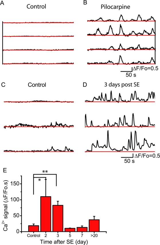
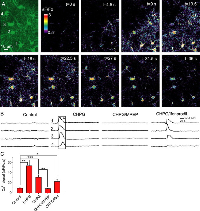
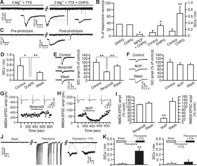
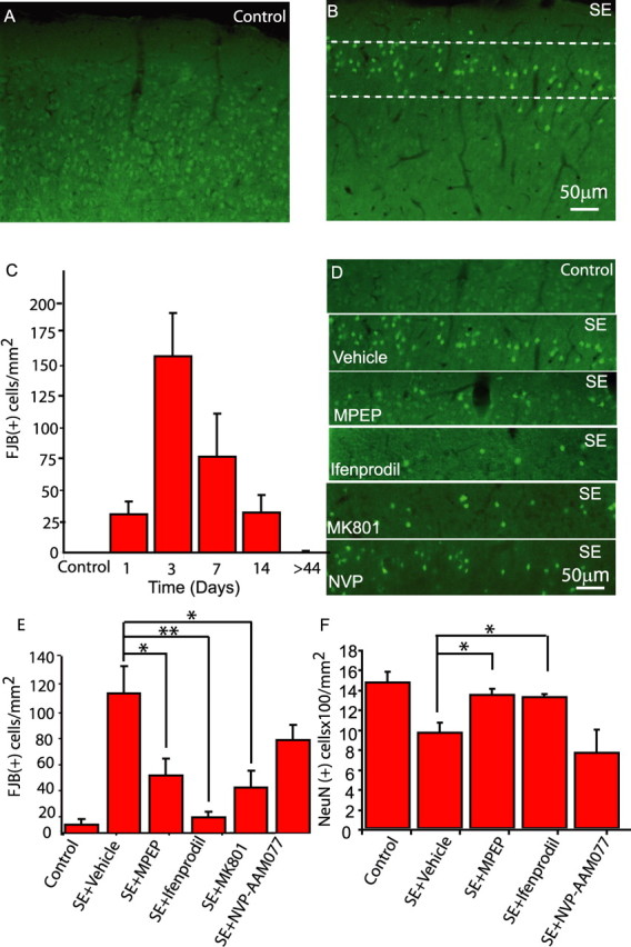
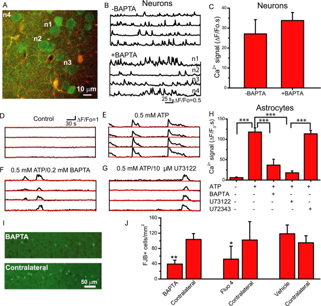
Status epilepticus (SE), an unremitting seizure, is known to cause a variety of traumatic responses including delayed neuronal death and later cognitive decline. Although excitotoxicity has been implicated in this delayed process, the cellular mechanisms are unclear. Because our previous brain slice studies have shown that chemically induced epileptiform activity can lead to elevated astrocytic Ca2+ signaling and because these signals are able to induce the release of the excitotoxic transmitter glutamate from these glia, we asked whether astrocytes are activated during status epilepticus and whether they contribute to delayed neuronal death in vivo. Using two-photon microscopy in vivo, we show that status epilepticus enhances astrocytic Ca2+ signals for 3 d and that the period of elevated glial Ca2+ signaling is correlated with the period of delayed neuronal death. To ask whether astrocytes contribute to delayed neuronal death, we first administered antagonists which inhibit gliotransmission: MPEP [2-methyl-6-(phenylethynyl)pyridine], a metabotropic glutamate receptor 5 antagonist that blocks astrocytic Ca2+ signals in vivo, and ifenprodil, an NMDA receptor antagonist that reduces the actions of glial-derived glutamate. Administration of these antagonists after SE provided significant neuronal protection raising the potential for a glial contribution to neuronal death. To test this glial hypothesis directly, we loaded Ca2+ chelators selectively into astrocytes after status epilepticus. We demonstrate that the selective attenuation of glial Ca2+ signals leads to neuronal protection. These observations support neurotoxic roles for astrocytic gliotransmission in pathological conditions and identify this process as a novel therapeutic target.


Figures

Figure 1.
Figure 1.
SE stimulates astrocytic Ca2+ signals. A, B, The time course of somatic Ca2+ signals recorded simultaneously in four astrocytes before (A) and after (B) acute injection of pilocarpine to the anesthetized mouse. Astrocytic Ca2+ signals (3 examples from 3 mice in each condition) recorded 3 d after a subthreshold injection of pilocarpine (Control) (C) or 3 d after SE (D). E, Time course of change in Ca2+ signals after SE reported as the integral of the ΔF/F0 signal (*p < 0.05; **p < 0.00005; n value is between 4 and 8 animals for each time point). Error bars indicate SEM.
Figure 2.
Figure 2.
mGluR5 stimulates astrocytic Ca2+ signals in vivo. A, Sequential ΔF/F0 images showing that cortical astrocytes exhibit propagating Ca2+ waves in vivo when stimulated by the mGluR5 agonist CHPG (1 mm). B, Time courses of somatic Ca2+ fluorescence changes (ΔF/F0; examples of 4 cells in each case) in control animals in the absence and presence CHPG (1 mm) as well as with CHPG in the presence of MPEP (30 μm) and ifenprodil (10 μm). Note that the mGluR5 antagonist MPEP and not the NR2B NMDA receptor antagonist, ifenprodil, attenuate CHPG-induced Ca2+ signaling. The boxed region corresponds to the images in A. C, Astrocytic Ca2+ signals are stimulated by DHPG (25 μm) and CHPG (1 mm). The mGluR5 antagonist MPEP, but not the NR2B NMDA receptor antagonist ifenprodil, significantly attenuates CHPG-induced Ca2+ signals (n = 4–8 animals per group). *p < 0.05; **p < 0.02; ***p < 0.01. Error bars indicate SEM.
Figure 3.
Figure 3.
Astrocytic Ca2+ signals in vivo in mice 3 d after SE are suppressed by the mGluR5 antagonist MPEP. A, Fluo-4-labeled astrocytes before (left) and after (right) the mouse was coinjected with MPEP and rhodamine-dextran through the tail vein. B, Calcium signals of a cell pair were inhibited after MPEP injection. C, Calcium signals before and after acute administration of MPEP (1 mg/kg tail vein injection), depicted as the ΔF/F0 integral. Also shown is the inhibition by four daily intraperitoneal administrations of MPEP (20 mg/kg). The NR2B NMDA receptor antagonist ifenprodil (20 μm) does not affect astrocytic Ca2+ signals. *p < 0.002 (n = 4 animals in each group except ifenprodil, n = 3). Error bars indicate SEM.
Figure 4.
Figure 4.
Gliotransmission but not intracortical synaptic transmission activates the NR2B-containing NMDA receptors of layer 2–3 pyramidal neurons. A, Whole-cell recording from a pyramidal neuron showing SICs induced by bath application of CHPG (0.5 mm). In these as well as in B–F, experiments are performed in the continuous presence of TTX (1 μm). B, Left, Percentage of neurons showing increased SIC frequency after application of DHPG (10–20 μm; 4 of 11 cells), CHPG (0.5–1 mm; 7 of 20), and CHPG and MPEP (0.5–1 mm and 50 μm, respectively; 1 of 17). In the latter experiments, the slice was preincubated with MPEP for 5 min before the application of CHPG and MPEP. The ability of MPEP to block actions of CHPG was tested using Fisher's exact test (p < 0.05). Right, Average frequency of SICs before and after DHPG (n = 4) and CHPG (n = 7) in the responsive neurons shown in the left panel. In this as well as in the other panels of this figure, *p < 0.05 and **p < 0.01. Error bars indicate SEM. C, Whole-cell recording from a pyramidal neuron showing SICs induced by photolysis of caged Ca2+ in single cortical astrocytes. D, Average frequency of SICs under control conditions, in the presence of d-AP5 (50 μm), and after d-AP5 washout in six cortical neurons showing SIC activity. SICs were stimulated by application of CHPG (0.5–1 mm) or low Ca2+ containing ACSF. E, F, Mean amplitude of SICs under control conditions, in the presence of ifenprodil (E; 3 μm) or NVP-AAM077 (F; 0.4 μm), and after drug washout. Data are normalized to the amplitude of SICs recorded under control conditions. See inset for representative SICs from the same cell under the different experimental conditions. Number of averaged SICs is 37, 48, and 70 from 9 cells for E, and 27, 25, and 13 from 7 cells for F, respectively. G, H, Representative experiments showing the time course of the NMDA EPSC-amplitude at basal condition (0 Mg2+-containing saline in the presence of 10 μm NBQX), during ifenprodil (G; 3 μm) and NVP-AAM077 (H; 0.4 μm) application, and after drug washout. Subsequent application of d-AP5 (50 μm) completely blocked the EPSC (data not shown). Inset, Average of 10 EPSCs in the different experimental conditions. EPSCs were evoked by positioning the stimulating electrode intracortically, 100–200 μm from the recording pipette. I, Ifenprodil (left; 3 μm) does not decrease the average amplitude of the NMDA EPSC (n = 6 cells), whereas NVP-AAM077 (right; 0.4 μm) drastically reduces its amplitude (n = 4 cells). Data are normalized to the average NMDA EPSC amplitude under control conditions. J, Epileptiform activity induced by acute application of pilocarpine (10 μm) triggers an increase in SIC frequency. Representative experiment showing membrane current under basal conditions (left), in the presence of pilocarpine (middle), and in the presence of pilocarpine and TTX (1 μm; right). Addition of TTX blocks pilocarpine-induced epileptiform activity and reveals the presence of SICs. Pilocarpine was always applied in the presence of 0 Mg2+ and 100 μm picrotoxin (see Materials and Methods). During epileptiform activity, unclamped action potentials were recorded and have been truncated for presentation purposes. K, Average SIC frequency under basal conditions in normal ACSF before perfusion of pilocarpine (left; open bar). After epileptiform activity was triggered by application of pilocarpine, 1 μm TTX was added to the saline to block neuronal activity. Thereafter, SIC frequency was measured again in 6 of 10 layer 2/3 neurons displaying SICs (left; filled bar). When TTX was added together with pilocarpine, no epileptiform activity was initiated in the slice and no significant increase in SIC frequency was observed in four of seven neurons displaying SICs (right).
Figure 5.
Figure 5.
MPEP and ifenprodil protect neurons from SE-evoked death. FJB-stained sections of murine cortex from control (A) and mice 3 d after SE (B) show FJB labeling in layer 2/3, the region in which in vivo imaging and slice electrophysiology were performed. C, Time course of FJB labeling. D, The region identified by the dashed lines in B is shown from animals treated with glutamate receptor antagonists, as labeled. E, MPEP (20 mg/kg), ifenprodil (20 mg/kg), and MK-801 (1 mg/kg) significantly reduce the number of FJB-labeled cells 3 d after SE, whereas NVP-AAM077 (2 mg/kg) is not protective. F, In parallel experiments, the number of NeuN-labeled neurons in cortical layers 2/3 7 d after SE were counted using an automated system. Similar to results obtained with FJB, MPEP and ifenprodil, but not NVP-AAM077, protected neurons from delayed death that normally follows SE. *p < 0.05; **p < 0.01; n = 4–7 animals in each condition. Error bars indicate SEM.
Figure 6.
Figure 6.
Attenuation of astrocytic Ca2+ signals by loading of Ca2+ chelators is neuroprotective. A, Two-photon overlay image (fluo-4, green; SR101, red; colocalization, yellow) of layer 2/3 neurons loaded with fluo-4 and astrocytes loaded with fluo-4 and SR101. Fluo-4 AM was injected into the cortex using the bulk-loading approach to label neurons and astrocytes, whereas SR101 was applied to the surface of the cortex to selectively label astrocytes. With neurons and astrocytes coloaded with fluo-4, BAPTA-AM was applied to the cortical surface to selectively load into astrocytes. Selectivity of loading was confirmed by monitoring neuronal and astrocytic Ca2+ signals. B, Spontaneous neuronal Ca2+ signals in control and BAPTA-loaded animals. C, Surface application of BAPTA-AM does not reduce neuronal Ca2+ signals. D–F, Application of ATP (0.5 mm; n = 4) to the cortex evokes robust Ca2+ signals in astrocytes (E), which are attenuated by previous application of BAPTA-AM to the cortical surface (n = 5; compare E, F). G, The phospholipase C inhibitor U73122 (10 μm; n = 4) attenuates ATP-induced Ca2+ signals, providing additional support for an astrocytic origin of these Ca2+ signals. H, Summary of ATP-induced Ca2+ signals. I, After status epilepticus, BAPTA-AM was applied to the cortical surface through a craniotomy to selectively attenuate astrocytic Ca2+ signals, and 3 d later, Fluoro-Jade B labeling of dying neurons was assessed both ipsilateral (BAPTA) and contralateral to the treatment. J, The selective loading of the Ca2+ chelator BAPTA into astrocytes resulted in fewer dying, FJB-labeled cells compared with contralateral untreated cortex (paired t test). Surface loading with the fluorescent Ca2+ indicator, fluo-4, caused similar neuroprotection, whereas application of vehicle did not. *p < 0.05; **p < 0.01; ***p < 0.001. Error bars indicate SEM.

Similar articles

Cited by

References

    1. Angulo MC, Kozlov AS, Charpak S, Audinat E. Glutamate released from glial cells synchronizes neuronal activity in the hippocampus. J Neurosci. 2004;24:6920–6927. - PMC - PubMed
    1. Araque A, Parpura V, Sanzgiri RP, Haydon PG. Glutamate-dependent astrocyte modulation of synaptic transmission between cultured hippocampal neurons. Eur J Neurosci. 1998;10:2129–2142. - PubMed
    1. Araque A, Carmignoto G, Haydon PG. Dynamic signaling between astrocytes and neurons. Annu Rev Physiol. 2001;63:795–813. - PubMed
    1. Auberson YP, Allgeier H, Bischoff S, Lingenhoehl K, Moretti R, Schmutz M. 5-Phosphonomethylquinoxalinediones as competitive NMDA receptor antagonists with a preference for the human 1A/2A, rather than 1A/2B receptor composition. Bioorg Med Chem Lett. 2002;12:1099–1102. - PubMed
    1. Babb TL, Brown WJ, Pretorius J, Davenport C, Lieb JP, Crandall PH. Temporal lobe volumetric cell densities in temporal lobe epilepsy. Epilepsia. 1984;25:729–740. - PubMed

Publication types

MeSH terms

LinkOut - more resources