. 2007 Oct 3;27(40):10685-94.
doi: 10.1523/JNEUROSCI.2624-07.2007.

Brain-derived neurotrophic factor rescues synaptic plasticity in a mouse model of fragile X syndrome

Affiliations

Brain-derived neurotrophic factor rescues synaptic plasticity in a mouse model of fragile X syndrome

Julie C Lauterborn et al. J Neurosci. .

Abstract

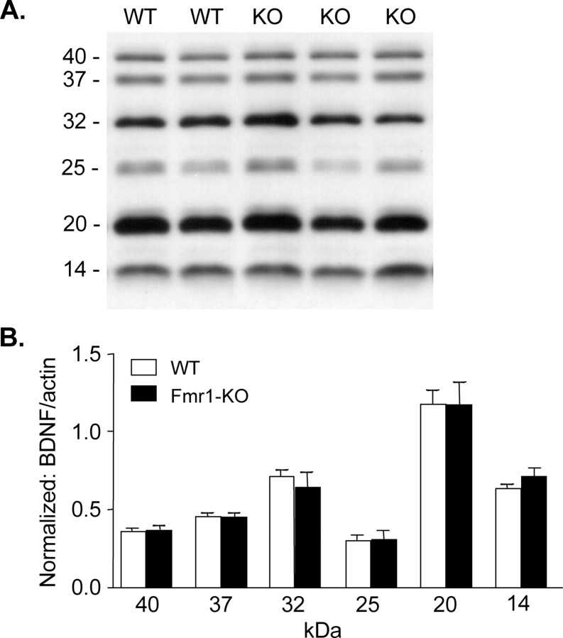
Mice lacking expression of the fragile X mental retardation 1 (Fmr1) gene have deficits in types of learning that are dependent on the hippocampus. Here, we report that long-term potentiation (LTP) elicited by threshold levels of theta burst afferent stimulation (TBS) is severely impaired in hippocampal field CA1 of young adult Fmr1 knock-out mice. The deficit was not associated with changes in postsynaptic responses to TBS, NMDA receptor activation, or levels of punctate glutamic acid decarboxylase-65/67 immunoreactivity. TBS-induced actin polymerization within dendritic spines was also normal. The LTP impairment was evident within 5 min of induction and, thus, may not be secondary to defects in activity-initiated protein synthesis. Protein levels for both brain-derived neurotrophic factor (BDNF), a neurotrophin that activates pathways involved in spine cytoskeletal reorganization, and its TrkB receptor were comparable between genotypes. BDNF infusion had no effect on baseline transmission or on postsynaptic responses to theta burst stimulation, but nonetheless fully restored LTP in slices from fragile X mice. These results indicate that the fragile X mutation produces a highly selective impairment to LTP, possibly at a step downstream of actin filament assembly, and suggest a means for overcoming this deficit. The possibility of a pharmacological therapy based on these results is discussed.


Figures

Figure 1.
Figure 1.
Hippocampal LTP is impaired in young adult Fmr1-KO mice. A, Plot of input–output curves generated from field responses to single pulse stimulation (duration increased in 0.02 ms steps) for Fmr1-KO (closed triangles) and WT (open circles) mice. B, A single train of 10 theta bursts was delivered (arrow, time 0) to the apical branch of the Schaffer commissural projections and fEPSP responses to single pulses were collected from field CA1b for 40 min. Group data (mean ± SEM) are expressed as the percentage of mean fEPSP slopes recorded during the baseline (pre-theta burst) period. There were no reliable differences between WT (open circles) and mutant (closed circles) slices. Inset, Overlaid representative fEPSP traces collected during baseline and for 35 min (arrows) post-TBS for WT and Fmr1-KO mice. Calibration: 0.5 mV, 10 ms. C, Same as B except that the theta train contained only five bursts. Fmr1-KO slices (closed circles) expressed comparable initial potentiation to WTs, but the effect decayed rapidly to baseline by 30 min. D, Representative traces of fEPSPs for time points denoted in C (a, baseline; b, 35 min post-TBS) for slices from WT and Fmr1-KO mice. Calibration: 0.5 mV, 10 ms.
Figure 2.
Figure 2.
The fragile X mutation does not impair events associated with the induction of LTP. A, The multiple fEPSPs in the composite response to a theta burst were normalized (by amplitude) to the first fEPSP in the first burst response. The responses for groups of slices were then averaged. A shows the averaged responses to the first and second theta bursts recorded from WT (n = 8) and Fmr1-KO (n = 7) slices. Note that the second burst response in each case is larger than the first and does not return as quickly to baseline (arrow in top trace). The superimposed traces (right side) indicate that the mutation does not affect the waveform of the composite response or its transformation within the train. B, Facilitation of burst responses within a theta train was estimated by expressing the area of responses 2–5 as a fraction of the first burst response. As shown, mean facilitation for both WT and Fmr1-KO slices was ∼80%. C, The size of the NMDA receptor contribution to the burst responses was estimated using the selective antagonist APV. A pair of theta bursts was delivered to the slice in the presence and absence of the compound. In WT slices, the effect of APV on the first burst response was limited to a slight reduction in the half-width of the fourth fEPSP, but on the second response it reduced the size of fEPSPs 2 through 4 (mean of 6 slices). Similar results were obtained in Fmr1-KO slices (mean of 7 slices).
Figure 3.
Figure 3.
TBS-induced p-cofilin immunoreactivity is normal in Fmr1-KOs. A, B, Laser confocal photomicrographs show p-cofilin-immunoreactivity in proximal CA1 stratum radiatum of hippocampal slices from WT (A) and Fmr1-KO (FX; B) mice that received either baseline low-frequency stimulation (lfs) or five TBSs; slices were collected 7 min after stimulation. Scale bar, 1 μm. C, Bar graph shows the number of p-cofilin-immunoreactive (ir) puncta (mean ± SEM) per 100 μm2 for fields receiving lfs (open bars) or TBS (closed bars) in WT and Fmr1-KO slices. Two-way ANOVA demonstrated a significant effect of TBS (p = 0.00096), but no effect of genotype on p-cofilin-ir puncta counts. Thus, numbers of p-cofilin-ir puncta were significantly greater in slices that received TBS than in those that received lfs for both WTs (**p = 0.0019, t test; lfs, n = 3 mice; TBS, n = 3 mice) and Fmr1-KOs (*p = 0.033, t test; lfs, n = 3 mice; TBS, n = 3 mice).
Figure 4.
Figure 4.
Fmr1-KO mice show normal activity-dependent actin polymerization in dendritic spines. Acute hippocampal slices prepared from Fmr1-KO or WT mice were processed for in situ Alexafluor 568-phalloidin labeling of filamentous actin after electrophysiological recording in hippocampal region CA1. LTP was induced by TBS; control slices received baseline, low-frequency stimulation (lfs). A, Photomicrographs of Fmr1-KO (left) and WT (right) hippocampal slices showing representative phalloidin labeling in the field of afferent stimulation in CA1 stratum radiatum after TBS or lfs. Scale bar, 10 μm. B, Plot summarizes group mean (±SEM) numbers of densely phalloidin-labeled spine-like puncta per sample field for control/lfs (white bars) and TBS (black bars) slices. As indicated, TBS induced similar increases in the numbers of densely labeled spines between genotypes (**p < 0.01 vs respective control group, Tukey's HSD after ANOVA). C, High-magnification photomicrographs show examples of densely phalloidin-labeled dendritic spines in CA1 stratum radiatum from Fmr1-KO and WT slices receiving TBS. Scale bar, 1 μm.
Figure 5.
Figure 5.
BDNF corrects the LTP deficit in fragile X hippocampus. A, Five theta bursts were delivered to the Schaffer commissural projections in WT and Fmr1-KO slices that had been treated with BDNF (2 nm) beginning 30 min before theta stimulation. Potentiation in the mutants did not decay rapidly toward baseline, as observed in untreated slices (Fig. 1 C), and did not differ in magnitude from the effect obtained in BDNF-treated WT slices. B, Mean fEPSP slope (average of 30–40 min post-TBS) expressed as a percentage of the last 10 min of baseline from Fmr1-KO slices either untreated (ACSF alone) or treated with BDNF or heat-inactivated BDNF. BDNF enhanced TBS-induced increases in the fEPSP slope compared with measures from the ACSF group (*p = 0.009), whereas heat-inactivated BDNF had no effect. C, Group input–output data from Fmr1-KO slices treated with BDNF and heat-inactivated BDNF showed no effect of BDNF on fEPSP amplitude. D, Averaged responses to the first and fourth (arrow) theta bursts recorded from Fmr1-KO slices infused with BDNF or with ACSF only. As shown, the response waveforms were comparable between the two groups. E, The effect of BDNF on burst response facilitation within a theta train in slices from fragile X mutant mice was estimated by expressing the area of responses 2–5 as a fraction of the first burst response. The mean degree of facilitation was similar in Fmr1-KO slices treated with BDNF and those bathed in ACSF alone.
Figure 6.
Figure 6.
Hippocampal BDNF levels are normal in Fmr1-KO mice. A, Representative Western blot showing pro-BDNF (40–20 kDa) and mature BDNF (14 kDa) bands in hippocampal homogenates from WT and Fmr1-KO mice; band sizes (in kilodaltons) are indicated on the left. B, Bar graph showing quantification of hippocampal BDNF bands ranging in mass from 14 to 40 kDa for samples from WTs and Fmr1-KOs (n = 6 per genotype). Plot shows BDNF band densities normalized to actin levels for the same sample. For each band, protein levels were equivalent between genotypes.

Similar articles

Cited by

References

    1. Aicardi G, Argilli E, Cappello S, Santi S, Riccio M, Thoenen H, Canossa M. Induction of long-term potentiation and depression is reflected by corresponding changes in secretion of endogenous brain-derived neurotrophic factor. Proc Natl Acad Sci USA. 2004;101:15788–15792. - PMC - PubMed
    1. Arai A, Lynch G. Factors regulating the magnitude of long-term potentiation induced by theta pattern stimulation. Brain Res. 1992;598:173–184. - PubMed
    1. Balkowiec A, Katz D. Cellular mechanisms regulating activity-dependent release of native brain-derived neurotrophic factor from hippocampal neurons. J Neurosci. 2002;22:10399–10407. - PMC - PubMed
    1. Biagini G, Avoli M, Marcinkiewicz J, Marcinkiewicz M. Brain-derived neurotrophic factor superinduction parallels anti-epileptic–neuroprotective treatment in the pilocarpine epilepsy model. J Neurochem. 2001;76:1814–1822. - PubMed
    1. Bramham C, Messaoudi E. BDNF function in adult synaptic plasticity: the synaptic consolidation hypothesis. Prog Neurobiol. 2005;76:99–125. - PubMed

Publication types

MeSH terms