. 2007 Oct 3;27(40):10703-13.
doi: 10.1523/JNEUROSCI.3102-07.2007.

Inflammation activates the interferon signaling pathways in taste bud cells

Affiliations

Inflammation activates the interferon signaling pathways in taste bud cells

Hong Wang et al. J Neurosci. .

Abstract

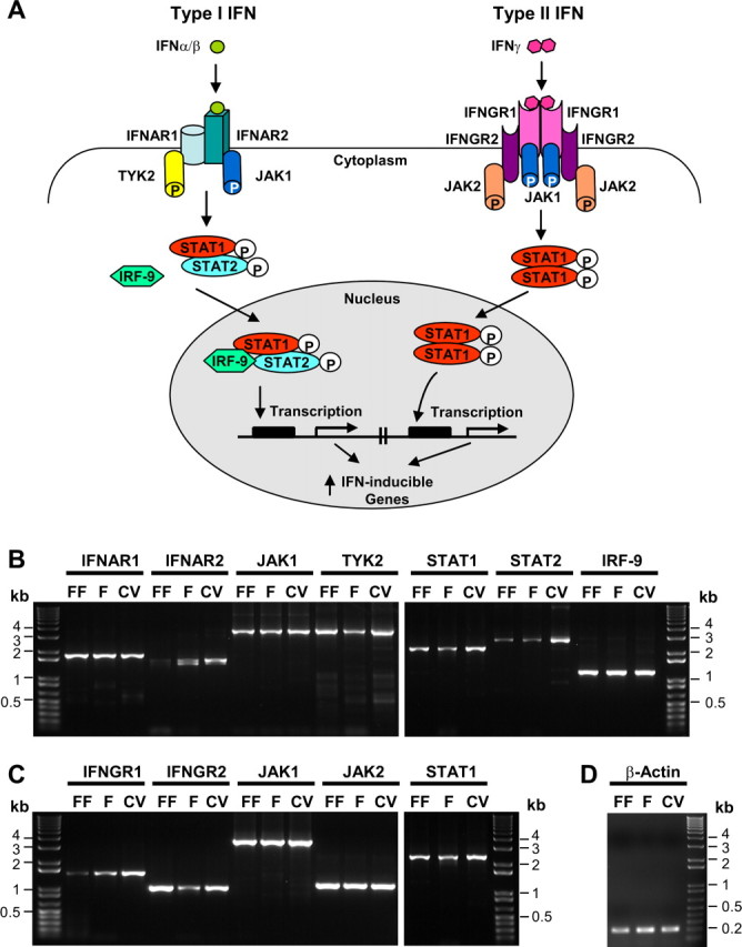
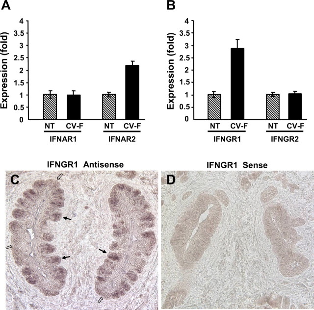
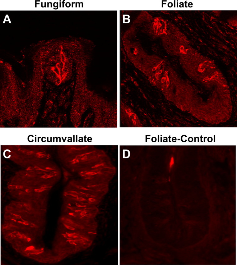
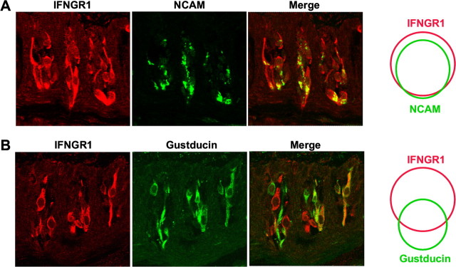
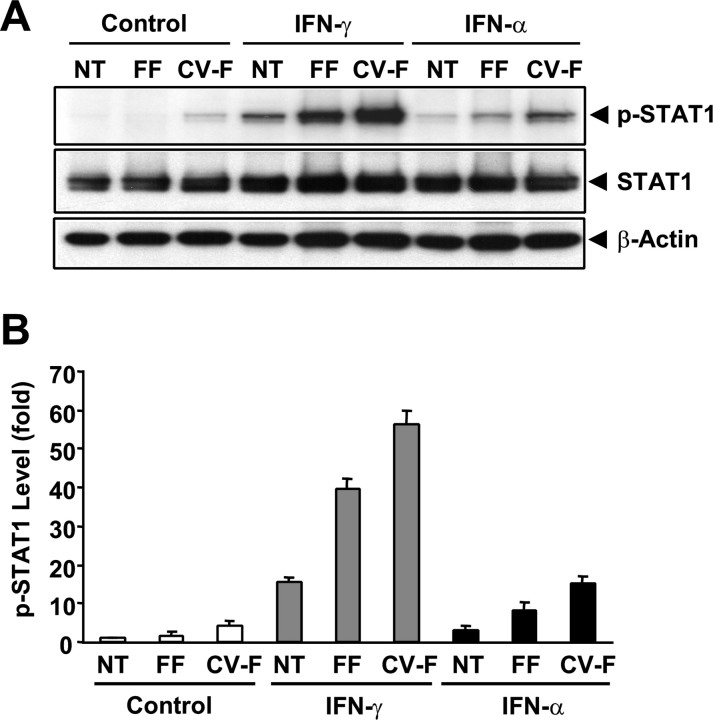
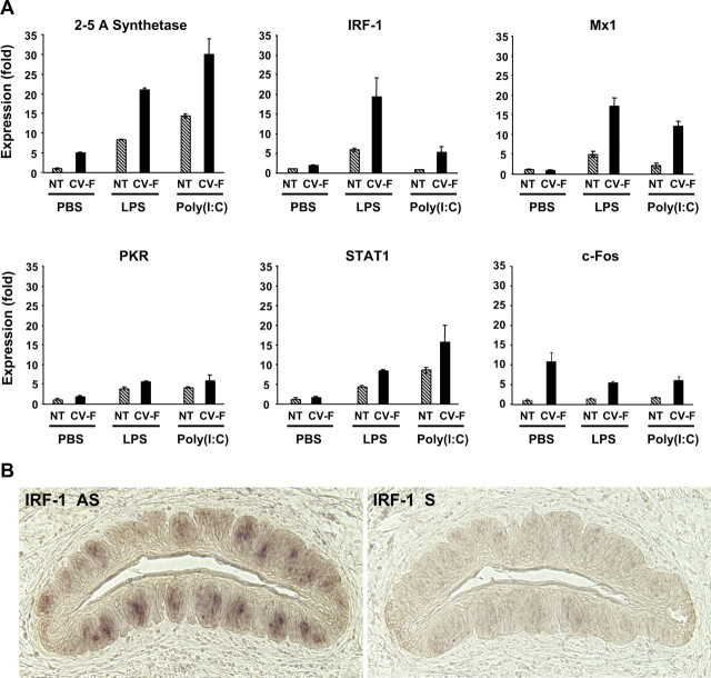
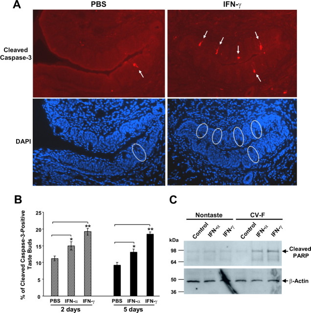
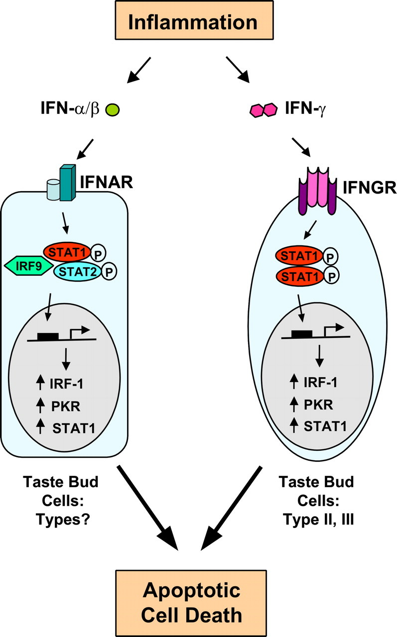
Patients with viral and bacterial infections or other inflammatory illnesses often experience taste dysfunctions. The agents responsible for these taste disorders are thought to be related to infection-induced inflammation, but the mechanisms are not known. As a first step in characterizing the possible role of inflammation in taste disorders, we report here evidence for the presence of interferon (IFN)-mediated signaling pathways in taste bud cells. IFN receptors, particularly the IFN-gamma receptor IFNGR1, are coexpressed with the taste cell-type markers neuronal cell adhesion molecule and alpha-gustducin, suggesting that both the taste receptor cells and synapse-forming cells in the taste bud can be stimulated by IFN. Incubation of taste bud-containing lingual epithelia with recombinant IFN-alpha and IFN-gamma triggered the IFN-mediated signaling cascades, resulting in the phosphorylation of the downstream STAT1 (signal transducer and activator of transcription protein 1) transcription factor. Intraperitoneal injection of lipopolysaccharide or polyinosinic:polycytidylic acid into mice, mimicking bacterial and viral infections, respectively, altered gene expression patterns in taste bud cells. Furthermore, the systemic administration of either IFN-alpha or IFN-gamma significantly increased the number of taste bud cells undergoing programmed cell death. These findings suggest that bacterial and viral infection-induced IFNs can act directly on taste bud cells, affecting their cellular function in taste transduction, and that IFN-induced apoptosis in taste buds may cause abnormal cell turnover and skew the representation of different taste bud cell types, leading to the development of taste disorders. To our knowledge, this is the first study providing direct evidence that inflammation can affect taste buds through cytokine signaling pathways.


Figures

Figure 1.
Figure 1.
The major components of both type I and type II IFN signaling pathways found in taste epithelium. A, Schematic drawing of IFN signaling pathways. On the left, the type I IFNs, such as IFN-α and IFN-β, bind to the IFNAR2 subunit of the heterodimeric receptor complex composed of IFNAR1 and IFNAR2. This binding triggers phosphorylation of JAK1 and TYK2 tyrosine kinases, which in turn phosphorylate STAT1 and STAT2 transcription factors. Phosphorylated STAT1 and STAT2 move into the nucleus and form a complex with IRF-9, which activates the transcription of many IFN-inducible genes. On the right, the type II IFN-γ binds to the IFNGR1 subunit of the tetrameric receptor complex composed of two IFNGR1 and two IFNGR2 subunits. This engagement activates JAK1 and JAK2 kinases, which subsequently phosphorylate STAT1. The phosphorylated STAT1 forms a homodimer, translocates to the nucleus, and induces the expression of IFN-γ-inducible genes. B, RT-PCR analyses of the expression of genes in type I IFN signaling pathway in taste epithelium. RNA from lingual epithelium containing fungiform (FF), foliate (F), or circumvallate (CV) taste buds was isolated, and the expression of IFNAR1, IFNAR2, JAK1, TYK2, STAT1, STAT2, and IRF-9 was analyzed by RT-PCR. C, RT-PCR analyses of the expression of the genes Ifngr1, Ifngr2, Jak1, Jak2, and Stat1 in type II IFN signaling pathway in taste epithelium. D, RT-PCR of β-actin was performed as a control. On the left and right in B–D, 1 Kb Plus DNA Ladders (Invitrogen) serve as references for DNA band size. RT-PCR primers and the sizes of the predicted PCR products are listed in Table 1.
Figure 2.
Figure 2.
The ligand-binding subunit of the IFN-α receptor complex IFNAR2 and that of the IFN-γ receptor complex IFNGR1 are expressed at higher levels in taste epithelium than in nontaste epithelium. A, Real-time RT-PCR analyses of the IFN-α receptor subunits IFNAR1 and IFNAR2 in nontaste (NT) versus circumvallate and foliate-containing (CV-F) lingual epithelium. Relative quantification of expression level was determined using glyceraldehyde-3-phosphate dehydrogenase (Gapdh) as the endogenous control gene, and the expression level in nontaste samples was arbitrarily set to 1. Error bars represent SEM. B, Real-time RT-PCR analyses of the IFN-γ receptor subunits IFNGR1 and IFNGR2 in nontaste (NT) verses circumvallate and foliate-containing (CV-F) lingual epithelium. Data were analyzed as described in A. Real-time PCR primers used in A and B are shown in Table 2. C, D, Mouse circumvallate sections processed for in situ hybridization with antisense (C) and sense (D) probes to IFNGR1. Filled arrows, Representative taste buds; open arrows, intragemmal regions lacking taste buds.
Figure 3.
Figure 3.
The IFN-γ receptor subunit IFNGR1 is expressed in a subset of taste bud cells. A–C, Immunofluorescence images of rat fungiform, foliate, and circumvallate papillae stained with antibody against IFNGR1, showing that a subset of taste bud cells were immunoreactive to the antibody. D, Peptide competition. A foliate papilla section was stained with IFNGR1 antibody that had been preincubated with the antigenic peptide.
Figure 4.
Figure 4.
IFNGR1 is partially coexpressed with NCAM and gustducin in taste bud cells. A, B, Coimmunofluorescence images of circumvallate papillae stained with antibodies against IFNGR1 (red), NCAM (green), and gustducin (green). Merged images show partial colocalization of IFNGR1 with NCAM and gustducin. Venn diagrams on the right represent colocalization of populations of IFNGR1-positive cells with NCAM- or gustducin-positive cells from circumvallate sections.
Figure 5.
Figure 5.
IFN-α and IFN-γ induce phosphorylation of STAT1 at Tyr701 in taste epithelium. Lingual epithelial pieces were excised from regions that either were devoid of any taste buds (NT) or contained fungiform (FF) or circumvallate and foliate (CV-F) taste buds. Three parallel sets of samples were treated with recombinant murine IFN-α (5 × 10³ units/ml), IFN-γ (1 × 10⁴ units/ml), or buffer only (control) for 1 h. A, Top, Western blot using phospho-STAT1 (p-STAT1, Y701)-specific antibody. Middle and bottom, The membrane used in the top was stripped and reblotted with a polyclonal antibody against total STAT1 covering both STAT1α and STATβ (STAT1, middle) and a monoclonal antibody against β-actin (bottom). B, Plot of signal intensities from Western blots in A analyzed using imaging software. The relative levels of p-STAT1 were normalized against the levels of β-actin in each sample. The level of p-STAT1 in the control nontaste sample was arbitrarily set to 1. Error bars represent SEM.
Figure 6.
Figure 6.
Inflammation induces the expression of IFN-inducible genes and decreases the level of c-fos mRNA in taste buds. A, Mice were injected intraperitoneally with either placebo (PBS) or inflammatory stimuli [LPS, 5 mg/kg in PBS, or poly(I:C), 25 mg/kg in PBS]. Six hours after injection, total RNA was isolated from nontaste (NT) and taste [circumvallate and foliate (CV-F)] lingual epithelium. Gene expression was analyzed by quantitative real-time RT-PCR. Data for five IFN-inducible genes (2–5A synthetase, Irf1, Mx1, Pkr, and Stat1) and the c-fos gene are shown. The relative expression levels of these genes were normalized using Gapdh as the endogenous control. The expression levels of these genes in PBS-treated nontaste samples were set to 1. Error bars represent SEM. The PCR primers used in this experiment are listed in Table 2. B, Expression of Irf1 in taste bud cells after LPS treatment. Mice were injected intraperitoneally with 5 mg/kg LPS. Six hours after injection, circumvallate tissues were processed for in situ hybridization with antisense (AS) or sense (S) probes to Irf1.
Figure 7.
Figure 7.
IFN treatments increase apoptotic cell death in taste buds. Mice were injected intraperitoneally with PBS, IFN-α (5 × 10³ units in PBS per mouse), or IFN-γ (2 × 10⁴ units in PBS per mouse) daily for 2 or 5 d as indicated. A, Representative immunofluorescence images of circumvallate sections from PBS-treated or IFN-γ-treated mice (5 d) stained with cleaved caspase-3 antibody (top) or corresponding images of DAPI staining (bottom). Arrows in the top point to cleaved caspase-3-positive taste cells; their corresponding taste buds in DAPI images of the bottom are marked by circles. B, The apoptotic index of circumvallate taste buds is represented as the percentage of cleaved caspase-3-positive taste buds. Error bars represent SEM; *p < 0.05; **p < 0.001. C, Peeled-off nontaste or taste [circumvallate and foliate (CV-F)] tongue epithelium was incubated for 48 h in culture medium only (control) or culture medium containing IFN-α (5 × 10³ units/ml) or IFN-γ (2 × 10⁴ units/ml). Cell lysates were assayed by Western blot analysis for cleaved PARP and β-actin. The positions of molecular weight markers are shown on the left.
Figure 8.
Figure 8.
A model for inflammation-induced taste disorders. Inflammation caused by immune responses to infection, tissue damage, or autoimmunity releases type I (IFN-α/β) and type II (IFN-γ) IFNs. IFNs bind to type I or II IFN receptors (IFNAR or IFNGR) on taste bud cells and trigger the phosphorylation of STAT proteins. Phosphorylated STAT proteins form active transcription complexes and stimulate the expression of IFN-inducible genes, including Irf1, Pkr, and Stat1. Highly induced expression of these genes increases apoptotic cell death in taste bud cells and results in a net loss of functional taste receptor cells.

Similar articles

Cited by

References

    1. Abdollahi M, Radfar M. A review of drug-induced oral reactions. J Contemp Dent Pract. 2003;4:10–31. - PubMed
    1. Aubert A, Dantzer R. The taste of sickness: lipopolysaccharide-induced finickiness in rats. Physiol Behav. 2005;84:437–444. - PubMed
    1. Balachandran S, Roberts PC, Brown LE, Truong H, Pattnaik AK, Archer DR, Barber GN. Essential role for the dsRNA-dependent protein kinase PKR in innate immunity to viral infection. Immunity. 2000;13:129–141. - PubMed
    1. Banchereau J, Pascual V. Type I interferon in systemic lupus erythematosus and other autoimmune diseases. Immunity. 2006;25:383–392. - PubMed
    1. Bartoshuk L, Desnoyers S, Hudson C, Marks L, O'Brien M. Tasting on localized areas. Ann NY Acad Sci. 1987;510:166–168.

Publication types

MeSH terms