. 2007 Oct 3;27(40):10832-9.
doi: 10.1523/JNEUROSCI.2588-07.2007.

Estradiol improves cerebellar memory formation by activating estrogen receptor beta

Affiliations

Estradiol improves cerebellar memory formation by activating estrogen receptor beta

Corina E Andreescu et al. J Neurosci. .

Abstract

Learning motor skills is critical for motor abilities such as driving a car or playing piano. The speed at which we learn those skills is subject to many factors. Yet, it is not known to what extent gonadal hormones can affect the achievement of accurate movements in time and space. Here we demonstrate via different lines of evidence that estradiol promotes plasticity in the cerebellar cortex underlying motor learning. First, we show that estradiol enhances induction of long-term potentiation at the parallel fiber to Purkinje cell synapse, whereas it does not affect long-term depression; second, we show that estradiol activation of estrogen receptor beta receptors in Purkinje cells significantly improves gain-decrease adaptation of the vestibulo-ocular reflex, whereas it does not affect general eye movement performance; and third, we show that estradiol increases the density of parallel fiber to Purkinje cell synapses, whereas it does not affect the density of climbing fiber synapses. We conclude that estradiol can improve motor skills by potentiating cerebellar plasticity and synapse formation. These processes may be advantageous during periods of high estradiol levels of the estrous cycle or pregnancy.


Figures

Figure 1.
Figure 1.
Scheme showing VOR circuitry. The VOR is an eye movement reflex that stabilizes retinal images during head movements (Collewijn and Grootendorst, 1979; Iwashita et al., 2001; Van Alphen and De Zeeuw, 2002; Boyden et al., 2004; Faulstich et al., 2004; Stahl, 2004). Primary afferents from the vestibular system [Scarpa's ganglion (Sg)] converge on second-order vestibular nuclei neurons (VN) that innervate the oculomotor nucleus (OMN) to control eye movements. Information about head movements reaches the cerebellar cortex via the mossy fibers (Mf) innervating the granule cells (GC) that generate parallel fibers (Pf). Information about retinal slip, processed by the accessory optic system (AOS) and inferior olive (IO), reaches the cerebellar cortex via the climbing fibers (Cf). This information is processed in the Purkinje cells (PC), which form the sole output of the cerebellar cortex. We tested the effect of E2 on motor performance and motor learning by investigating the VOR in the dark, before and after visuovestibular training. This reflex can be altered by the cerebellar side loop that can modulate the activity of the vestibular nuclei.
Figure 2.
Figure 2.
E2 enhances LTP but not LTD. A, B, Induction of parallel fiber-LTP by parallel fiber (PF) stimulation at 1 Hz for 5 min results in a more robust response in the slices from Eovx mice (square) compared with those from Covx (triangle) and male (M; diamond) mice. C, D, Induction of parallel fiber-LTD by parallel fiber (PF) and climbing fiber (CF) stimulation at 1 Hz for 5 min generated the same response in slices from Eovx (square), Covx (triangle), and male (diamond) mice. Traces show superimposed PF-EPSCs from Purkinje cells from a Covx (left) and an Eovx (right) mouse recorded before conjunctive stimulation (black) and 25 min after conjunctive stimulation (blue and red, respectively); each trace represents an average of 30 traces. *p < 0.05. All values are mean ± SEM.
Figure 3.
Figure 3.
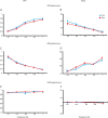
E2 does not affect motor performance. Robust compensatory eye movements are generated by head rotations in the dark (VOR) (A, B), by environment rotations in the light (OKR) (C, D), and by head rotations in light (VVOR) (E, F) with frequencies ranging from 0.2 to 1 Hz at an amplitude of 5°. The amplitude (gain) was computed as the ratio of eye velocity to stimulus velocity; timing (phase) was expressed as the difference (in degrees) between the eye velocity and stimulus velocity. In all conditions, no differences were observed in gain or phase between mice with low (Covx; triangle) and high (Eovx; square) levels of E2 (all p > 0.05, 2-way ANOVA). Error bars indicate SEM.
Figure 4.
Figure 4.
E2 enhances gain-decrease VOR learning. A, B, Normalized VOR gain and phase before training, after 50 min of training, and 24 h later in Covx mice (open bar) and Eovx mice (filled bar). Note that Eovx mice learned significantly better than Covx mice. Twenty-four hours in darkness reduced the effect of the training on gain, but the difference between groups persisted. No differences were observed in phase (*p < 0.05). C, D, No differences were noticed in gain-increase OKR learning between Eovx mice and Covx mice either after 50 min of training or 24 h later, either in gain or phase. Error bars indicate SEM.
Figure 5.
Figure 5.
Motor learning is enhanced by endogenous high levels of the E2. A, B, Thirty minutes of training induced a higher VOR gain-decrease in female mice with natural high levels of E2 [proestrus (P); squares] than in female mice with natural low levels of E2 [diestrus (D); triangles] or than in male mice (M; diamonds). No differences were observed in phase. *p < 0.05. All values are mean ± SEM.
Figure 6.
Figure 6.
E2 receptor expression in the vestibulocerebellum. A, B, ERα immunoreactivity in sagittal cerebellar slices counterstained with thionin reveals no ERα in any of the cerebellar neurons. C, D, ERβ immunoreactivity in sagittal cerebellar slices shows a high expression of the ERβ. ERβ is present in Purkinje cells (arrows) and Golgi cells (arrowheads) in the flocculus and paraflocculus. Scale bars: A, C, 400 μm; B, D, 100 μm.
Figure 7.
Figure 7.
Motor learning is enhanced by the presence of the ERβ in the Purkinje cells. A, B, Thirty minutes of training induced a higher VOR gain-decrease in L7-ERβ+/+ littermates (triangles) than in L7-ERβ−/− (diamonds). No significant differences were observed in phase. *p < 0.05. All values are mean ± SEM.
Figure 8.
Figure 8.
E2 increases synaptic density in the vestibulocerebellum. Electron micrographs of the molecular layer of the flocullus that are labeled with antibody against calbindin showing ultrastructural characteristics of Purkinje cell synapses in OVX mice that received oil (A) or E2 (B). Arrows mark synapses between Purkinje cell and parallel fibers. Scale bar, 0.5 μm.

References

    1. Adams MM, Shah RA, Janssen WG, Morrison JH. Different modes of hippocampal plasticity in response to estrogen in young and aged female rats. Proc Natl Acad Sci USA. 2001;98:8071–8076. - PMC - PubMed
    1. Albus JS. A theory of cerebellar function. Math Biosci. 1971;10:25–61.
    1. Andreescu CE, De Ruiter MM, De Zeeuw CI, De Jeu MT. Otolith deprivation induces optokinetic compensation. J Neurophysiol. 2005;94:3487–3496. - PubMed
    1. Barski JJ, Dethleffsen K, Meyer M. Cre recombinase expression in cerebellar Purkinje cells. Genesis. 2000;28:93–98. - PubMed
    1. Belmeguenai A, Hansel C. A role for protein phosphatases 1, 2A, and 2B in cerebellar long-term potentiation. J Neurosci. 2005;25:10768–10772. - PMC - PubMed

Publication types

MeSH terms