. 2007 Oct 3;27(40):10849-59.
doi: 10.1523/JNEUROSCI.2152-07.2007.

Moderate reduction of gamma-secretase attenuates amyloid burden and limits mechanism-based liabilities

Affiliations

Moderate reduction of gamma-secretase attenuates amyloid burden and limits mechanism-based liabilities

Tong Li et al. J Neurosci. .

Abstract

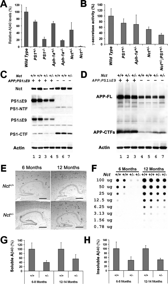
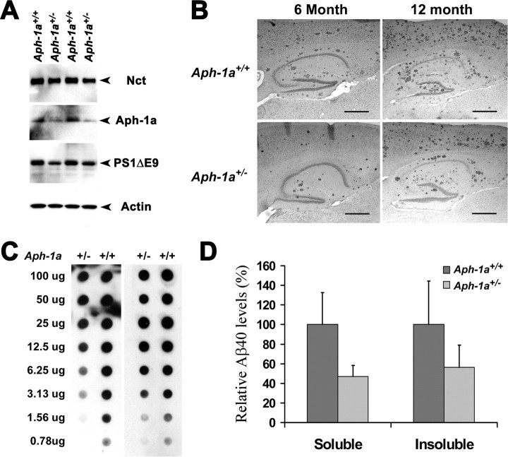
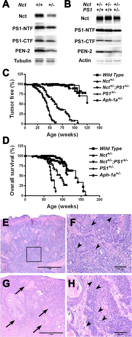
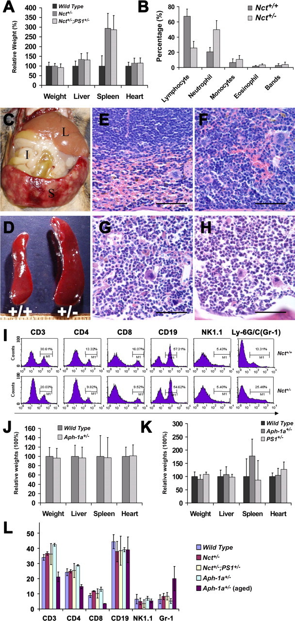
Although gamma-secretase is recognized as a therapeutic target for Alzheimer's disease, side effects associated with strong inhibition of this aspartyl protease raised serious concerns regarding this therapeutic strategy. However, it is not known whether moderate inhibition of this enzyme will allow dissociation of beneficial effects in the CNS from mechanism-based toxicities in the periphery. We tested this possibility by using a series of mice with genetic reduction of gamma-secretase (levels ranging from 25 to 64% of control mice). Here, we document that even 30% reduction of gamma-secretase can effectively ameliorate amyloid burden in the CNS. However, global reduction of this enzyme below a threshold level increased the risk of developing squamous cell carcinoma as well as abnormal proliferation of granulocytes in a gamma-secretase dosage-dependent manner. Importantly, we demonstrate that there exists a critical gamma-secretase level that reduces the risk of amyloidosis in the CNS and limits tumorigenesis in epithelia. Our findings suggest that moderate inhibition of gamma-secretase represents an attractive anti-amyloid therapy for Alzheimer's disease.


Figures

Figure 1.
Figure 1.
Reduction of γ-secretase activity attenuates Aβ deposition in brains of APPswe/PS1ΔE9;Nct+/− mice. A, ELISA analysis of secreted Aβ40 peptides in the conditional medium of fibroblasts that have been infected with adenovirus-expressing APPswe protein. The data were averages from four parallel experiments (p = 0.002). B, In vitro γ-secretase assay of brain extracts of wild-type, PS1+/−, Aph1a+/−, Nct+/−, and Nct+/−;PS1+/− mice. The data were averages from four parallel experiments (p = 0.002). C, Protein extracts (40 μg each) from brains of APPswe;PS1ΔE9 (lanes 1, 2), Nct+/−;APPswe;PS1ΔE9 (lanes 3, 4), Nct+/− (lanes 7,8), and nontransgenic control (lanes 5,6) mice were immunoblotted with antisera specific to Nct, PS1–NTF, PS1–CTF, or actin. D, Protein extracts (40 μg each) from brains of APPswe;PS1ΔE9 (lane 1), Nct+/−;APPswe;PS1ΔE9 (lanes 2, 3), Nct+/− (lanes 6,7), and nontransgenic control (lanes 4, 5) mice were immunoblotted with antisera that recognize full-length APP (APP-FL) and APP–CTFs. The same blot was stripped and reblotted using antiserum specific to actin as loading control. E, Sagittal brain sections (10 μm) of 6- and 12-month-old APPswe;PS1ΔE9;Nct+/+ and APPswe;PS1ΔE9;Nct+/− female mice. The Aβ plaques were visualized by immunostaining with antibodies specific to ubiquitin. Scale bars, 0.5 mm. F, Quantitative analysis of the levels of Aβ aggregation in the brains of APPswe;PS1ΔE9;Nct+/− and APPswe;PS1ΔE9;Nct+/+ mice at 6 and 12 months of age using the filter trap assay. G, H, ELISA of soluble (PBS extraction, G, n = 6; p = 0.05) and insoluble (formic acid extraction, H; n = 6; p = 0.001) Aβ40 peptides extracted from the brains of female APPswe;PS1ΔE9;Nct+/+ and APPswe;PS1ΔE9;Nct+/− mice ranging from 6 and 12 months of age.
Figure 2.
Figure 2.
Reduction of γ-secretase activity attenuates Aβ deposition in brains of APPswe/PS1ΔE9;Aph-1a+/− mice. A, Protein extracts (40 μg each) from brains of APPswe;PS1ΔE9;Aph-1a+/+ and APPswe;PS1ΔE9;Aph-1a+/− were immunoblotted with antisera specific to Nct, Aph-1aL, PS1–NTF, and actin. B, Sagittal brain sections (10 μm) of 6- and 12-month-old APPswe;PS1ΔE9;Aph-1a+/+ and APPswe;PS1ΔE9;Aph-1a+/− female mice. The Aβ plaques were visualized by immunostaining with antibodies specific to ubiquitin. Scale bars, 0.5 mm. C, Filter trap analysis of Aβ aggregation in the brains of APPswe;PS1ΔE9;Nct+/− and APPswe;PS1ΔE9;Nct+/+ mice at 12 months of age. D, ELISA of insoluble (formic acid extraction) and soluble (PBS extraction) Aβ40 peptides extracted from the brains of female APPswe;PS1ΔE9;Aph-1a+/+ and APPswe;PS1ΔE9;Aph-1a+/− mice at 12 months of age (n = 5; p = 0.034).
Figure 3.
Figure 3.
Mice with reduced γ-secretase activity develop spontaneous tumors. A, Protein extracts (40 μg each) from skin of Nct+/+ and Nct+/− mice were immunoblotted with antisera specific to Nct, PS1–NTF, PS1–CTF, or PEN-2. Immunoblots were striped and reprobed using antiserum against β-tubulin as loading control. B, Protein extracts (40 μg each) from skins of Nct+/− and Nct+/−;PS1+/− mice were immunoblotted with antisera specific to Nct, PS1–NTF, PS1–CTF, or PEN-2. Immunoblots were striped and reprobed using antiserum against actin as loading control. C, Kaplan–Meyer plots of tumor development in Nct+/+, Nct+/−, and Nct+/−;PS1+/− mice. Kaplan–Meyer plots of tumor development in Nct+/+ (n = 96) and Nct+/− (n = 89) mice. Note that Nct+/− mice developed tumors with median age of ∼60 weeks, and >90% of Nct+/− mice developed skin tumors by 100 weeks of age; none of Nct+/+ mice developed skin tumors by this age. The median age of tumor development in Nct+/−;PS1+/− mice is greatly accelerated when compared with Nct+/− mice (45 vs 60 weeks), and >90% of Nct+/−;PS1+/− mice developed skin tumors by 60 weeks of age (p < 0.0001). D, Kaplan–Meyer survival plots for Nct+/+, Nct+/−, and Nct+/−;PS1+/− mice. Compared with wild-type mice, Nct+/− mice have a significantly shorter lifespan, with a median life expectancy of 94 weeks, whereas Nct+/−;PS1+/− mice have a further shortened lifespan, with median life expectancy of 71 weeks (p < 0.0001). Death is defined as the time when mice became moribund. E, SCC near the tail of Nct+/− mice (33 weeks of age) shows moderately well differentiated invasive clusters of squamous type epithelial cells (4×). F, Higher magnification (20×) of E (boxed area) showing moderately well differentiated invasive clusters of squamous type epithelial cells with cornification, disorganization, atypia, and mitotic figures (arrowheads). G, Squamous cell carcinoma (arrows) metastasized to the renal cortex in an Nct+/− mouse (98 weeks of age) (4×). H, Higher magnification (40×) of skin lesions in Nct+/−;PS1+/− mice (21 weeks of ages) showing clusters of squamous epithelial cells with disorganization, atypia, and mitotic figures (arrowheads). All sections (10 μm) were stained with hematoxylin and eosin.
Figure 4.
Figure 4.
Nct+/− mice exhibit abnormalities in spleen. A, Comparison of the size of organs between Nct+/−, Nct+/−;PS1+/−, and wild-type mice. The average weight of spleen in Nct+/− and Nct+/−;PS1+/− mice is approximately threefold larger than wild-type control mice. Wet weight of organs was determined immediate after necropsy. Samples were collected from 53 Nct+/− mice, 35 Nct+/−;PS1+/− mice, and 66 age-matched wild-type controls (p < 0.0001). B, Cell counts of peripheral blood; percentage of various cell types from Nct+/− (n = 8) and Nct+/+ (n = 8) mice are plotted (p = 0.01). C, The spleen can weigh >2 g in some Nct+/− mice. Organs indicated are liver (L), intestine (I), and spleen (S). D, Examples of spleens from an Nct+/− (right) and its littermate Nct+/+ mice (left) at 56 weeks of age. E, F, Section (10 μm) of spleen from an Nct+/− mouse (F) and an Nct+/+ littermate (E) at 51 weeks of age stained with hematoxylin and eosin. Note that subcapsular red pulp is expanded and filled almost exclusively by granulocytes and granulocyte precursors in Nct+/− mouse (F), whereas in spleen of Nct+/+ mouse, normal subcapsular red pulp with abundant red blood cells and normal white pulp (top half of image) are observed (60×). Scale bars, 50 μm. G, H, Section (10 μm) of bone marrow from an Nct+/− mouse (H) and a wild-type mouse (G) at 51 weeks of age stained with hematoxylin and eosin; note that bone marrow from Nct+/− mouse composed almost exclusively of granulocyte precursors with irregular segmented nuclear morphology (H). The bone marrow from wild-type mouse showed ample erythroid precursors with more regularly rounded nuclei (G) (60×). Scale bars, 50 μm. I, Phenotypic analysis of splenocytes from Nct+/+ and Nct+/− mice. Single splenocyte suspensions were prepared and stained with antibodies indicated. The cells were then analyzed using FACSCalibur and CELLQuest software. Note that the percentage of T-cell population is decreased, whereas granulocytes are increased in spleen of Nct+/− mice. J, Comparison of the size of organs between Aph-1a+/− and wild-type mice at older than 60 weeks of age or younger. Samples were collected from 25 Aph-1a+/− mice and 48 age-matched wild-type controls. K, Comparison of the size of organs between Aph-1a+/−, PS1+/−, and wild-type mice at older than 60 weeks of age. Samples were collected from 18 Aph-1a+/− mice, 7 PS1+/− mice, and 18 age-matched wild-type controls. L, Analysis of splenocytes from young Aph-1a+/−, Nct+/−, Nct+/−;PS1+/−, and wild-type mice (n = 4 for each genotype, 3 months of age) compared with aged Aph-1a+/− mice at 15 months of age (n = 4). Note that the percentage of T-cell population is decreased, whereas percentage of granulocytes is increased only in the spleen of aging Aph-1a+/− mice.
Figure 5.
Figure 5.
Sizes of spleens in different lines of mice. The weights of spleen were plotted against the age of the mice. Spleen in mice with splenomegaly weighed >0.2 g, which is more than twofold compared with that of wild-type mice. Note that risk of developing splenomegaly is dramatically increased in Nct+/−, Nct+/−;PS1+/−, and Aph-1a+/− mice that are >1 year of age (arrow) but not in PS1+/− mice even at older ages.
Figure 6.
Figure 6.
Normal cognition in Nct+/− mice. A, Learning in the Morris water maze reference memory task was equivalent in wild-type and Nct+/− mice at 3 months of age as determined by the percentage of time spent in annulus 40 on the probe trial (40 cm diameter around the platform position; see inset). B, C, No difference in anxiety was observed between 3-month-old wild-type and Nct+/− mice as assessed in an open-field test as determined by the percentage of time spent in each zone (B) and on the elevated plus maze as determined by the number of entries in closed and opened arm (C). D, Performance in Y-maze memory task was comparable between wild-type and Nct+/− mice at 6 months of age. There was no significant difference in spontaneous alternation (SA, inner box; p = 0.29) and number across the sessions between two groups (p = 0.32).

Comment in

References

    1. Allman D, Punt JA, Izon DJ, Aster JC, Pear WS. An invitation to T and more: Notch signaling in lymphopoiesis. Cell. 2002;109:S1–S11. - PubMed
    1. Andreasson KI, Savonenko AV, Vidensky S, Goellner JJ, Zhang Y, Shaffer A, Kaufmann WI, Worley PF, Isakson P, Markowska AL. Age-dependent cognitive deficits and neuronal loss cyclooxygenase-2 transgenic mice. J Neurosci. 2001;21:8198–8209. - PMC - PubMed
    1. Artavanis-Tsakonas S, Rand MD, Lake RJ. Notch signaling: cell fate control and signal integration in development. Science. 1999;284:770–776. - PubMed
    1. Barten DM, Guss VL, Corsa JA, Loo A, Hansel SB, Zheng M, Munoz B, Srinivasan K, Wang B, Robertson BJ, Polson CT, Wang J, Roberts SB, Hendrick JP, Anderson JJ, Loy JK, Denton R, Verdoorn TA, Smith DW, Felsenstein KM. Dynamics of β-amyloid reductions in brain, cerebrospinal fluid, and plasma of β-amyloid precursor protein transgenic mice treated with a γ-secretase inhibitor. J Pharmacol Exp Ther. 2005;312:635–643. - PubMed
    1. Birchler JA, Riddle NC, Auger DL, Veitia RA. Dosage balance in gene regulation: biological implications. Trends Genet. 2005;21:219–226. - PubMed

Publication types

MeSH terms