Bim expression indicates the pathway to retinal cell death in development and degeneration
- PMID: 17913922
- PMCID: PMC6672824
- DOI: 10.1523/JNEUROSCI.0903-07.2007
Bim expression indicates the pathway to retinal cell death in development and degeneration
Abstract
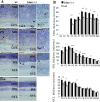
Programmed cell death (PCD) during development of the mouse retina involves activation of the mitochondrial pathway. Previous work has shown that the multidomain Bcl-2 family proteins Bax and Bak are fundamentally involved in this process. To induce mitochondrial membrane permeabilization, Bax and Bak require that prosurvival members of the family be inactivated by binding of "BH3-only" members. We showed previously that the BH3-only protein BimEL is highly expressed during postnatal retinal development but decreases dramatically thereafter. The purpose of this study was to investigate a possible role for Bim, in retinal development and degeneration, upstream of Bax and Bak. Bim-/- mice analyzed for defective retinal development exhibit an increase in retinal thickness and a delay in PCD, thereby confirming a role for Bim. We also demonstrate that in response to certain death stimuli, bim+/+ retinal explants upregulate BimEL leading to caspase activation and cell death, whereas bim-/- explants are resistant to apoptosis. Finally, we analyzed Bim expression in the retinal degeneration (rd) mouse, an in vivo model of retinal degeneration. Bim isoforms, which decrease during development, are not reexpressed during retinal degeneration and ultimately photoreceptor cells die by a caspase-independent mechanism. Thus, we conclude that in cases in which BimEL is reexpressed during pathological cell death, developmental cell death pathways are reactivated. However, the absence of BimEL expression correlates with caspase-independent death in the rd model.
Figures


Similar articles
-
A Critical role for Bim in retinal ganglion cell death.J Neurochem. 2007 Aug;102(3):922-30. doi: 10.1111/j.1471-4159.2007.04573.x. Epub 2007 Apr 17. J Neurochem. 2007. PMID: 17442051
-
The role of BH3-only protein Bim extends beyond inhibiting Bcl-2-like prosurvival proteins.J Cell Biol. 2009 Aug 10;186(3):355-62. doi: 10.1083/jcb.200905153. Epub 2009 Aug 3. J Cell Biol. 2009. PMID: 19651893 Free PMC article.
-
BH3-only protein BIM mediates heat shock-induced apoptosis.PLoS One. 2014 Jan 10;9(1):e84388. doi: 10.1371/journal.pone.0084388. eCollection 2014. PLoS One. 2014. PMID: 24427286 Free PMC article.
-
Bax activation by Bim?Cell Death Differ. 2009 Sep;16(9):1187-91. doi: 10.1038/cdd.2009.83. Epub 2009 Jun 26. Cell Death Differ. 2009. PMID: 19557009 Review.
-
Transforming growth factor beta (TGFbeta)-induced apoptosis: the rise & fall of Bim.Cell Cycle. 2009 Jan 1;8(1):11-7. doi: 10.4161/cc.8.1.7291. Epub 2009 Jan 30. Cell Cycle. 2009. PMID: 19106608 Free PMC article. Review.
Cited by
-
Apoptotic cell death in disease-Current understanding of the NCCD 2023.Cell Death Differ. 2023 May;30(5):1097-1154. doi: 10.1038/s41418-023-01153-w. Epub 2023 Apr 26. Cell Death Differ. 2023. PMID: 37100955 Free PMC article. Review.
-
Prolonged expression of Puma in cholinergic amacrine cells during the development of rat retina.J Histochem Cytochem. 2012 Oct;60(10):777-88. doi: 10.1369/0022155412452737. Epub 2012 Jun 26. J Histochem Cytochem. 2012. PMID: 22736709 Free PMC article.
-
Mouse Retinal Organoid Growth and Maintenance in Longer-Term Culture.Front Cell Dev Biol. 2021 Apr 27;9:645704. doi: 10.3389/fcell.2021.645704. eCollection 2021. Front Cell Dev Biol. 2021. PMID: 33996806 Free PMC article.
-
Photoreceptor cell death and rescue in retinal detachment and degenerations.Prog Retin Eye Res. 2013 Nov;37:114-40. doi: 10.1016/j.preteyeres.2013.08.001. Epub 2013 Aug 28. Prog Retin Eye Res. 2013. PMID: 23994436 Free PMC article. Review.
-
Human retinal transmitochondrial cybrids with J or H mtDNA haplogroups respond differently to ultraviolet radiation: implications for retinal diseases.PLoS One. 2014 Jun 11;9(2):e99003. doi: 10.1371/journal.pone.0099003. eCollection 2014. PLoS One. 2014. PMID: 24919117 Free PMC article.
References
-
- Bahi N, Zhang J, Llovera M, Ballester M, Comella JX, Sanchis D. Switch from caspase-dependent to -independent death during heart development: essential role of EndoG in ischemia-induced DNA processing of differentiated cardiomyocytes. J Biol Chem. 2006;281:22943–22952. - PubMed
-
- Biswas SC, Greene LA. Nerve growth factor (NGF) down-regulates the Bcl-2 homology 3 (BH3) domain-only protein Bim and suppresses its proapoptotic activity by phosphorylation. J Biol Chem. 2002;277:49511–49516. - PubMed
-
- Bouillet P, Metcalf D, Huang DC, Tarlinton DM, Kay TW, Kontgen F, Adams JM, Strasser A. Proapoptotic Bcl-2 relative Bim required for certain apoptotic responses, leukocyte homeostasis, and to preclude autoimmunity. Science. 1999;286:1735–1738. - PubMed
-
- Chahory S, Padron L, Courtois Y, Torriglia A. The LEI/L-DNase II pathway is activated in light-induced retinal degeneration in rats. Neurosci Lett. 2004;367:205–209. - PubMed
-
- Chen L, Willis SN, Wei A, Smith BJ, Fletcher JI, Hinds MG, Colman PM, Day CL, Adams JM, Huang DC. Differential targeting of prosurvival Bcl-2 proteins by their BH3-only ligands allows complementary apoptotic function. Mol Cell. 2005;17:393–403. - PubMed
Publication types
MeSH terms
Substances
LinkOut - more resources
Full Text Sources
Molecular Biology Databases
Research Materials
Miscellaneous
