. 2008 Jan 15;111(2):624-32.
doi: 10.1182/blood-2007-04-084533. Epub 2007 Oct 3.

Membrane cofactor protein mutations in atypical hemolytic uremic syndrome (aHUS), fatal Stx-HUS, C3 glomerulonephritis, and the HELLP syndrome

Affiliations

Membrane cofactor protein mutations in atypical hemolytic uremic syndrome (aHUS), fatal Stx-HUS, C3 glomerulonephritis, and the HELLP syndrome

Celia J Fang et al. Blood. .

Abstract

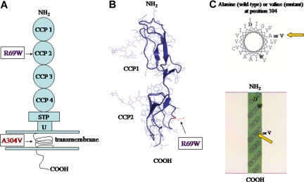
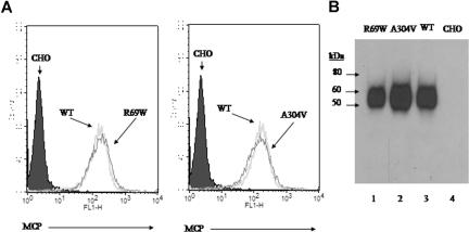
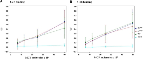
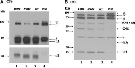
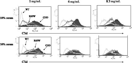
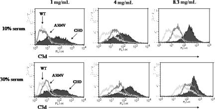
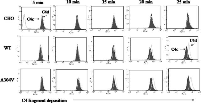
The hemolytic uremic syndrome (HUS) is a triad of microangiopathic hemolytic anemia, thrombocytopenia, and renal impairment. Genetic studies demonstrate that heterozygous mutations of membrane cofactor protein (MCP;CD46) predispose to atypical HUS (aHUS), which is not associated with exposure to Shiga toxin (Stx). Among the initial 25 MCP mutations in patients with aHUS were 2, R69W and A304V, that were expressed normally and for which no dysfunction was found. The R69W mutation is in complement control protein module 2, while A304V is in the hydrophobic transmembrane domain. In addition to 3 patients with aHUS, the A304V mutation was identified in 1 patient each with fatal Stx-HUS, the HELLP (hemolysis, elevated liver enzymes, and low platelets) syndrome, and glomerulonephritis with C3 deposits. A major goal was to assess if these putative mutations lead to defective complement regulation. Permanent cell lines expressing the mutated proteins were complement "challenged," and membrane control of C3 fragment deposition was monitored. Both the R69W and A304V MCP mutations were deficient in their ability to control the alternative pathway of complement activation on a cell surface, illustrating the importance of modeling transmembrane proteins in situ.


Figures

Figure 1
Figure 1
MCP structure. (A) Schematic diagram of protein structure. R69W mutation is in CCP 2. The A304V mutation is in the transmembrane domain. (B) Crystal structure of CCP 2 highlighting the location of R69W. (C) Helical wheel diagram of predicted segments and transmembrane representation by SOSUI (http://bp.nuap.nagoya-u.ac.jp/sosui) of A304V MCP mutation versus that of wild type. Yellow arrows indicate mutation of MCP at position 304 from alanine (left) to valine (right).
Figure 2
Figure 2
Expression profile and electrophoretic mobility of R69W and A304V mutants. (A) Flow cytometry of MCP expression on CHO cell lines. Stable transfectants R69W and A304V express similar copy number of MCP/cell (approximately 100 000) compared with wild type. (B) Western blot of these same CHO cell lysates probed with a rabbit polyclonal Ab to MCP. R69W and A304V have similar Mr to wild type. Representative experiment of 3.
Figure 3
Figure 3
Analysis of MCP binding to C3b and C4b. C3b and C4b binding assays were performed at 25 mM (Figure S1) and 50 mM (shown here) salt concentration. CHO cell lysates (5 × 10⁶ to 25 × 106 MCP molecules as measured by ELISA) of R69W, A304V, and wild type from both transient transfections and stable cell lines were used. Data represent means (± SD) for 4 experiments at 50 mM.
Figure 4
Figure 4
Analysis of C3b and C4b cofactor activity. Lysates of CHO cells expressing R69W, A304V, or wild-type MCP were incubated at 50 mM NaCl for 90 minutes with C3b or C4b and factor I. Cleavage of the α′ chain and generation of the α1 and α2 by the mutants are comparable with that of wild type. For the Western blot C3b cofactor activity (A), α′ and β represent the 2 chains of C3b. α1 corresponds to the larger proteolytic product of the α′ chain of C3b. The bottom portion of panel A is a 3-fold longer exposure of the same gel to show the smaller α2. SDS-PAGE, 10% gel, reducing conditions. Representative experiment of 3. For the Western blot of C4b cofactor activity (B), cleavage fragments C4d and α4 are similar between the mutants and wild type. The α3 is poorly biotinylated and not seen. The light band at approximately 60 kDa is most likely C4d (45 kDa) + α4 (15 kDa) and again is similar for the mutants in comparison with wild type. SDS-PAGE, 4% to 10% gel, reducing conditions. Representative experiment of 3.
Figure 5
Figure 5
Comparison of R69W mutant versus wild-type MCP in inhibiting the alternative complement pathway. FACS analysis of C3d deposition using 1, 4, or 8.3 mg/mL of sensitizing Ab with 10% or 30% C7d serum for 60 minutes in Mg2+-EGTA. Representative experiment of 3. See Table 3 for MFIs.
Figure 6
Figure 6
Comparison of A304V mutant versus wild-type MCP in inhibiting the alternative pathway. FACS analysis of C3d deposition using 1, 4, or 8.3 mg/mL of sensitizing Ab with 10% or 30% C7d serum for 60 minutes in Mg2+-EGTA. Representative experiment of 3. See Table 4 for MFIs.
Figure 7
Figure 7
Kinetic analysis of cofactor activity for C4b of wild type versus the A304V mutant MCP. Control profile was nearly identical for cells exposed to sensitizing antibody but not to serum or to serum alone. Antibody, 1.0 mg/mL; serum concentration, 10%. Representative experiment of 3.

Similar articles

Cited by

References

    1. Moake JL. Thrombotic microangiopathies. N Engl J Med. 2002;347:589–600. - PubMed
    1. Warwicker P, Goodship THJ, Donne RL, et al. Genetic studies into inherited and sporadic hemolytic uremic syndrome. Kidney Int. 1998;53:836–844. - PubMed
    1. Perez-Caballero D, Gonzalez-Rubio C, Gallardo ME, et al. Clustering of missense mutations in the C-terminal region of factor H in atypical hemolytic uremic syndrome. Am J Hum Genet. 2001;68:478–484. - PMC - PubMed
    1. Richards A, Buddles MR, Donne RL, et al. Factor H mutations in hemolytic uremic syndrome cluster in exons 18–20, a domain important for host cell recognition. Am J Hum Genet. 2001;68:485–490. - PMC - PubMed
    1. Caprioli J, Bettinaglio P, Zipfel PF, et al. The molecular basis of familial hemolytic uremic syndrome: mutation analysis of factor H gene reveals a hot spot in short consensus repeat 20. J Am Soc Nephrol. 2001;12:297–307. - PubMed

Publication types

MeSH terms