. 2007 Dec;18(12):4979-91.
doi: 10.1091/mbc.e07-06-0622. Epub 2007 Oct 3.

The golgin GCC88 is required for efficient retrograde transport of cargo from the early endosomes to the trans-Golgi network

Affiliations

The golgin GCC88 is required for efficient retrograde transport of cargo from the early endosomes to the trans-Golgi network

Zi Zhao Lieu et al. Mol Biol Cell. 2007 Dec.

Abstract

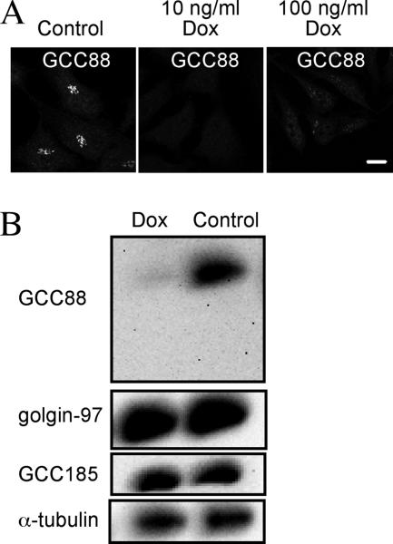
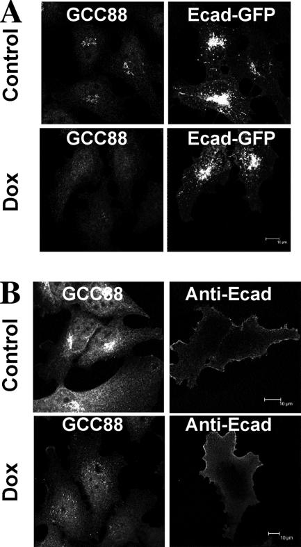
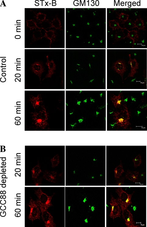
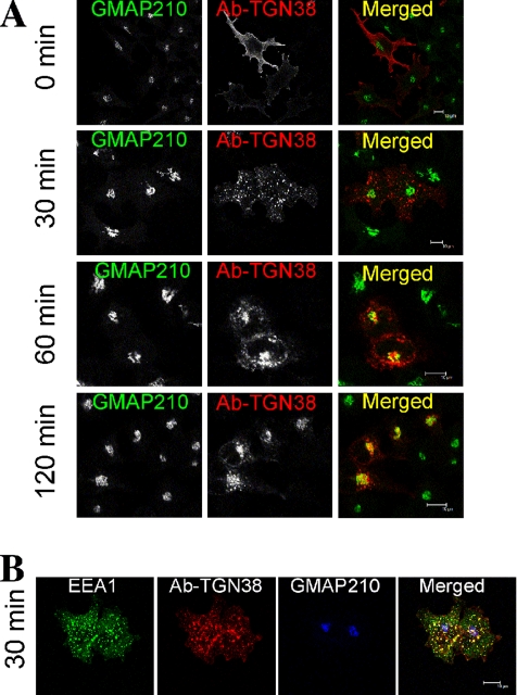
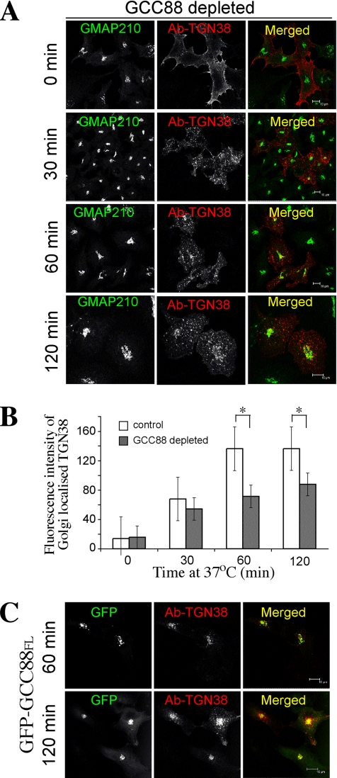
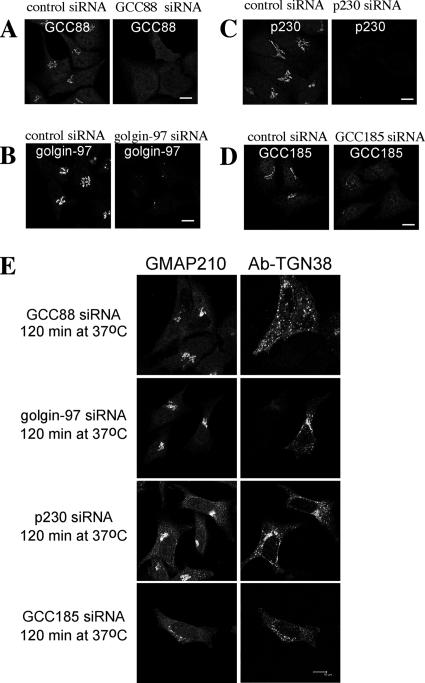
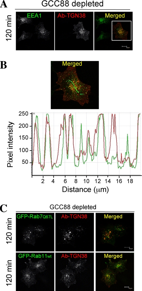
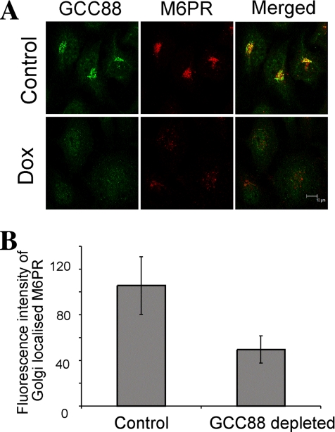
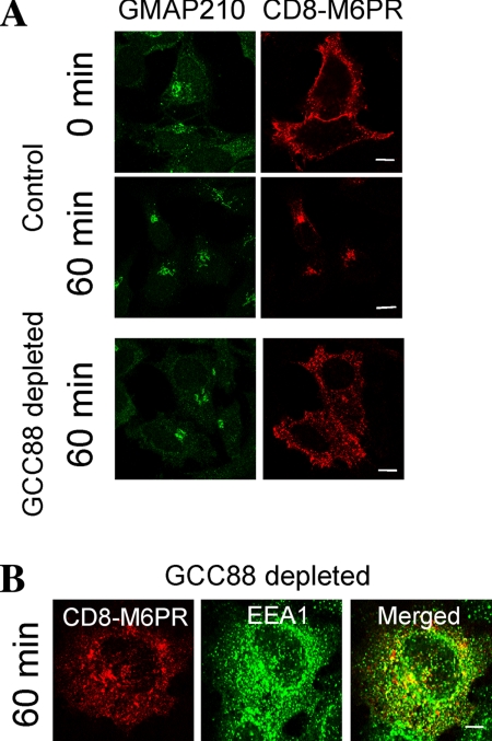
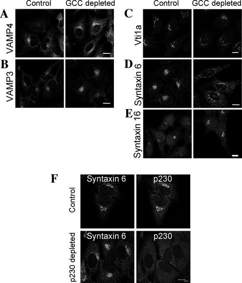
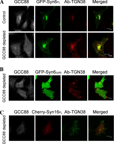
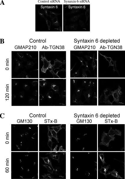
Retrograde transport pathways from early/recycling endosomes to the trans-Golgi network (TGN) are poorly defined. We have investigated the role of TGN golgins in retrograde trafficking. Of the four TGN golgins, p230/golgin-245, golgin-97, GCC185, and GCC88, we show that GCC88 defines a retrograde transport pathway from early endosomes to the TGN. Depletion of GCC88 in HeLa cells by interference RNA resulted in a block in plasma membrane-TGN recycling of two cargo proteins, TGN38 and a CD8 mannose-6-phosphate receptor cytoplasmic tail fusion protein. In GCC88-depleted cells, cargo recycling was blocked in the early endosome. Depletion of GCC88 dramatically altered the TGN localization of the t-SNARE syntaxin 6, a syntaxin required for endosome to TGN transport. Furthermore, the transport block in GCC88-depleted cells was rescued by syntaxin 6 overexpression. Internalized Shiga toxin was efficiently transported from endosomes to the Golgi of GCC88-depleted cells, indicating that Shiga toxin and TGN38 are internalized by distinct retrograde transport pathways. These findings have identified an essential role for GCC88 in the localization of TGN fusion machinery for transport from early endosomes to the TGN, and they have allowed the identification of a retrograde pathway which differentially selects TGN38 and mannose-6-phosphate receptor from Shiga toxin.


Figures

Figure 1.
Figure 1.
Depletion of endogenous GCC88 by an inducible shRNA. HeLa cells (clone A8) stably expressing a tetracyclineon inducible shRNA to GCC88 (tetRGCC88KD A8) were either untreated (control) or treated with 10 or 100 ng/ml doxycycline (Dox) for 96 h, and monolayers were fixed with 4% paraformaldehyde. (A) Endogenous GCC88 was detected with rabbit anti-GCC88 antibodies followed by Alexa 488-conjugated anti-rabbit IgG. (B) HeLa A8 cells were incubated with 10 ng/ml doxycycline for 96 h, lysed in SDS-PAGE reducing buffer, and then extracts were subjected to SDS-PAGE on a 7.5% polyacrylamide gel. Proteins were transfer to a polyvinylidene difluoride membrane and probed with rabbit anti-GCC88 antibodies using a chemiluminescence detection system. The membrane were then stripped and reprobed with anti-α-tubulin, followed by anti-GCC185 and anti-golgin-97 antibodies. Bar, 10 μm.
Figure 2.
Figure 2.
Anterograde transport of E-cadherin in GCC88-depleted cells is unaffected. HeLa A8 cells were either untreated (control) or incubated with 10 ng/ml Dox for 72 h and then transfected with Ecad-GFP for 24 h before staining. (A) Monolayers were fixed with 4% paraformaldehyde, permeabilized, and endogenous GCC88 was detected with rabbit anti-GCC88 antibodies followed by Alexa 568-conjugated anti-rabbit IgG and Ecad-GFP by GFP fluorescence. (B) Monolayers were fixed and stained with monoclonal anti-Ecad antibodies followed by Alexa 568-conjugated goat ant-mouse IgG. Fixed monolayers were then permeabilized and stained with rabbit anti-GCC88 antibodies followed by Alexa 647-conjugated goat anti-rabbit IgG. Bars, 10 μm.
Figure 3.
Figure 3.
Shiga toxin is transported efficiently to the Golgi in GCC88-depleted cells. HeLa A8 cells were either untreated (control) (A) or incubated with 10 ng/ml doxycycline for 96 h (GCC88 depleted) (B). Monolayers were incubated with Cy3-conjugated STx-B for 45 min on ice, and then they were either fixed immediately (0 min) or incubated at 37°C for either 20 or 60 min, followed by fixation in 4% paraformaldehyde. Cells were stained with monoclonal antibodies to GM130 followed by Alexa-conjugated anti-mouse IgG. Bars, 10 μm.
Figure 4.
Figure 4.
Retrograde transport of TGN38 in noninduced HeLa A8 cells. HeLa A8 cells were transfected with TGN38-CFP and 24 h later they were incubated with monoclonal mouse anti-TGN38 antibodies for 30 min on ice. After washing, cells were either fixed directly (0 min) or incubated at 37°C in serum-free media for the indicated time to internalize the antibody–TGN38 complex. (A) Monolayers were fixed in 4% paraformaldehyde, permeabilized, and stained with Alexa568-conjugated anti-mouse IgG for 60 min. Endogenous GMAP-210 was stained with rabbit anti-GMAP-210, followed by Alexa488-conjugated anti-rabbit IgG. (B) Monolayers were fixed and stained with anti-mouse IgG as in A and also stained with rabbit anti-GMAP-210, followed by Alexa647-conjugated anti-rabbit IgG and for EEA1 with human anti-EEA1 antibodies followed by FITC conjugated anti-human IgG. Bars, 10 μm.
Figure 5.
Figure 5.
TGN38 trafficking is impaired in GCC88 depleted cells. (A) HeLa A8 cells were treated with 10 ng/ml doxycycline for 72 h, transfected with TGN38-CFP, and 24 h later they were incubated with anti-TGN38 antibodies at 0°C. After washing unbound antibodies, monolayers were either fixed directly (0 min) or incubated at 37°C for the indicated time to internalize the antibody–TGN38 complex, and then they were fixed in 4% paraformaldehyde and permeabilized. Cells were stained with Alexa568-conjugated anti-mouse IgG. Endogenous GMAP-210 was stained with rabbit anti-GMAP-210, followed by Alexa488-conjugated anti-rabbit IgG. (B) GCC88-depleted cells were stained as described above, and TGN38 staining intensity within the Golgi region was determined using GMAP-210 staining to mark the Golgi location. Fluorescence intensity was quantified using Leica imaging software (n = 25 for each time point). Results are means and error bars represent standard deviation. *P < 0.001. (C) HeLa A8 cells were incubated with 10 ng/ml doxycycline for 96 h, and then they were transfected with TGN38 together with GFP-GCC88FL 24 h before an internalization assay. Monolayers were incubated with monoclonal anti-TGN38 antibodies for 30 min on ice, washed in PBS, and then incubated at 37°C in serum-free media for either 60 or 120 min, as indicated. Monolayers were fixed in 4% paraformaldehyde, permeabilized, and stained with Alexa568-conjugated anti-mouse IgG. GFP-GCC88FL was detected by GFP fluorescence. Bars, 10 μm.
Figure 6.
Figure 6.
TGN38 trafficking in inhibited by silencing GCC88 but not the other TGN golgins. HeLa cells were transfected with GCC88 siRNA (A), golgin-97 siRNA (B), p230 siRNA (C), and GCC185 siRNA (D) for 72 h, fixed in 4% paraformaldehyde, and stained with rabbit anti-GCC88 antibodies, monoclonal mouse anti-golgin-97 antibodies, human anti-p230 antibodies, and rabbit anti-GCC185 antibodies, respectively, followed by Alexa568-conjugated IgG. (E) HeLa cells transfected with siRNA as described above for 48 h and then transfected a second time with TGN38-CFP for a further 24 h. Monolayers were then incubated with monoclonal mouse anti-TGN38 antibodies on ice for 30 min, washed with PBS, and incubated in serum-free media at 37°C for 120 min. Monolayers were fixed in 4% paraformaldehyde, permeabilized, and stained with Alexa568-conjugated anti-mouse IgG. Endogenous GMAP-210 was stained with rabbit anti-GMAP-210, followed by Alexa488-conjugated anti-rabbit IgG. Bars, 10 μm.
Figure 7.
Figure 7.
Internalized TGN38 accumulates in an EEA1-positive compartment in GCC88-depleted cells. HeLa A8 were treated with 10 ng/ml doxycycline for 72 h and then cotransfected with TGN38 alone (A and B) or with TGN38 and GFP-Rab7(Q67L) or GFP-Rab11wt (C), as indicated, 24 h before an internalization assay. Monolayers were incubated with monoclonal mouse anti-TGN38 antibodies for 30 min on ice, washed in PBS, and then incubated at 37°C in serum-free media for 120 min to internalize the antibody–TGN38 complex. Monolayers were fixed in 4% paraformaldehyde, permeabilized, and stained with Alexa568-conjugated anti-mouse IgG for 60 min. (A) Endogenous EEA1 was stained with human anti-EEA1 antibodies, followed by an FITC-conjugated anti-human IgG. (B) Magnification of the boxed image in A. The fluorescence intensity linescan profile was generated along the green line indicated in the merged panel, demonstrating the coincidence of the two fluorescent populations. Bars, 10 μm.
Figure 8.
Figure 8.
Intracellular distribution of endogenous CI-M6PR is disrupted in GCC88-depleted cells. (A) HeLa A8 were either untreated (control) or incubated in 10 ng/ml Dox for 96 h and then fixed in 4% paraformaldehyde and permeablized. Cell monolayers were stained with rabbit anti-GCC88 antibodies followed by Alexa488-conjugated anti-rabbit IgG, and monoclonal anti-M6PR followed by Alexa568-conjugated anti-mouse IgG. (B) For quantification of M6PR levels within the Golgi apparatus, fixed cell monolayers from A were stained with monoclonal anti-M6PR followed by Alexa568-conjugated anti-mouse IgG and rabbit anti-GMAP-210 followed by Alexa488-conjugated anti-rabbit IgG and analyzed for M6PR staining intensity within the Golgi, defined by the Golgi marker GMAP-210. Fluorescence intensity was quantified using Leica imaging software (n = 25). Results are means and error bars represent standard deviation (P < 0.001). Bars, 10 μm.
Figure 9.
Figure 9.
Trafficking of CD8-M6PR in GCC88 depleted cells. (A and B) HeLa A8 were either untreated (control) or incubated in 10 ng/ml doxycycline for 72 h (GCC88 depleted) and transfected with CD8-M6PR 24 h before an internalization assay. Transfected monolayers were incubated with monoclonal mouse anti-CD8α antibodies on ice for 30 min, washed in PBS, and then either fixed directly (0 min) or incubated in serum-free media for 60 min at 37°C. Monolayers were fixed in 4% paraformaldehyde, permeabilized, and stained with Alexa568-conjugated anti-mouse IgG for 60 min. Monolayers were stained with rabbit anti-GMAP-210 antibodies and Alexa488-conjugated anti-rabbit IgG. (B) Monolayers were also stained with human anti-EEA1 antibodies and Alexa647-conjugated antihuman IgG for 60 min. Bars, 10 μm.
Figure 10.
Figure 10.
Depletion of GCC88 results in mislocalization of syntaxin 6. (A–E) HeLa A8 were either untreated (control) or incubated in 10 ng/ml doxycycline for 96 h (GCC88 depleted) and then fixed in 4% paraformaldehyde and permeablized. Fixed monolayers were stained for endogenous VAMP4 (A), VAMP3 (B), Vti1a (C), syntaxin 6 (D), and syntaxin 16 (E), with rabbit polyclonal antibodies to VAMP4, VAMP3, and syntaxin 16, respectively, followed by Alexa 568-conjugated rabbit IgG and mouse monoclonal antibodies to vti1a and syntaxin 6, respectively, followed by Alexa 568-conjugated anti-mouse IgG. (F) Untransfected HeLa cells (control) or HeLa cells transfected with p230 siRNA (p230 depleted) for 72 h were fixed in 4% paraformaldehyde and saponin permeabilized. Endogenous p230 was stained with human anti-p230 antibodies followed by FITC-conjugated anti-human IgG and syntaxin 6 stained with monoclonal mouse anti-syntaxin 6, followed by Alexa 568-conjugated anti-mouse IgG. Bars, 10 μm.
Figure 11.
Figure 11.
Defect in TGN38 recycling is rescued by expression of wild-type syntaxin 6. HeLa A8 were either untreated (control) or incubated in 10 ng/ml doxycycline for 96 h (GCC88 depleted) and cotransfected with TGN38 and either GFP-syntaxin6FL (GFP-Syn6FL) (A), GFP-syntaxin6cyto (GFP-Syn6cyto) (B), or cherry-syntaxin 16FL (Cherry-Syn16FL) (C) for 24 h before the internalization assay. Monolayers were incubated with monoclonal mouse anti-TGN38 antibodies for 30 min on ice, washed in PBS, and then incubated at 37°C in serum-free media 120 min to internalize the antibody–TGN38 complex. Monolayers were fixed in 4% paraformaldehyde, permeabilized, and stained with Alexa 568-conjugated anti-mouse IgG for 60 min. Endogenous GCC88 was stained with rabbit anti-GCC88 antibodies, followed by Alexa 647-conjugated anti-rabbit IgG. Cells with no GCC88 staining and perinuclear level of syntaxin 6 or syntaxin 16 expression were analyzed (n = 15, in duplicate). Bars, 10 μm.
Figure 12.
Figure 12.
Syntaxin 6 depletion impairs TGN38, but not Shiga toxin, trafficking to the Golgi apparatus. (A) HeLa cells were transfected with syntaxin 6 siRNA for 72 h, fixed in 4% paraformaldehyde, saponin permeablized, and stained with monoclonal anti-syntaxin 6 antibodies followed by Alexa-conjugated mouse IgG. (B) HeLa cells transfected with siRNA as described above for 48 h, and then they were transfected a second time with CFP-TGN38 for a further 24 h. Monolayers were then incubated with monoclonal mouse anti-TGN38 antibodies on ice for 30 min, washed in PBS, and incubated in serum-free media at 37°C for 120 min. Monolayers were fixed in 4% paraformaldehyde, permeabilized, and stained with Alexa-conjugated anti-mouse IgG. Endogenous GMAP-210 was stained with rabbit anti-GMAP-210, followed by Alexa-conjugated anti-rabbit IgG. C) HeLa cells were transfected with syntaxin 6 siRNA for 72 h and incubated with Cy3-conjugated STx-B for 45 min on ice and then either fixed immediately (0 min) or incubated at 37°C for 60 min followed by fixation. Cells were stained with monoclonal antibodies to GM130 followed by Alexa 647-conjugated mouse IgG. Bars, 10 μm.

Similar articles

Cited by

References

    1. Allan B. B., Moyer B. D., Balch W. E. Rab1 recruitment of p115 into a cis-SNARE complex: Programming budding COPII vesicles for fusion. Science. 2000;289:444–448. - PubMed
    1. Amessou M., Fradagrada A., Falguieres T., Lord J. M., Smith D. C., Roberts L. M., Lamaze C., Johannes L. Syntaxin 16 and syntaxin 5 are required for efficient retrograde transport of several exogenous and endogenous cargo proteins. J. Cell Sci. 2007;120:1457–1468. - PMC - PubMed
    1. Arighi C. N., Hartnell L. M., Aguilar R. C., Haft C. R., Bonifacino J. S. Role of the mammalian retromer in sorting of the cation-independent mannose 6-phosphate receptor. J. Cell Biol. 2004;165:123–133. - PMC - PubMed
    1. Barr F. A. A novel Rab6-interacting domain defines a family of Golgi-targeted coiled-coil proteins. Curr. Biol. 1999;9:381–384. - PubMed
    1. Bock J. B., Klumperman J., Davanger S., Scheller R. H. Syntaxin 6 functions in trans-Golgi network vesicle trafficking. Mol. Biol. Cell. 1997;8:1261–1271. - PMC - PubMed

Publication types

MeSH terms