. 2007 Dec;18(12):4957-68.
doi: 10.1091/mbc.e07-04-0368. Epub 2007 Oct 3.

Synaptotagmin C2A loop 2 mediates Ca2+-dependent SNARE interactions essential for Ca2+-triggered vesicle exocytosis

Affiliations

Synaptotagmin C2A loop 2 mediates Ca2+-dependent SNARE interactions essential for Ca2+-triggered vesicle exocytosis

K L Lynch et al. Mol Biol Cell. 2007 Dec.

Abstract

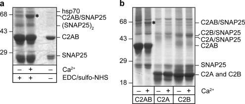
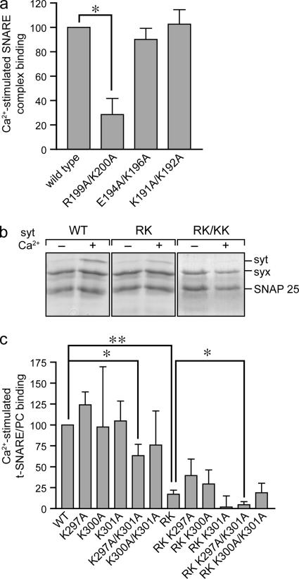
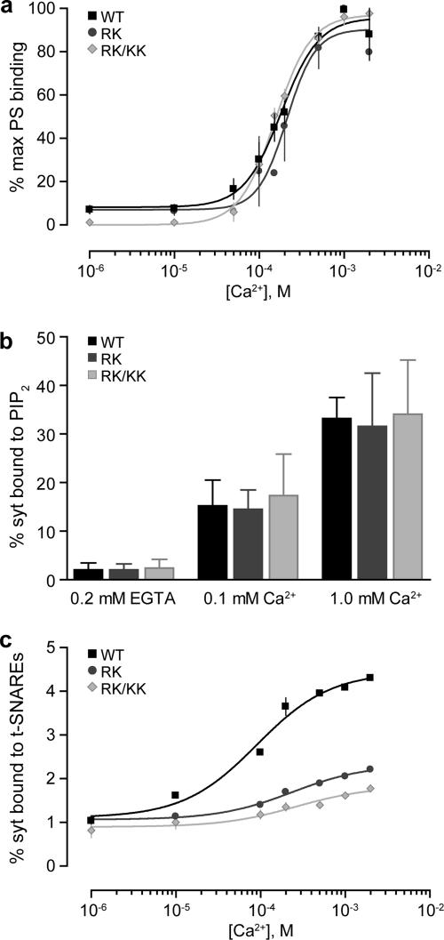
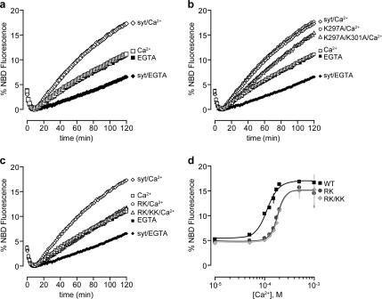
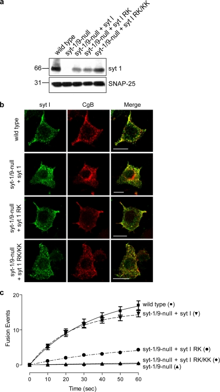
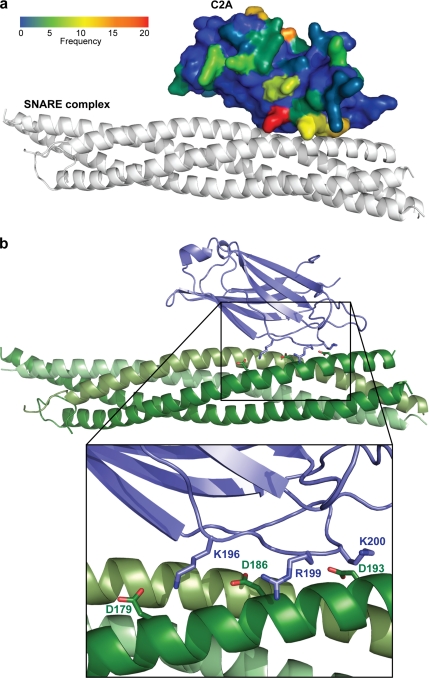
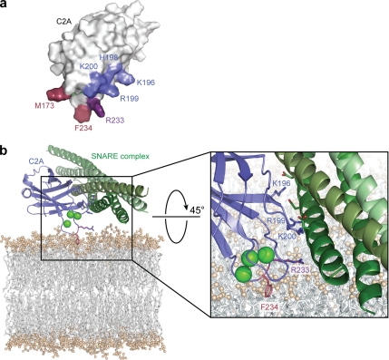
Synaptotagmins contain tandem C2 domains and function as Ca(2+) sensors for vesicle exocytosis but the mechanism for coupling Ca(2+) rises to membrane fusion remains undefined. Synaptotagmins bind SNAREs, essential components of the membrane fusion machinery, but the role of these interactions in Ca(2+)-triggered vesicle exocytosis has not been directly assessed. We identified sites on synaptotagmin-1 that mediate Ca(2+)-dependent SNAP25 binding by zero-length cross-linking. Mutation of these sites in C2A and C2B eliminated Ca(2+)-dependent synaptotagmin-1 binding to SNAREs without affecting Ca(2+)-dependent membrane binding. The mutants failed to confer Ca(2+) regulation on SNARE-dependent liposome fusion and failed to restore Ca(2+)-triggered vesicle exocytosis in synaptotagmin-deficient PC12 cells. The results provide direct evidence that Ca(2+)-dependent SNARE binding by synaptotagmin is essential for Ca(2+)-triggered vesicle exocytosis and that Ca(2+)-dependent membrane binding by itself is insufficient to trigger fusion. A structure-based model of the SNARE-binding surface of C2A provided a new view of how Ca(2+)-dependent SNARE and membrane binding occur simultaneously.


Figures

Figure 1.
Figure 1.
Ca2+ stimulates the formation of a C2AB-SNAP25 cross-linked adduct. (a) Wild-type C2AB (not shown) or C2AB K326A/K327A (1 μM) and SNAP25 (1 μM) were incubated in the absence (−) or presence (+) of 1 mM CaCl2 before the addition of cross-linker (EDC/sulfo-NHS) and analyzed by SDS PAGE with Coomassie staining. C2AB K326A/K327A was used to minimize oligomerization (Chapman et al., 1998), but identical results were obtained with wild-type C2AB and no self cross-linking of C2AB was observed. Bands corresponding to SNAP25, C2AB, SNAP25 dimer, C2AB-SNAP25 adduct (*), and hsp70 are indicated. Lane on right was loaded with 10% input lacking cross-linker. (b) Both C2 domains are required for optimal cross-linking of C2AB to SNAP25. C2AB, C2A, or C2B fusion proteins (1 μM) were incubated with SNAP25 (1 μM) in the absence (−) or presence (+) of 1 mM Ca2+ before cross-linking. Bands corresponding to C2AB-SNAP25 (*), C2B-SNAP25, C2A-SNAP25, or free proteins are indicated.
Figure 2.
Figure 2.
Schematic summary of C2AB interactions with SNAP25. The presence of Ca2+ altered interactions between C2A and C2B domains with SNAP25. For C2A, residues 191-196 bound the C terminal portion of the N terminal helix of SNAP25 (residues 77-83) in the absence but not presence of Ca2+ (orange). In the presence of Ca2+, C2A residues 191–200 bound the C terminal helix of SNAP25 (residues 176-201; blue). For C2B, residues 301-313 bound to the N terminus of SNAP25 (residues 32-40; green) in the absence of Ca2+ but shifted to C2B residues 289-297 in the presence of Ca2+.
Figure 3.
Figure 3.
C2A loop 2 and C2B loop 1 mutations reduce Ca2+-stimulated binding of C2AB to SNARE complexes. (a) Wild-type C2AB, C2AB(R199A/K200A), C2AB (E194A/K196A) or C2AB(K191A/K192A), all at 1 μM, were incubated with 1 μM SNARE complex immobilized on glutathione-Sepharose beads in the absence (−) or presence (+) of 1 mM CaCl2. Binding of C2AB was determined by Western blotting after SDS PAGE. Mean values ± SE are shown (n = 3). (b) C2AB, C2AB(R199A/K200A) (RK) or C2AB (R199A/K200A/K297A/K301A) (RK/KK), all at 10 μM, were incubated with SNAP25/syntaxin-containing PC liposomes in the absence or presence of 1 mM Ca2+. Liposome-bound C2AB was isolated from Accudenz gradients and analyzed by SDS-PAGE and Coomassie Blue staining. (c) C2AB and indicated mutants were incubated with SNAP25/syntaxin-containing liposomes in the presence or absence of 1 mM Ca2+. RK corresponds to R199A/K200A. Bound C2AB was analyzed as in panel b, quantified by densitometry, and normalized to syntaxin in each gradient. Ca2+-dependent binding by C2AB mutants was expressed as the difference bound in the presence or absence of Ca2+ and normalized to binding by wild-type C2AB. Mean values ± SE are shown (n = 3). Differences in binding between wild-type C2AB and RK or K297A/K301A mutants, or between RK and RK/K297A/K301A mutants were significant (**p < 0.01; *p < 0.05).
Figure 4.
Figure 4.
C2A loop 2 and C2B loop 1 mutants exhibit decreased Ca2+-dependent SNARE binding but normal Ca2+-dependent phosphatidylserine binding. (a) C2AB, C2AB(R199A/K200A) (RK) or C2AB(R199A/K200A/K297A/K301A) (RK/KK) were incubated with protein-free PC liposomes containing 15% PS at the indicated Ca2+ concentrations and bound C2AB was determined as in Figure 3b. Maximal bound C2AB was set equal to 100% and plotted (±SE, n = 3) as a function of [Ca2+]. (b) C2AB, C2AB(R199A/K200A) (RK), or C2AB(R199A/K200A/K297A/K301A) (RK/KK) were incubated with protein-free PC liposomes containing 5% PIP2 at the indicated Ca2+ concentrations and bound C2AB was determined as in Figure 3b. Fractional bound C2AB was plotted (±SE, n = 3) as a function of [Ca2+]. (c) C2AB and indicated mutants were incubated with PC liposomes containing SNARE complexes at the indicated Ca2+ concentrations, and bound C2AB was determined as in Figure 3b. The fraction of C2AB bound was plotted (±SE, n = 3) as a function of [Ca2+].
Figure 5.
Figure 5.
C2A loop 2 and C2B loop 1 mutations reduce Ca2+ stimulation of liposome fusion mediated by C2AB. (a) C2AB, 10 μM, stimulated the fusion of v-SNARE with t-SNARE liposomes in the presence of 100 μM Ca2+ but not in the presence of 0.2 mM EGTA. NBD fluorescence was expressed as percentage maximum fluorescence after 1% dodecyl maltoside addition. (b) C2AB, C2AB(K297A), or C2AB(K297A/K301A), all at 10 μM, were tested in the liposome fusion assay. C2AB(K297A/K301A) exhibited partial loss of function in conferring Ca2+ stimulation. (c) C2AB(R199A/K200A) (RK) or C2AB (R199A/K200A/K297A/K301A) (RK/KK) mutants failed to stimulate liposome fusion in the presence of 100 μM Ca2+. (d) C2AB, 10 μM, and mutants were incubated in the liposome fusion assay with indicated [Ca2+]. % maximal NBD fluorescence at 120 min was plotted as a function of [Ca2+] (±SE, n = 3). EC50 values for Ca2+ were 113 μM (±4 μM) for C2AB, 181 μM (±9 μM) for C2AB(R199A/K200A) (RK), and 187 μM (±9 μM) for C2AB (R199A/K200A/K297A/K301A) (RK/KK).
Figure 6.
Figure 6.
Synaptotagmin−1 mutants fail to restore Ca2+-dependent exocytosis in synaptotagmin-depleted PC12 cells. (a) Immunoblot analysis of wild-type PC12 cells, synaptotagmin−1/9-null cells, and null cells expressing wild-type synaptotagmin−1 or R199A/K200A (RK) or R199A/K200A/K297A/K301A (RK/KK) mutants. SNAP25 was used as a loading control. (b) Wild-type and synaptotagmin−1/9-null cells expressing synaptotagmin−1 or indicated mutants were fixed and stained with synaptotagmin−1 (green channel) and CgB (red channel) antibodies, and colocalization was determined (yellow). The synaptotagmin−1 mutants were localized to dense-core vesicles. (c) Cells expressing ANF-EGFP were stimulated with depolarization medium, and images were acquired at 0.25-s intervals. Fusion events were counted as a flash/puff of fluorescence for each cell type, and the sum of fusion events in 10-s intervals was determined. The sums of the average number of fusion events per cell over time (mean value ± SD) for wild-type (n = 25), synaptotagmin−1/9-null (n = 25), synaptotagmin−1/9-null + synaptotagmin−1 (n = 20), synaptotagmin−1/9-null + synaptotagmin−1 RK (n = 15), and synaptotagmin−1/9-null + synaptotagmin−1 RK/KK (n = 15) cells were plotted.
Figure 7.
Figure 7.
Ca2+-dependent C2A/SNARE binding interface derived from computational modeling. (a) Using ZDOCK, each of 20 NMR structures of C2A was computationally docked to the atomic coordinates of the SNARE complex crystal structure. The frequency of occurrence of individual C2A residues within 5 Å of acidic C-terminal SNAP25 residues D179, D186, and D193 is depicted. C2A is shown in a space filling model docked to a ribbon model of the SNARE complex. C2A residues are colored according to their frequency of proximity to SNAP25 D179, D186, or D193 from 0 (blue) to 20 (red) of 59. (b) The model shows the predicted low-energy docking orientation for Ca2+-dependent C2A-SNARE binding. The model corresponds to complex 1–1826 shown in Supplementary Tables S1 and S2. Close-up view depicts the binding interface for synaptotagmin−1 residues (K196, R199, and K200) with SNAP 25 residues (D179, D186, and D193).
Figure 8.
Figure 8.
Membrane-docked model for Ca2+-dependent C2A-SNARE interaction. (a) Space-filling model of C2A depicting clustered basic residues of β4-loop 2 (K196, H198, R199, and K200) positioned orthogonally to membrane-inserting (M173 and F234) residues of loops 1 and 3. R233 is shown as positioned between both interfaces. (b) Model depicts simultaneous interactions of C2A with SNARE complex and membrane. Ca2+ ions (green spheres) bound by loops 1 and 3 with F234 penetrating the bilayer are shown. The orthogonal face containing K196, R199, and K200 interacting with SNAP25 C-terminal helix is depicted. R233 is shown between membrane- and SNARE-binding interfaces.

Similar articles

Cited by

References

    1. Arac D., Murphy T., Rizo J. Facile detection of protein-protein interactions by one-dimensional NMR spectroscopy. Biochemistry. 2003;42:2774–2780. - PubMed
    1. Bai J., Wang C. T., Richards D. A., Jackson M. B., Chapman E. R. Fusion pore dynamics are regulated by synaptotagmin*t-SNARE interactions. Neuron. 2004;41:929–942. - PubMed
    1. Bai J., Wang P., Chapman E. R. C2A activates a cryptic Ca2+-triggered membrane penetration activity within the C2B domain of synaptotagmin I. Proc. Natl. Acad. Sci. USA. 2002;99:1665–1670. - PMC - PubMed
    1. Bennett M. K., Calakos N., Scheller R. H. Syntaxin: a synaptic protein implicated in docking of synaptic vesicles at presynaptic active zones. Science. 1992;257:255–259. - PubMed
    1. Bhalla A., Chicka M. C., Tucker W. C., Chapman E. R. Ca2+-synaptotagmin directly regulates t-SNARE function during reconstituted membrane fusion. Nat. Struct. Mol. Biol. 2006;13:323–330. - PubMed

Publication types

MeSH terms