. 2007 Dec;18(12):4911-20.
doi: 10.1091/mbc.e07-03-0243. Epub 2007 Oct 3.

Caenorhabditis elegans prom-1 is required for meiotic prophase progression and homologous chromosome pairing

Affiliations

Caenorhabditis elegans prom-1 is required for meiotic prophase progression and homologous chromosome pairing

Verena Jantsch et al. Mol Biol Cell. 2007 Dec.

Abstract

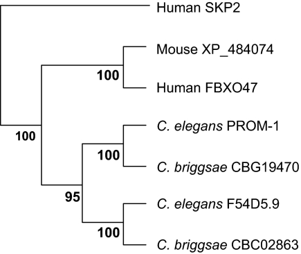
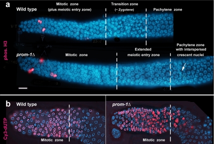
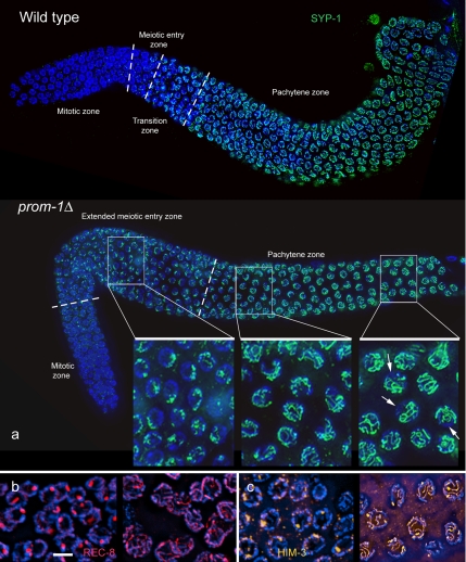
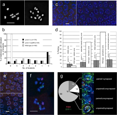
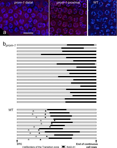
A novel gene, prom-1, was isolated in a screen for Caenorhabditis elegans mutants with increased apoptosis in the germline. prom-1 encodes an F-box protein with limited homology to the putative human tumor suppressor FBXO47. Mutations in the prom-1 locus cause a strong reduction in bivalent formation, which results in increased embryonic lethality and a Him phenotype. Furthermore, retarded and asynchronous nuclear reorganization as well as reduced homologous synapsis occur during meiotic prophase. Accumulation of recombination protein RAD-51 in meiotic nuclei suggests disturbed repair of double-stranded DNA breaks. Nuclei in prom-1 mutant gonads timely complete mitotic proliferation and premeiotic replication, but they undergo prolonged delay upon meiotic entry. We, therefore, propose that prom-1 regulates the timely progression through meiotic prophase I and that in its absence the recognition of homologous chromosomes is strongly impaired.


Figures

Figure 1.
Figure 1.
Phylogenetic tree of C. elegans PROM-1 and related proteins. The human F-box protein SKP2 is displayed as a member of the outgoup. The phylogenetic tree was derived from a multiple protein sequence alignment by the ClustalX program (Thompson et al., 1997). The resulting tree was tested by resampling (bootstrapping) by using the programs sequboot, protpars, and consense from the PHYLIP package (Felsenstein, 1989), executed on a server of the Department of Molecular Evolution, Uppsala University (http://artedi.ebc.uu.SE/programs/). The numbers at the forks indicate in how many trees (out of 100) the grouping to the right of the fork occurred.
Figure 2.
Figure 2.
Organization of gonads in the wild type and in prom-1Δ (ok1140). (a) In the wild type, the mitotic zone of the germline (with mitotic nuclei immunostainined for phosphorylated histone H3 depicted in red) and a narrow meiotic entry zone (see text) is followed by the transition zone containing crescent-shaped nuclei and then by the pachytene zone. In the prom-1Δ mutant, the mitotic zone is followed by an extended meiotic entry zone with large nuclei that are not crescent shaped. The pachytene zone contains some crescent-shaped nuclei (arrow). The vertical broken lines indicate the boundaries between different regions, defined by DAPI staining (see text). (b) Incorporation of Cy3-labeled dUTP (red) is limited to the mitotic zone in wild-type and mutant gonads. Chromatin is stained blue with DAPI. Bar, 10 μm.
Figure 3.
Figure 3.
(a) Immunostaining of SC component SYP-1 (green) in the wild type and in the prom-1Δ mutant. In the images of whole ovaries, the broken lines mark the borders between the zones with distinguishable nuclear morphologies (see Figure 2a), namely, the mitotic zone, the meiotic entry zone and the pachytene zone. In the wild type, small spots of SYP-1 first occur in a narrow meiotic entry zone just upstream of the transition zone, and then they develop into continuous lines along the pachytene zone. In the prom-1Δ mutant, the meiotic entry zone with SYP-1 spots is greatly expanded; SYP-1 starts to form lines in the distal pachytene zone. Insets are enlarged details of the regions indicated. By the end of the pachytene zone, SC formation in some nuclei is quite extensive, as is evident from the formation of long SYP-1 lines, but incomplete (arrows denote DAPI-positive chromatin regions devoid of SYP-1). (For blow up images of corresponding regions of the wild-type gonad, see Supplemental Figure S4.) (b) REC-8 in the extended meiotic entry zone (left) and the pachytene zone (right) of the prom-1Δ mutant showing increasing formation of linear structures. (c) HIM-3 in the extended meiotic entry zone (left) where it only forms spots, and the pachytene zone (right) where in some nuclei thick HIM-3 lines suggest the presence of synapsed regions in the prom-1Δ mutant. Bar, 5 μm (b, c, and insets in a are shown at the same magnification).
Figure 4.
Figure 4.
Reduced homologous pairing and bivalent formation in the prom-1Δ mutant. (a) A variable number of univalents is formed in the prom-1Δ mutant. Although six bivalents are formed in diakinesis in the wild type (left), univalents are abundant in the mutant. In this particular prom-1Δ mutant cell (right), 10 univalents and one bivalent are present (DAPI staining). (b) Bivalent frequencies in prom-1Δ mutant, prom-1Δ/qDf8 heterozygous, and wild-type diakineses. Shown is the percentage of diakinesis nuclei with the number of bivalents indicated. n = number of diakineses scored. (c and d) Homologous pairing was tested by FISH of a locus near the left end of chromosome I (Cy3, red) and of the 5S rDNA region on the right arm of chromosome V (FITC, green). (c) In the pachytene zone of the wild type (left), a single signal or a closely associated pair of signals is mostly observed for both loci. In the pachytene zone of the prom-1Δ mutant (right), homologous loci on autosomes remain mostly unpaired. (d) Homologous pairing frequencies of the rDNA locus in different zones of the wild-type and prom-1Δ gonads as indicated by the association of FISH signals. The percentages of nuclei with paired loci (represented by a single dot or two dots touching each other) are shown. The raw data for this figure are given in Supplemental Table S6. (e) Immunostaining of the X chromosome-associated protein HIM-8 (orange) produces mostly a single signal in wild-type (top) and in prom-1Δ mutant (bottom) pachytenes. (f) Examples of prom-1Δ mutant diakinesis nuclei with X chromosomes (red, FISH signal) and chromosomes V (green, FISH signal). The X chromosomes are more often involved in bivalent formation (as indicated by signals sharing a DAPI-positive entity) than the autosomes (see text). (g) Frequencies of homologous pairing and synapsis of the 5S rDNA locus determined by simultaneous FISH and SYP-1 staining. This experiment shows that most of the time synapsis is nonhomologous. Bars, 5 μm.
Figure 5.
Figure 5.
The abundance and distribution of recombination sites visualized by immunostaining of RAD-51. (a) RAD-51 foci (red) in the prom-1Δ mutant and wild-type (WT) animals. For the prom-1Δ mutant, the distal and the proximal border of a RAD-51–positive region are shown, where RAD-51 foci appear and are most abundant, respectively. In the wild type, there is not very much variation in the number of RAD-51 foci along the RAD-51–positive region of which a representative section is shown. (b) The position and extent of the RAD-51–positive region (black bars) in the gonads (shaded bars) of prom-1Δ mutant and wild-type (WT) animals. Gonads were measured from the DTC to the end of continuous cell rows (roughly coinciding with the end of the pachytene zone) and normalized, and the borders of the RAD-51–positive region were entered. This region is striking due to the abundance of RAD-51 foci. In quantitative terms, it is defined as the zone in which >90% of nuclei have more than two prominent foci by RAD-51 immunostaining. The open and solid arrowheads indicate the distal and proximal borders of the transition zone of the wild type, respectively (for a classification of meiotic stages in the gonad; see MacQueen et al., 2002), whereas the transition zone is absent from the prom-1Δ mutant gonads. Bar, 10 μm.
Figure 6.
Figure 6.
Analysis of the epistatic relationship between prom-1 and htp-1. Dissected prom-1, htp-1 and prom-1 htp-1 gonads are shown. MZ, mitotic zone; TZ, transition zone; MEZ, meiotic entry zone; PZ, pachytene zone. In the prom-1 mutant, a transition zone is missing, but there is an extended meiotic entry zone. This zone is characterized by the expression of SC proteins, but they do not organize into axial elements but form nuclear spots, such as seen here for REC-8. In the pachytene zone axial elements and SCs do form but some of the spots remain. The pachytene zone is interspersed with nuclei with chromatin arranged in a crescent shape. In the htp-1 mutant, there is onset of axial element formation and precocious synapsis immediately downstream of a narrow and poorly developed transition zone. There are no nuclei with crescent-shaped chromatin in the pachytene zone. In the prom-1 htp-1 double mutant, the extended meiotic entry zone is present and spots of SC proteins remain in pachytene. Nuclei with crescent-shaped chromatin are no longer found.

Similar articles

Cited by

References

    1. Alpi A., Pasierbek P., Gartner A., Loidl J. Genetic and cytological characterization of the recombination protein RAD-51 in Caenorhabditis elegans. Chromosoma. 2003;112:6–16. - PubMed
    1. Baarends W. M., Wassenaar E., Hoogerbrugge J. W., van Cappellen G., Roest H. P., Vreeburg J., Ooms M., Hoeijmakers J.H.J., Grootegoed J. A. Loss of HR6B ubiquitin-conjugating activity results in damaged synaptonemal complex structure and increased crossing-over frequency during the male meiotic prophase. Mol. Cell Biol. 2003;23:1151–1162. - PMC - PubMed
    1. Bhalla N., Dernburg A. F. A conserved checkpoint monitors meiotic chromosome synapsis in Caenorhabditis elegans. Science. 2005;310:1683–1686. - PubMed
    1. Borts R. H., Lichten M., Haber J. E. Analysis of meiosis-defective mutations in yeast by physical monitoring of recombination. Genetics. 1986;113:551–567. - PMC - PubMed
    1. Brenner S. The genetics of Caenorhabditis elegans. Genetics. 1974;77:71–94. - PMC - PubMed

Publication types