. 2007 Oct 31;26(21):4423-32.
doi: 10.1038/sj.emboj.7601868. Epub 2007 Oct 4.

HCN4 provides a 'depolarization reserve' and is not required for heart rate acceleration in mice

Affiliations

HCN4 provides a 'depolarization reserve' and is not required for heart rate acceleration in mice

Stefan Herrmann et al. EMBO J. .

Abstract

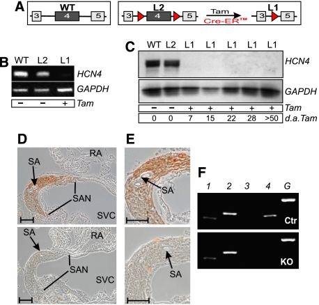
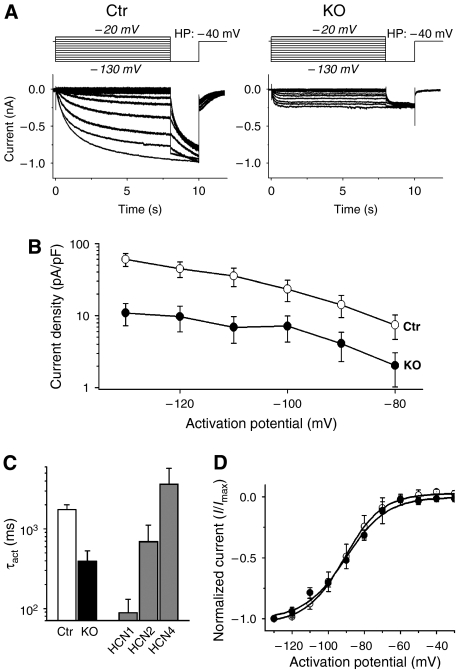
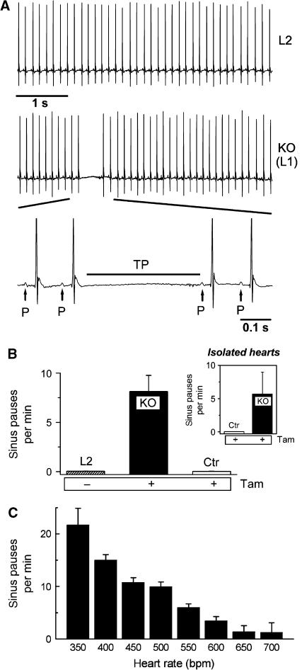
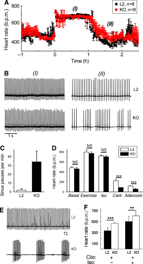
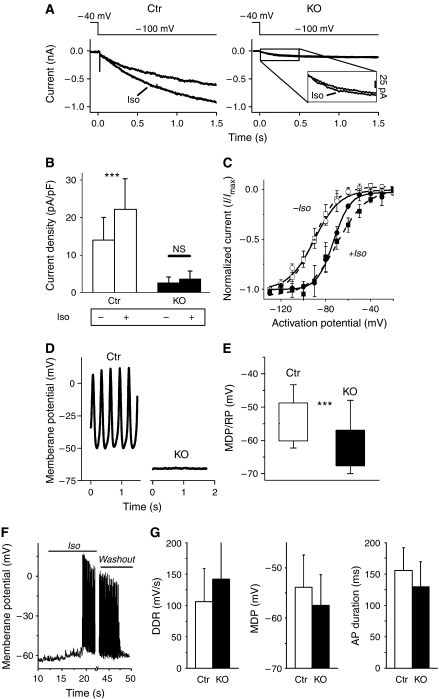
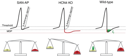
Cardiac pacemaking involves a variety of ion channels, but their relative importance is controversial and remains to be determined. Hyperpolarization-activated, cyclic nucleotide-gated (HCN) channels, which underlie the I(f) current of sinoatrial cells, are thought to be key players in cardiac automaticity. In addition, the increase in heart rate following beta-adrenergic stimulation has been attributed to the cAMP-mediated enhancement of HCN channel activity. We have now studied mice in which the predominant sinoatrial HCN channel isoform HCN4 was deleted in a temporally controlled manner. Here, we show that deletion of HCN4 in adult mice eliminates most of sinoatrial I(f) and results in a cardiac arrhythmia characterized by recurrent sinus pauses. However, the mutants show no impairment in heart rate acceleration during sympathetic stimulation. Our results reveal that unexpectedly the channel does not play a role for the increase of the heart rate; however, HCN4 is necessary for maintaining a stable cardiac rhythm, especially during the transition from stimulated to basal cardiac states.


Figures

Figure 1
Figure 1
Temporally controlled deletion of HCN4 in the primary cardiac conduction system. (A) Schematic representation of wild-type (WT), floxed (L2) and HCN4-knockout (L1) alleles. Numbered boxes indicate exons 3–5. LoxP sites are represented by red triangles. Activation of the Tam-inducible Cre recombinase (Cre-ER™) results in the deletion of exon 4, encoding pore and S6 segment of the channel. (BE) Tam-induced disruption of HCN4 in the sinoatrial node. (B) RT–PCR analysis. Total RNA was prepared from sinoatrial node tissue of wild-type and double-transgenic animals before (L2) and after (L1) Tam treatment. HCN4, amplicon corresponding to HCN4 wild-type RNA; GAPDH, internal control. (C) Immunoblot analysis. At different time points, sinoatrial proteins were isolated from wild-type and double transgenic animals before (L2) and after (L1) injection of Tam. The blot was probed with an anti-HCN4 antibody. A GAPDH antibody was used to check for equal loading. d.a.Tam., days after last injection of Tam. (D) Immunohistochemical detection of HCN4. Transverse sections through the sinoatrial node region of a control (upper panel) and a Tam-treated double-transgenic (lower panel) animal labeled with anti-HCN4 antibody. RA, right atrium; SA, sinoatrial node artery; SAN, sinoatrial node; SVC, superior vena cava. (E) Higher-magnification images of the sections shown in (D). Scale bars in (D, E), 100 μm. (F) RT–PCR using RNA prepared from the SAN area shows no change in the expression level of the remaining HCN isoforms in knockout compared to control animals. 1, HCN1; 2, HCN2; 3, HCN3; 4, HCN4; G, GAPDH.
Figure 2
Figure 2
Analysis of If in isolated knockout (L1) sinoatrial node cells. (A) Example traces of If recorded from a control (Ctr) and knockout (KO=L1) sinoatrial cell. Currents were activated by stepping from a holding potential of −40 mV to test potentials ranging from −130 mV to −20 mV for 8 s (in 10 mV steps), followed by −130 mV for 2 s before stepping back to the holding potential (protocol above current traces). Cells were held at −40 mV for 30 s before applying each new activation. Control (Ctr): HCN4L2/+, CAGGCre-ER™tg/0, 4–6 weeks after Tam; knockout (KO): HCN4L1/L2, CAGGCre-ER™tg/0, 4–6 weeks after Tam. (B) Means of voltage-dependent current densities from control (white symbols) and HCN4-knockout cells (black symbols) at activation potentials from −130 to −80 mV. The current density of the knockout cells (n=23) was reduced by 75% on average compared to control cells (n=32). The absolute current amplitudes showed equal percentages of reduction, see Supplementary Figure S6. (C) Time constants of activation (τact) at −100 mV of control and knockout cells compared to τact of the murine HCN channel isoforms 1, 2 and 4 expressed in HEK293 cells. Please refer to Supplementary Figure S6 for the analysis over the full range of activation potentials. The activation kinetic of If in knockout cells (black column) is between the ones of HCN1 and HCN2, whereas control cells (white column) are close to HCN4. (D) Means of normalized If from control (white symbols) and HCN4-knockout cells (black symbols) at activation potentials from −130 to −30 mV. Boltzman fits of the means are used to display the voltage-dependent activation curves. There are no significant differences between the curves.
Figure 3
Figure 3
HCN4-knockout mice show recurrent sinus pauses. (A) The ECG of the same mouse before (L2) and after (L1 or KO) Tam injection demonstrates a typical sinus pause occurring after deletion of HCN4. During the pauses, no P-waves are detected (bottom, enlarged ECG segment); the pauses are characterized by lack of electrical activity ranging from the end of the preceding T-wave to the beginning of the next regular P-wave (TP). (B) Quantitative analysis of the number of sinus pauses. L2 and KO represent the same animals before and after Tam injection, Control (Ctr) indicates HCN4L2/+ animals after Tam injection (n=8 for each group). (Inset) Isolated spontaneously contracting hearts from knockout, but not control animals display similar sinus pauses (n=4 for each group). (C) Mean sinus pause frequency plotted against the heart rate. Uninterrupted recordings over 7 days were used for this analysis. Heart rates were grouped into 50 b.p.m. intervals, numbers indicate the upper range limit (e.g. 350 refers to the interval of 300–350 b.p.m.).
Figure 4
Figure 4
HCN4 is not required for upregulation of the heart rate. (A) Isoproterenol (0.5 mg/kg) injected at t=0 increased the heart rate of control (L2, n=8) and knockout (L1 or KO, n=8) animals to similar maximum levels. (B) Example ECG traces from control and knockout mice at (I), 0.5 h and (II), 1.5 h after isoproterenol injection. All traces are displayed at the same scale. See Supplementary Figures S5 and S6 for enlarged ECGs. (C) Quantitative analysis of the sinus pauses in phase (II). The number of sinus pauses increased fourfold compared to the basal level (Figure 3B). (D) Mean heart rates of control and knockout mice during stimulation by an exercise protocol (running on a treadmill) or isoproterenol (iso, 0.5 mg/kg) did not differ significantly. In contrast, carbachol (carb, 0.5 mg/kg) and the A1 adenosine receptor agonist CCPA (adenosine, 0.3 mg/kg) induced highly significant lower heart rates in knockouts (n=8 for both genotypes, ***P<0.001). (E) Example ECG traces recorded 30 min after the injection of 0.5 mg/kg cilobradine of the same mouse before (L2) and after (KO) Tam injection. Both traces are displayed at the same scale (bar=1 s). (F) Analysis of heart rates (n=8 mice) reveals a changed effect of cilobradine in knockouts. The decrease of the heart rate by cilobradine (excluding sinus pauses in the calculation) is less in knockout than in control animals. Additional stimulation by isoproterenol leads to a similar heart rate increase in both groups. cilo, cilobradine; iso, isoproterenol. ***P<0.001; **P<0.01.
Figure 5
Figure 5
Isoproterenol does not significantly increase the residual If in HCN4-deficient sinoatrial cells, but induces the spontaneous discharge of action potentials. (A) Examples of If traces elicited at −100 mV from control (left panel) and knockout (right panel) SAN cells before and after application of 1 μM isoproterenol (iso). An enlargement of If in HCN4-knockout cells is shown in the inset (right panel). (B) If in knockout cells (black columns, n=15) was only slightly modulated by isoproterenol resulting in a tendency towards an increased current density after 0.5 s (P=0.057). In contrast, If in control cells (white columns, n=14) was readily accelerated resulting in a significant current increase after 0.5 s (P<0.05). (C) Voltage-dependent activation curves of both control (squares) and HCN4-knockout (circles) If are shifted towards more-positive activation potentials by 1 μM isoproterenol (+iso, filled symbols; −iso, open symbols). V1/2 and slope factors were calculated using the Boltzman fit of the means, here displayed as open and broken lines. (DG) Membrane potential recordings from isolated sinoatrial node cells. MDP/RP refers to the maximum diastolic potential (MDP) of spontaneously firing cells and the resting membrane potential (RP) of silent cells, respectively. (D) Example traces of membrane potential recordings from control and knockout cells. Typical spontaneous SAN action potentials were readily recorded from control cells (11 out of 13 cells), but rarely from knockout cells (3 out of 27 cells). The majority of these cells (24/27) were electrically silent. (E) Analysis of membrane potentials revealed a significant more negative membrane potential of knockout cells. Boxes indicate mean±s.d. of all measured cells (27 KO, 13 Ctr cells), error bars represent the absolute range. ***P<0.001. (F) Example trace of a membrane potential recording from an HCN4-deficient SAN cell that was ‘silent' at first. Superfusion with 1 μM isoproterenol (‘iso') induced a spontaneous discharge of action potentials. After 30 s, superfusion was switched back to iso-free bath solution (‘washout'), which terminated the firing after about 20 s. (G) Action potential parameters of iso-stimulated knockout cells (black columns, n=7) were not significantly different from control pacemaker cells. In addition to the parameters displayed here, no significant differences were also found with upstroke velocity, overshoot potential and repolarization velocity.
Figure 6
Figure 6
Proposed role for HCN4 channels in sinoatrial node cells. top, Spontaneous action potentials under basal (solid line) and beta-adrenergic-stimulated (broken line) conditions. MDP, maximum diastolic potential. Lower part, scale illustrating the overall relationship between depolarizing and repolarizing currents. Left, regular SAN action potential (SAN-AP). To ensure stable pacemaking over time, depolarizing currents are in a dynamic balance with repolarizing currents (indicated by balanced scale). In most situations, and particularly during sympathetic stimulation, HCN4 is not required to promote stable pacemaking. Middle and right, SAN action potentials after an increase in repolarizing currents (e.g. muscarinic stimulation or transition from activated to basal cardiac state; symbolized by red weight). In wild-type cells (right), HCN4 is activated and provides a depolarizing current (green) keeping the system well-balanced. In HCN4-deficient cells (middle), the membrane potential tends to remain sub-threshold fostering the appearance of sinus pauses.

Similar articles

Cited by

References

    1. Akagi K, Sandig V, Vooijs M, Van der Valk M, Giovannini M, Strauss M, Berns A (1997) Cre-mediated somatic site-specific recombination in mice. Nucleic Acids Res 25: 1766–1773 - PMC - PubMed
    1. Biel M, Schneider A, Wahl C (2002) Cardiac HCN channels: structure, function, and modulation. Trends Cardiovasc Med 12: 206–212 - PubMed
    1. Bois P, Bescond J, Renaudon B, Lenfant J (1996) Mode of action of bradycardic agent, S 16257, on ionic currents of rabbit sinoatrial node cells. Br J Pharmacol 118: 1051–1057 - PMC - PubMed
    1. Brown HF, DiFrancesco D, Noble SJ (1979) How does adrenaline accelerate the heart? Nature 280: 235–236 - PubMed
    1. Bruning JC, Michael MD, Winnay JN, Hayashi T, Horsch D, Accili D, Goodyear LJ, Kahn CR (1998) A muscle-specific insulin receptor knockout exhibits features of the metabolic syndrome of NIDDM without altering glucose tolerance. Mol Cell 2: 559–569 - PubMed

Publication types

Substances