. 2007 Oct 2;5(10):e268.
doi: 10.1371/journal.pbio.0050268.

Force-induced unfolding of fibronectin in the extracellular matrix of living cells

Affiliations

Force-induced unfolding of fibronectin in the extracellular matrix of living cells

Michael L Smith et al. PLoS Biol. .

Abstract

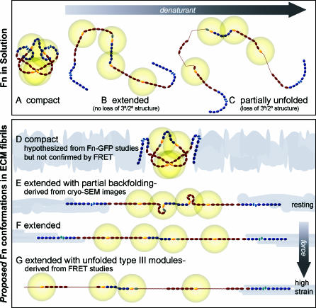
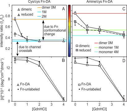
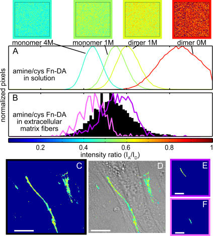
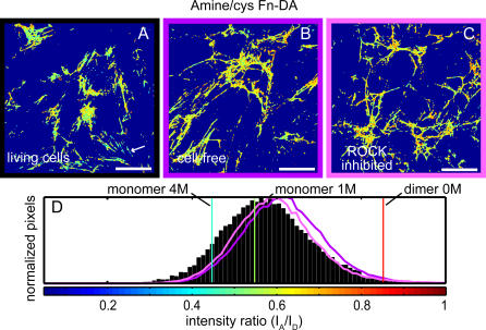
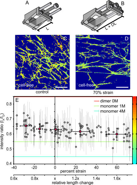
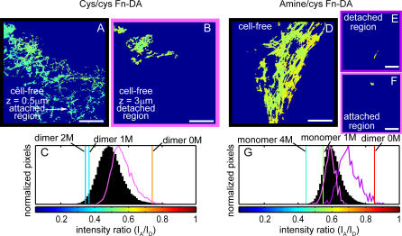
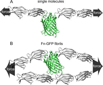
Whether mechanically unfolded fibronectin (Fn) is present within native extracellular matrix fibrils is controversial. Fn extensibility under the influence of cell traction forces has been proposed to originate either from the force-induced lengthening of an initially compact, folded quaternary structure as is found in solution (quaternary structure model, where the dimeric arms of Fn cross each other), or from the force-induced unfolding of type III modules (unfolding model). Clarification of this issue is central to our understanding of the structural arrangement of Fn within fibrils, the mechanism of fibrillogenesis, and whether cryptic sites, which are exposed by partial protein unfolding, can be exposed by cell-derived force. In order to differentiate between these two models, two fluorescence resonance energy transfer schemes to label plasma Fn were applied, with sensitivity to either compact-to-extended conformation (arm separation) without loss of secondary structure or compact-to-unfolded conformation. Fluorescence resonance energy transfer studies revealed that a significant fraction of fibrillar Fn within a three-dimensional human fibroblast matrix is partially unfolded. Complete relaxation of Fn fibrils led to a refolding of Fn. The compactly folded quaternary structure with crossed Fn arms, however, was never detected within extracellular matrix fibrils. We conclude that the resting state of Fn fibrils does not contain Fn molecules with crossed-over arms, and that the several-fold extensibility of Fn fibrils involves the unfolding of type III modules. This could imply that Fn might play a significant role in mechanotransduction processes.


Conflict of interest statement

Competing interests. The authors have declared that no competing interests exist.

Figures

Figure 1
Figure 1. Schematic Sketch of Putative Fn Conformations in Solution and within ECM Fibrils
Fn consists of tandem repeats of type I (dark blue ovals), II (narrow, light blue ellipses), and III modules (dark red ovals). Average end-to-end lengths of each module type are drawn to scale using lengths of 2.5 nm for Fn type I [51], 0.7 nm for Fn type II [52], and 3.2 nm for FnIII modules [53]. Two free cysteines are present on each monomer within FnIII7 and III15 (yellow). Energy transfer between donors and acceptors bound to free cysteines are limited to within approximately double the Förster radius (∼12 nm), denoted by gold circles around III7 and III15. Fn in solution assumes a compact conformation stabilized through ionic interactions between dimer arms (A) [7]. Denaturant destabilizes these ionic interactions, leading to an extended conformation (B), and higher denaturant concentrations lead to loss of tertiary/secondary structure of FnIII modules (C). The quaternary model for fibril elongation [11,12] predicts a compact conformation with crossover of opposing arms in the absence of tension (D). In contrast, high-resolution cryo-scanning electron microscopic images of Fn fibrils [39,40], taken together with our FRET studies, suggest that fully relaxed fibers do not contain the compact quaternary structure, but are composed of Fn in an extended conformation with partial backfolding of its arms upon themselves (nodules; E). Cell-generated tensile forces first extend Fn fibrils (F) and finally unfold FnIII modules (G).
Figure 2
Figure 2. Fn-DA Unfolding in Denaturant Solution Probed by FRET and Circular Dichroism
Dimeric Fn-DA (open symbols) or monomeric Fn-DA generated by reduction in 50 mM DTT for 60 min (shaded symbols) with donors and acceptors labeled exclusively on cysteines (cys/cys Fn-DA; A and B) or on amines and cysteines (amine/cys Fn-DA; C and D) was unfolded in 0, 0.6, 1, 2, and 4 M GdnHCl. I A/I D was quantified by calculating the ratio of measured acceptor and donor intensities using 12-nm bandwidths from 566–572 nm and 514–526 nm, respectively (A and C). Mean residue ellipticity was measured for labeled Fn-DA and Fn-u (black circles) in GdnHCl (B and D). Horizontal colored lines for dimeric amine/cys Fn-DA in 0 M GdnHCl, monomeric amine/cys Fn-DA in 1 and 4 M GdnHCl, and dimeric cys/cys Fn-DA in 0, 1, and 2 M GdnHCl are shown based on an artificial color code ranging from 0.05 to 1.0 I A/I D units that is identical to that used in Figures 3–6. Bar width corresponds to the standard deviation of the mean values from three different measurements in each denaturant concentration.
Figure 3
Figure 3. Fn Conformation in Matrix Fibrils
Spatial ratiometric images and histograms of all pixels within each field of view are shown for dimeric amine/cys Fn-DA in 0 and 1 M GdnHCl and monomeric amine/cys Fn-DA in 1 and 4 M GdnHCl (A). Amine/cys Fn-DA was added to the culture medium of fibroblasts for 24 h, and excess Fn-u was added to suppress intermolecular energy transfer. Confocal microscopic images of acceptor and donor peak intensities taken 1 μm above the glass–cell interface were background subtracted, averaged, and thresholded, and the I A/I D ratiometric image of acceptor to donor was color-coded within the range of 0.05 to 1.0. A histogram (B) for all pixels of amine/cys Fn-DA–containing ECM (C) and an overlay of I A/I D on the DIC image (D) are shown in a region in which the matrix showed a transition from low to intermediate I A/I D within a single Fn fiber. Histograms are overlaid in (B) for regions of extended (E; purple) and unfolded Fn (F; pink). Histograms were generated with 0.01-ratio-unit bin widths. Scale bars = 25 μm.
Figure 4
Figure 4. Matrix Refolding after Cell Extraction or ROCK Inhibition
Amine/cys Fn-DA and excess Fn-u were added to the culture medium of fibroblasts for 24 h. Color-coded I A/I D ratiometric images are shown for control cells (A), extracted cell-free matrix (B), and fibroblast cells after 60 min exposure to the ROCK inhibitor Y-27632 (C). Histograms with 0.01-ratio-unit bin widths for all pixels of control (black), cell-free (purple), and ROCK-inhibited matrix (pink) were derived from three random fields of view each from three separate experiments in each group (D). Solution denaturation values for dimeric Fn-DA in 0 M GdnHCl and monomeric Fn-DA in 1 and 4 M GdnHCl are shown as red, green, and blue lines, respectively. Scale bars = 50 μm.
Figure 5
Figure 5. Fn Conformation after Application of Exogenous Strain
A schematic of the strain device is shown in the relaxed configuration with length L before (A) and length L + DL after (B) application of strain. PDMS sheets were covalently modified with Fn-u as described in Materials and Methods, and fibroblast cells were cultured for 24 h in the presence of amine/cys Fn-DA and excess Fn-u. Cells were extracted in mild detergent. Color-coded I A/I D ratiometric images are shown for a field of view without application of stretch (C) and after application of 70% elongation strain with 28% transverse compression (D). Region of interest analysis on individual fibrils was used to determine the impact of elongation on I A/I D on a per fibril basis (circles, mean ± standard deviation), and binned averages were calculated for fibrils between −37% and −20%, −20% and −10%, −10% and 10%, 10% and 40%, and 40% and 73% strain (red squares, mean ± standard deviations) (E). Abscissa is also plotted as relative length change. Solution values for dimeric Fn-DA in 0 M GdnHCl and monomeric Fn-DA in 1 and 4 M GdnHCl are shown as horizontal red, green, and blue lines, respectively. Scale bars = 50 μm.
Figure 6
Figure 6. Fully Relaxed Fn-DA Does Not Assume the Fully Compact Conformation
Cys/cys Fn-DA (A–C) or amine/cys Fn-DA (D–G) was incorporated into fibroblast matrix on Fn-u that was adsorbed to plasma cleaned PDMS, and after cell extraction the substrate was relaxed to 4/5 (A and B; 3.7% transverse stretch) or 3/5 the starting length (D–F; 10% transverse stretch). I A/I D ratiometric images of cys/cys Fn-DA–containing matrix are shown at the PDMS–ECM interface (A), where a portion of the cell-free fibers are still attached to the substrate, and from the same field of view but acquired 3 μm above the PDMS surface (B), where the strain-free Fn mat randomly diffused around its points of attachment to the underlying ECM. Histograms are shown for all pixels within the field of view at the substrate (C; black) and from the upper, strain-free confocal slice (C; pink). An I A/I D ratiometric image of amine/cys Fn-DA is shown with both detached (E) and still-attached (F) regions of matrix within the same confocal slice. Region of interest analysis was used to generate histograms (G0 for all pixels within the detached (E and G; purple) and attached (F and G; pink) regions of matrix, which were overlaid on a histogram of all pixels in the field of view (black). Scale bars = 50 μm.
Figure 7
Figure 7. Restricting GFP Movement if Embedded in a Fn Fibris
Cartoons of FnIII modules (gray) and GFP (green) were generated to scale using Tachyon renderer and implemented in VMD version 8.5 to illustrate the degree of protrusion from the central axis of an extended Fn-GFP molecule. In single-molecule pulling experiments, GFP has the freedom to align along the direction of force (A). If embedded into a densely packed Fn fiber that is stretched by mechanical force, the rotational motion of GFP is restricted (B), which could lead to unfolding along a different pathway.

Similar articles

Cited by

References

    1. Vogel V, Sheetz M. Local force and geometry sensing regulate cell functions. Nat Rev Mol Cell Biol. 2006;7:265–275. - PubMed
    1. Vogel V. Mechanotransduction involving multimodular proteins: Converting force into biochemical signals. Annu Rev Biophys Biomol Struct. 2006;35:459–488. - PubMed
    1. Pankov R, Yamada KM. Fibronectin at a glance. J Cell Sci. 2002;115:3861–3863. - PubMed
    1. Geiger B, Bershadsky A, Pankov R, Yamada KM. Transmembrane crosstalk between the extracellular matrix and the cytoskeleton. Nat Rev Mol Cell Biol. 2001;2:793–805. - PubMed
    1. Mao Y, Schwarzbauer JE. Fibronectin fibrillogenesis, a cell-mediated matrix assembly process. Matrix Biol. 2005;24:389–399. - PubMed

Publication types

MeSH terms