. 2007 Dec;85(6):762-71.
doi: 10.1016/j.exer.2007.08.010. Epub 2007 Aug 24.

VEGF modulation of retinal pigment epithelium resistance

Affiliations

VEGF modulation of retinal pigment epithelium resistance

Zsolt Ablonczy et al. Exp Eye Res. 2007 Dec.

Abstract

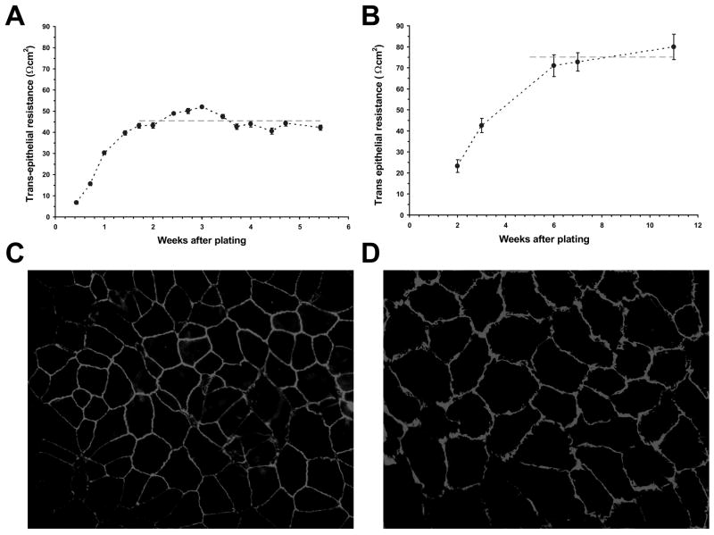
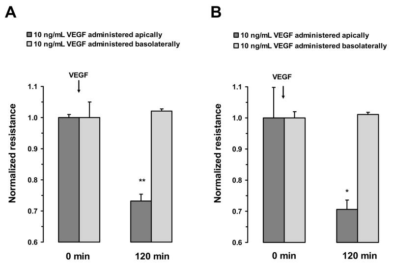
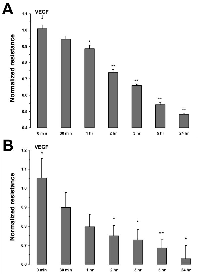
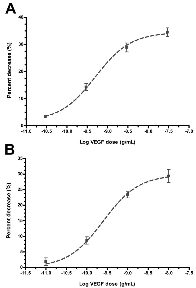
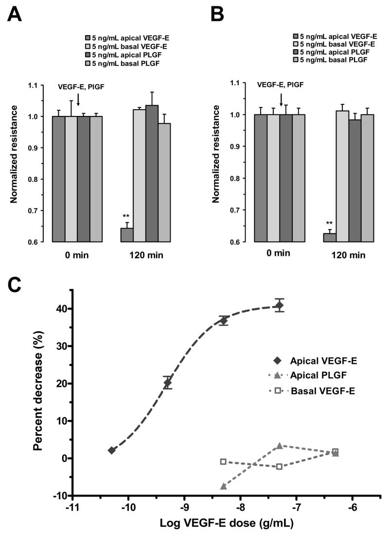
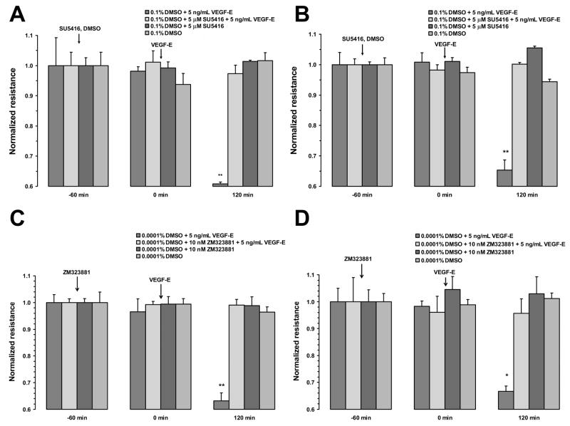
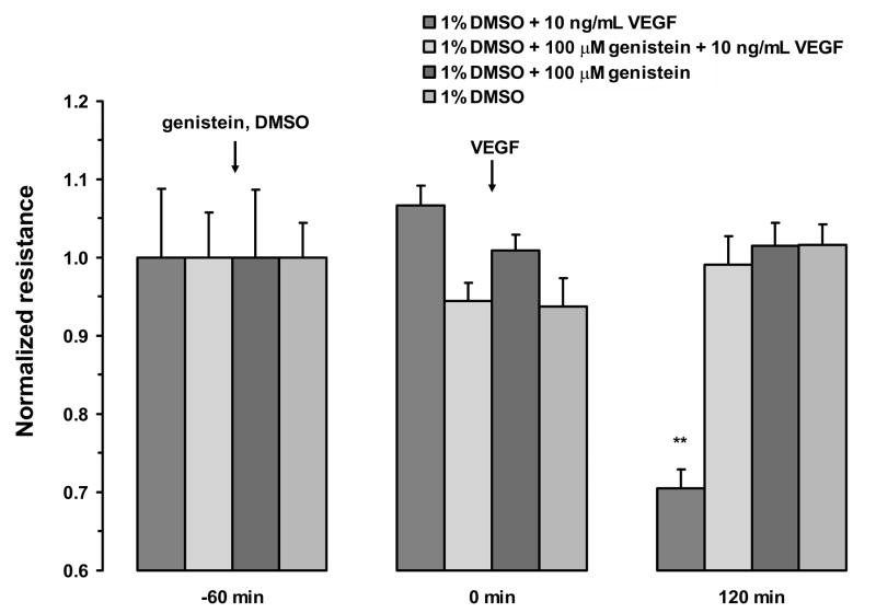
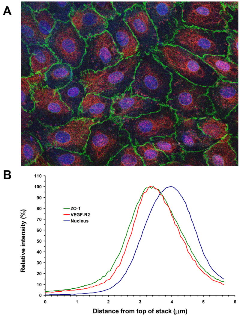
Fluid accumulation into the subretinal space and the development of macular edema is a common condition in age-related macular degeneration, diabetic retinopathy, and following ocular surgery, or injury. Vascular endothelial growth factor (VEGF) and other cytokines have been implicated in the disruption of retinal pigment epithelium (RPE) barrier function and a reduction in the regulated removal of subretinal fluid; however, the cellular and molecular events linking these agents to the disruption of barrier function have not been established. In the current study, cultures of ARPE-19 and primary porcine retinal pigment epithelium (RPE) cells were utilized to investigate the effects of the VEGF-induced modifications to the barrier properties of the RPE. The barrier function was determined by transepithelial resistance (TER) measurements and morphology of the RPE monolayers. In both ARPE-19 and primary porcine RPE cells the administration of VEGF produced a significant drop in TER, and this response was only observed following apical administration. Maximum reduction in TER was reached 5h post VEGF administration. These responses were concentration-dependent with an EC(50) of 502pg/mL in ARPE-19 cells and 251pg/mL in primary porcine cells. In both ARPE-19 and primary RPE cells, the response to VEGF was blocked by pretreatment with the relatively selective VEGF-R2 antagonists, SU5416 or ZM323881, or the protein tyrosine kinase inhibitor, genistein. Administration of the relatively selective VEGF-R2 agonist, VEGF-E, also reduced TER in a concentration-dependent manner (EC(50) of 474pg/mL), while VEGF-R1 agonist, placental growth factor (PlGF), did not significantly alter the TER. Immunolocalization studies demonstrated that confluent monolayers exhibited continuous cell-to-cell ZO-1 protein contacts and apical localization of the VEGF-R2 receptors. These data provide evidence that the VEGF-induced breakdown of RPE barrier function is mediated by the activation of apically-oriented VEGF-R2 receptors. Thus, VEGF-mediated increases in RPE permeability are initiated by a rise in intraocular levels of VEGF.


Figures

Figure 1
Figure 1
Resistance and morphology of RPE cell monolayers. A, Increase in transepithelial resistance of ARPE-19 cell monolayers following cell plating on transwell inserts. The dashed line represents the average resistance (45 ± 4 Ωcm2) for confluent monolayers between weeks 2 and 5. Cultures were tested for up to 8 weeks; however, routine experiments were performed following three weeks in culture. B, Increase in transepithelial resistance of porcine primary RPE cell monolayers following cell plating on transwell inserts. The dashed line represents the average resistance (76 ± 9 Ωcm2) achieved for confluent monolayers between weeks 5 and 11. Values are means ±SE. Immunohistochemistry of confluent ARPE-19 cells (C) and porcine primary RPE cells (D) with mouse anti-ZO-1 as primary, and FITC-conjugated anti-mouse as secondary antibodies. The magnification was ×200.
Figure 2
Figure 2
Polarized response of RPE cell monolayer to VEGF-A165. In both ARPE-19 cells (A) and porcine primary RPE cells (B), the treatment with VEGF-A165 (10 ng/mL) to the apical side significantly decreased TER, while the treatment of the basal side did not significantly alter resistance measurements. Values are means ±SE of individual measurements normalized to the average TER at t = 0. (* P <0.05, ** P <0.01).
Figure 3
Figure 3
Time course of TER changes in response to VEGF-A165. The apical side of confluent monolayers were treated with 10 ng/mL of VEGF-A165 for 30 minutes to 24 hours. Addition of 10 ng/mL VEGF-A165 ARPE-19 cells reduced TER to approximately 50% of its original value within 5 hours (A). The addition of VEGF-A-165 to primary porcine RPE cells reduced TER by 70% within 5 hours (B). Values are means ±SE of individual measurements normalized to the average TER values at t = 0. (* P <0.05, ** P <0.01).
Figure 4
Figure 4
Concentration-dependent reduction in TER induced by VEGF-A165. In both ARPE-19 cells (A) and porcine primary RPE cells (B), the percent decrease in resistance was concentration dependent with an EC50 of 502 pg/mL (LogEC50 = -9.30 ±0.09) for ARPE-19 cells and 251 pg/mL (LogEC50 = -9.60 ±0.05) for porcine primary RPE cells. The Hill coefficients were not significantly different from 1.0. Values are means ±SE of individual measurements normalized to the average TER values at t = 0.
Figure 5
Figure 5
Responses of RPE monolayers following VEGF-E and PlGF administration. In both ARPE-19 cells (A) and porcine primary RPE cells (B), apically administered VEGF-E (5 ng/mL) significantly reduced TER; however, apically-administration of PlGF (5 ng/mL) did not significantly alter resistance. The administration of either agonist to the basolateral side did not significantly alter TER. C, Concentration-response curves of the ARPE-19 cells to apical VEGF-E and PlGF and basal VEGF-E. The apical administration of VEGF-E induced a concentration dependent decrease in TER. The EC50 to this response was 474 pg/mL (LogEC50 = -9.32 ±0.04). The Hill coefficient was not significantly different from 1.0. Apically-applied PlGF or basolaterally-applied VEGF-E were not effective in significantly altering TER. Values are means ±SE normalized to the average resistance at t = 0. (** P <0.01)
Figure 6
Figure 6
Inhibition of VEGF-E165 response by VEGF-R2 antagonists. Pretreatment of ARPE-19 (A) or primary porcine RPE (B) cells with with 5 μM SU5416 blocked the response to VEGF-E. In monolayers pretreated with vehicle, VEGF-E induced a significant drop in TER within 120 minutes after treatment. Pretreatment of ARPE-19 cells (C) or primary porcine RPE cells (D) with the more selective VEGF-R2 antagonist, ZM323881, also blocked the response to VEGF-E. Again, vehicle pretreatment did not significantly alter VEGF-E-induced reduction in TER. Values are means ±SE of individual measurements normalized to the average TER values at t = 0. (* P <0.05, ** P <0.01).
Figure 7
Figure 7
Inhibition of VEGF-A165 response by Genistein. Pretreatment of ARPE-19 cells with genistein (100 μM) for 1 hour blocked the reduction in TER induced by VEGF-A165. The addition of Genistein alone or vehicle did not significantly alter TER. Values are means ±SE normalized to the average resistance at t = 0. (** P <0.01)
Figure 8
Figure 8
Apical localization of VEGF-R2 in confluent RPE monolayers. A, Immunohistochemical staining of the porcine primary RPE cell monolayer with mouse anti-ZO-1 and rabbit anti-VEGF-R2 as primary antibodies, and FITC-conjugated goat anti-mouse (green) and rhodamine-conjugated goat anti-rabbit (red) as secondary antibodies, respectively. The cell nuclei were visualized with DRAQ5 (blue). The image was obtained as a Z-stack on the confocal microscope. The VEGF-R2 staining was only observed in the apical regions and was absent at depths below the nuclei. The magnification was ×400. B, Color profile in the Z-stack images. The figure shows the profile for a representative region, containing a single RPE cell. Optical sectioning revealed that single color profiles represent the localization of the individual stains in the depth of the image. Analysis of color profiles revealed the close co-localization of VEGF-R2 receptor and ZO-1 protein in the apical region of the RPE cell.

References

    1. Antcliff RJ, Marshall J. The pathogenesis of edema in diabetic maculopathy. Seminars in ophthalmology. 1999;14(4):223–232. - PubMed
    1. Autiero M, Waltenberger J, Communi D, Kranz A, Moons L, Lambrechts D, Kroll J, Plaisance S, De Mol M, Bono F, Kliche S, Fellbrich G, Ballmer-Hofer K, Maglione D, Mayr-Beyrle U, Dewerchin M, Dombrowski S, Stanimirovic D, Van Hummelen P, Dehio C, Hicklin DJ, Persico G, Herbert JM, Shibuya M, Collen D, Conway EM, Carmeliet P. Role of PlGF in the intra- and intermolecular cross talk between the VEGF receptors Flt1 and Flk1. Nat Med. 2003;9(7):936–943. - PubMed
    1. Barber AJ, Antonetti DA. Mapping the blood vessels with paracellular permeability in the retinas of diabetic rats. Invest Ophthalmol Vis Sci. 2003;44(12):5410–5416. - PubMed
    1. Becker PM, Waltenberger J, Yachechko R, Mirzapoiazova T, Sham JS, Lee CG, Elias JA, Verin AD. Neuropilin-1 regulates vascular endothelial growth factor-mediated endothelial permeability. Circulation research. 2005;96(12):1257–1265. - PubMed
    1. Blaauwgeers HG, Holtkamp GM, Rutten H, Witmer AN, Koolwijk P, Partanen TA, Alitalo K, Kroon ME, Kijlstra A, van Hinsbergh VW, Schlingemann RO. Polarized vascular endothelial growth factor secretion by human retinal pigment epithelium and localization of vascular endothelial growth factor receptors on the inner choriocapillaris. Evidence for a trophic paracrine relation. Am J Pathol. 1999;155(2):421–428. - PMC - PubMed

Publication types

MeSH terms