

Shear-induced capping of L-selectin on the neutrophil surface during centrifugation
- PMID: 17915247
- PMCID: PMC2121613
- DOI: 10.1016/j.jim.2007.08.011
Shear-induced capping of L-selectin on the neutrophil surface during centrifugation
Abstract
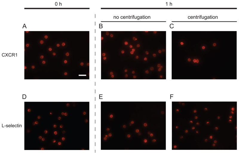
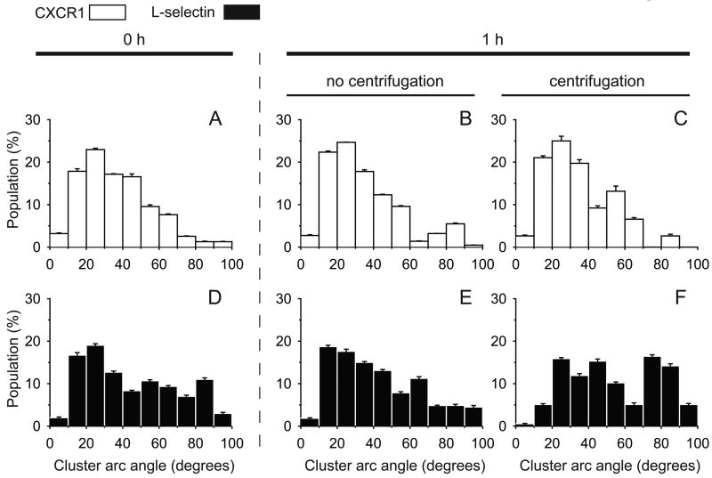
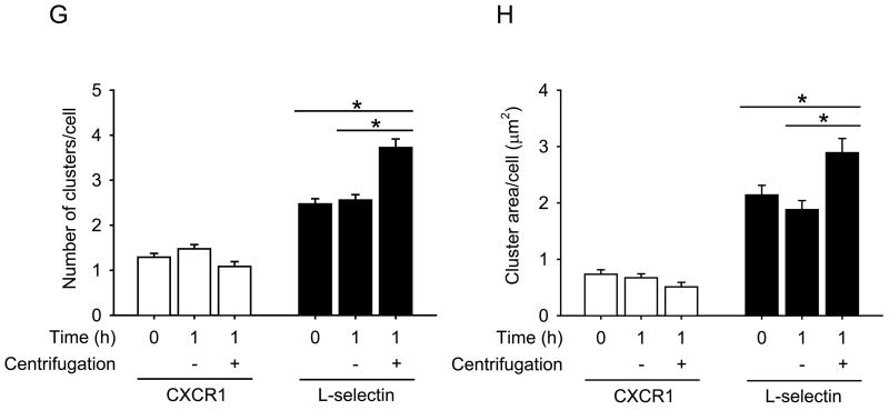
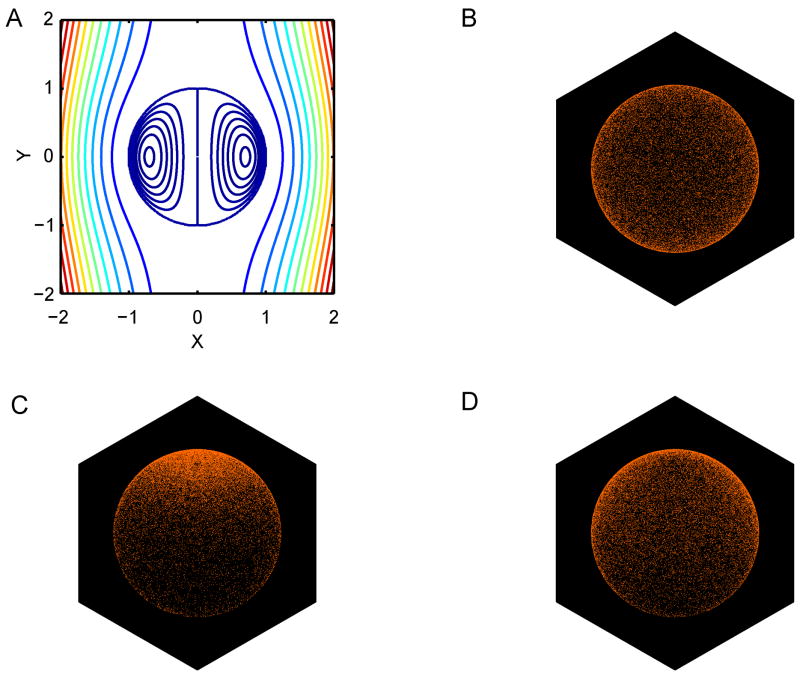
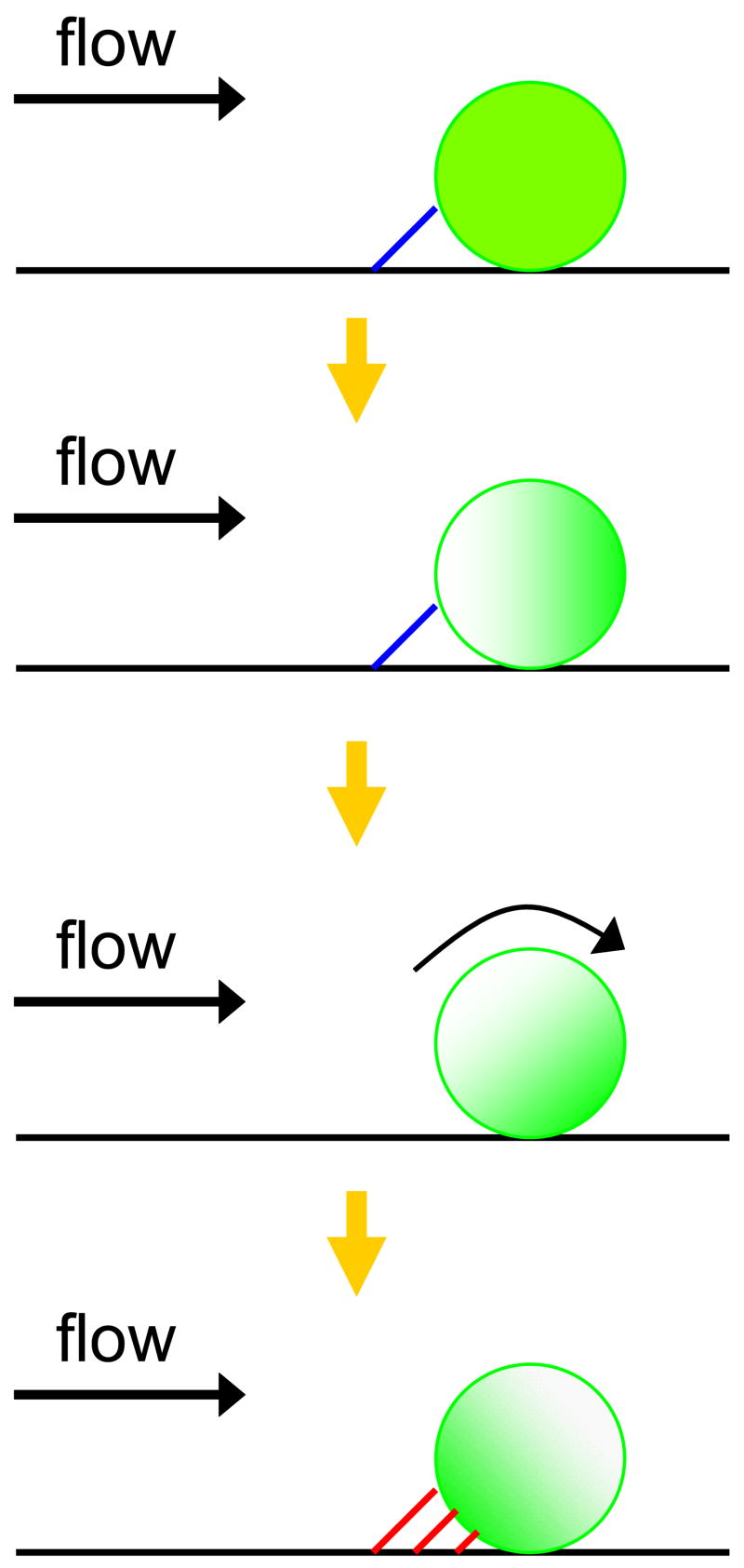
L-selectin on leukocytes is critical in leukocyte tethering and adhesion to inflamed endothelium and lymphocyte homing to lymphoid organs. The spatial distribution of L-selectin on leukocytes controls cellular adhesive function in hydrodynamic shear. How L-selectin changes its position on the cell membrane remains an open question, but a possible candidate is shear stress encountered on the cell surface. Here we demonstrate shear-induced L-selectin polarization on the membrane during the process of centrifugation of resting neutrophils via immunofluorescent microscopy. It was found that randomly distributed L-selectin on neutrophils moves to a polar cap at one end of the cell after centrifugation (300 x g for 2 min) without inflammatory stimuli. This L-selectin redistribution under shear was predicted by Monte Carlo simulations that show how convection dominates over diffusion, leading to L-selectin cap formation during centrifugation at 280 x g or during leukocyte adhesion to the endothelial wall at 1 dyn/cm(2). Those results point to a role for shear stress in the modulation of L-selectin distribution, and suggest a possible alternate mechanism and reinterpretation of previous in vitro studies of L-selectin mediated adhesion of neutrophils isolated via centrifugation.
Figures
Similar articles
-
Adhesive dynamics simulations of the mechanical shedding of L-selectin from the neutrophil surface.J Theor Biol. 2009 Sep 7;260(1):27-30. doi: 10.1016/j.jtbi.2009.05.014. Epub 2009 May 30. J Theor Biol. 2009. PMID: 19486904 Free PMC article.
-
Neutrophil-neutrophil interactions under hydrodynamic shear stress involve L-selectin and PSGL-1. A mechanism that amplifies initial leukocyte accumulation of P-selectin in vitro.J Clin Invest. 1996 Sep 1;98(5):1081-7. doi: 10.1172/JCI118888. J Clin Invest. 1996. PMID: 8787668 Free PMC article.
-
Selectin catch-bonds mechanotransduce integrin activation and neutrophil arrest on inflamed endothelium under shear flow.Blood. 2017 Nov 9;130(19):2101-2110. doi: 10.1182/blood-2017-05-783027. Epub 2017 Aug 15. Blood. 2017. PMID: 28811304 Free PMC article. Clinical Trial.
-
Neutrophil rolling at high shear: flattening, catch bond behavior, tethers and slings.Mol Immunol. 2013 Aug;55(1):59-69. doi: 10.1016/j.molimm.2012.10.025. Epub 2012 Nov 9. Mol Immunol. 2013. PMID: 23141302 Free PMC article. Review.
-
Targeting Neutrophil Adhesive Events to Address Vaso-Occlusive Crisis in Sickle Cell Patients.Front Immunol. 2021 Apr 28;12:663886. doi: 10.3389/fimmu.2021.663886. eCollection 2021. Front Immunol. 2021. PMID: 33995392 Free PMC article. Review.
Cited by
-
Vascular Recruitment of Human Retinoblastoma Cells by Multi-Cellular Adhesive Interactions with Circulating Leukocytes.Cell Mol Bioeng. 2010 Dec 1;3(4):361-368. doi: 10.1007/s12195-010-0145-8. Cell Mol Bioeng. 2010. PMID: 25110524 Free PMC article.
-
Halloysite Nanotube Coatings Suppress Leukocyte Spreading.Langmuir. 2015 Dec 22;31(50):13553-60. doi: 10.1021/acs.langmuir.5b03288. Epub 2015 Dec 9. Langmuir. 2015. PMID: 26605493 Free PMC article.
References
-
- Bevilacqua MP, Stengelin S, Gimbrone MA, Jr, Seed B. Endothelial leukocyte adhesion molecule 1: an inducible receptor for neutrophils related to complement regulatory proteins and lectins. Science. 1989;243:1160. - PubMed
-
- Burns MP, Depaola N. Flow-conditioned HUVECs support clustered leukocyte adhesion by coexpressing ICAM-1 and E-selectin. Am J Physiol Heart Circ Physiol. 2005;288:H194. - PubMed
-
- Carman CV, Springer TA. Integrin avidity regulation: are changes in affinity and conformation underemphasized? Curr Opin Cell Biol. 2003;15:547. - PubMed
-
- Chen C, Ba X, Xu T, Cui L, Hao S, Zeng X. c-Abl is involved in the F-actin assembly triggered by L-selectin crosslinking. J Biochem (Tokyo) 2006;140:229. - PubMed
-
- Chen HQ, Tian W, Chen YS, Li L, Raum J, Sung KL. Effect of steady and oscillatory shear stress on F-actin content and distribution in neutrophils. Biorheology. 2004;41:655. - PubMed
Publication types
MeSH terms
Substances
Grants and funding
LinkOut - more resources
Full Text Sources
Miscellaneous
