. 2007 Oct 30;46(43):12100-10.
doi: 10.1021/bi7007728. Epub 2007 Oct 4.

Solution structure of polymerase mu's BRCT Domain reveals an element essential for its role in nonhomologous end joining

Affiliations

Solution structure of polymerase mu's BRCT Domain reveals an element essential for its role in nonhomologous end joining

Eugene F DeRose et al. Biochemistry. .

Abstract

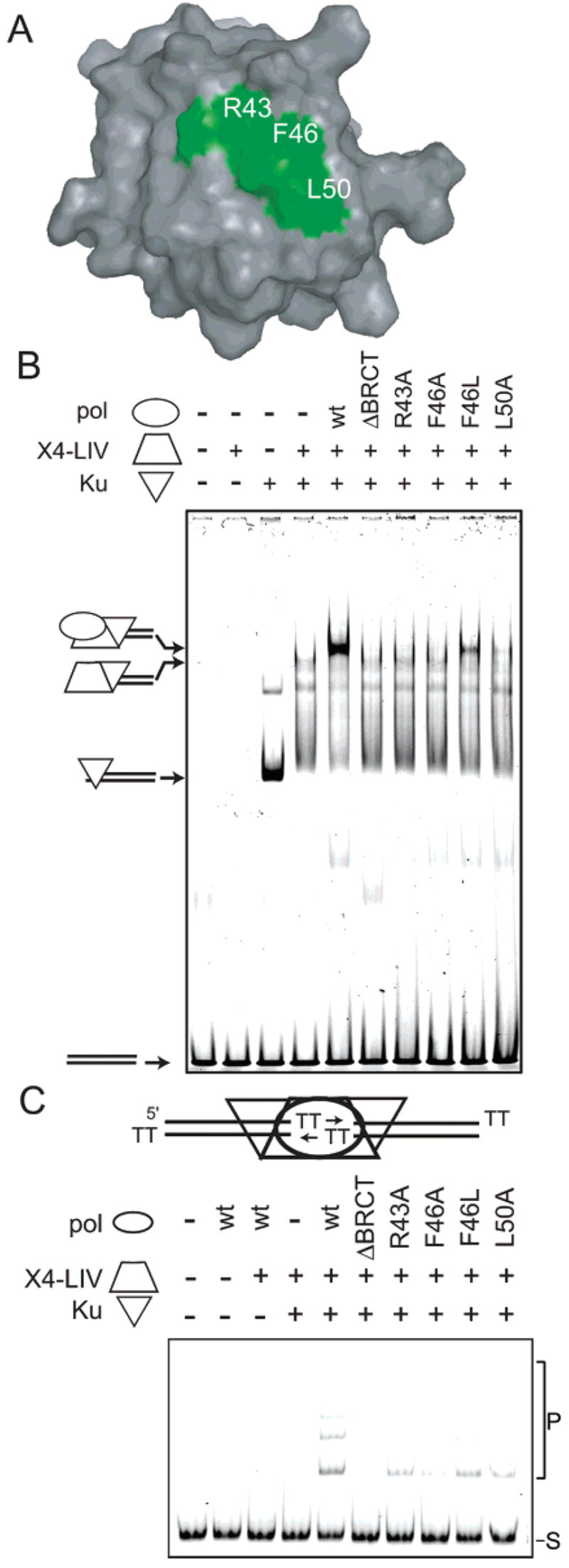
The solution structure and dynamics of the BRCT domain from human DNA polymerase mu, implicated in repair of chromosome breaks by nonhomologous end joining (NHEJ), has been determined using NMR methods. BRCT domains are typically involved in protein-protein interactions between factors required for the cellular response to DNA damage. The pol mu BRCT domain is atypical in that, unlike other reported BRCT structures, the pol mu BRCT is neither part of a tandem grouping, nor does it appear to form stable homodimers. Although the sequence of the pol mu BRCT domain has some unique characteristics, particularly the presence of >10% proline residues, it forms the characteristic alphabetaalpha sandwich, in which three alpha helices are arrayed around a central four-stranded beta-sheet. The structure of helix alpha1 is characterized by two solvent-exposed hydrophobic residues, F46 and L50, suggesting that this element may play a role in mediating interactions of pol mu with other proteins. Consistent with this argument, mutation of these residues, as well as the proximal, conserved residue R43, specifically blocked the ability of pol mu to efficiently work together with NHEJ factors Ku and XRCC4-ligase IV to join noncomplementary ends together in vitro. The structural, dynamic, and biochemical evidence reported here identifies a functional surface in the pol mu BRCT domain critical for promoting assembly and activity of the NHEJ machinery. Further, the similarity between the interaction regions of the BRCT domains of pol mu and TdT support the conclusion that they participate in NHEJ as alternate polymerases.


Figures

Figure 1
Figure 1
Alignment of pol X BRCT domains. Secondary structure elements (blue cartoons) in the human pol μ BRCT domain are aligned relative to its amino acid sequence and five other pol X BRCT domain sequences. Following their names and species, positions are numbered according to locations in their respective full length proteins. Positions conserved in all six members are highlighted in yellow. Residues conserved in more than four pol X members are identified in red (identical) or green (similar). Sequences were aligned using AlignX, a clustalW-based module of Vector NTI (Invitrogen). The first residue in human pol μ (G20) represents a substitution relative to the published sequence (S20).
Figure 2
Figure 2
Solution structure of the BRCT domain of pol μ.. (A) The 10 lowest energy structures after water refinement superimpose with a backbone rmsd of 0.70 ± 0.12 Å. The alpha helices are shown in red; the beta strands are shown in cyan. (B) The average, energy-minimized structure computed from the 10 lowest energy structures is shown. The central β sheet is shown with α1 and α3 packed against one side of the sheet and α2 packed against the opposite side of the sheet.
Figure 3
Figure 3
Structurally important residues of the BRCT domain of pol μ Positions of fully conserved residues, salt bridges, and exposed hydrophobic residues of the BRCT domain of pol μ (A) The average, energy-minimized structure is depicted with the side chains of residues that are fully conserved between, pol μ, TdT, and pol λ. The conserved residues H68, V70, L99, W104, L105, and V115 form part of the hydrophobic core of the domain. (B) The side chains of arginine, aspartate, and glutamate residues involved in salt-bridge formation and the exposed hydrophobic residues, F46 and L50 are shown.
Figure 4
Figure 4
Activity of pol μ BRCT domain mutants. (A) A surface representation of the pol μ BRCT domain, with the solvent exposed area of R43, F46, and L50 highlighted in green. (B) EMSA analysis was performed in the presence of a 60 bp DNA duplex, Ku, XRCC4-ligase IV (X4-LIV), and full length wild type pol μ (wt), pol μ with the BRCT domain deleted (ΔBRCT), or full length pol μ with BRCT domain substitution mutations as noted. The composition of each species of distinct mobility has previously been determined (Mahajan MCB 2002) and is noted with cartoons at the left of the panel. (C) Joining of a 300 bp substrate with two nucleotide TT 3′ overhangs was performed in the presence of Ku, XRCC4-ligase IV, and various pol μ constructs as noted. S denotes substrate, and P denotes joined concatemer products.
Figure 5
Figure 5
Regions of flexibility in the BRCT domain of pol μ. Backbone flexibility by three different measures are shown as a function of residue number. The 15N transverse relaxation rate, R2, is shown in filled circles. Increases from the mean are indicative of “slow” motions on the μs-ms time scale. These rates are typically inversely proportional to the intensity of peaks in a 1H–15N HSQC, as shown in the filled triangles. Lipari-Szabo model-free order parameters, S2, are shown in open circles. S2 can take on values from 0 to 1, with 0 corresponding to the limit of complete flexibility and 1 corresponding to complete rigidity of an N–H bond vector. Order parameters report on motion on the ps-ns time scale.
Figure 6
Figure 6
Comparison of pol μ and TdT BRCT domains. (A) The average, energy-minimized structure of the BRCT domain of polμ (cyan) and the BRCT domain of TdT (yellow, PDB ID 2COE model 1) superimpose with a backbone rmsd of 2.1 Å. (B, C) The positions of the aromatic side chains in the hydrophobic core are conserved between the two domains, as well as the positions of exposed hydrophobic residues on 1 and 2.
Figure 7
Figure 7
Electrostatic surfaces of the BRCT domains of pol μ and TdT. (A) A ridge of positively charged residues on the surface of the BRCT domain of pol μ may be the site of phosphopeptide or DNA binding. (B) Electrostatic surface of the BRCT domain of pol μ rotated 180° about the vertical direction with respect to part A, showing a large patch of negatively charged residues. (C) As in the pol μ domain, a ridge of positively charged residues on the surface of the TdT domain may be the site of phospho-peptide or DNA binding. (D) Electrostatic surface of the BRCT domain of TdT rotated 180° about the vertical direction with respect to part C, showing a large patch of negatively charge residues.

References

    1. Aoufouchi S, Flatter E, Dahan A, Faili A, Bertocci B, Storck S, Delbos F, Cocea L, Gupta N, Weill JC, Reynaud CA. Two novel human and mouse DNA polymerases of the polX family. Nucleic Acids Res. 2000;28:3684–3693. - PMC - PubMed
    1. Dominguez O, Ruiz JF, Lain de Lera T, Garcia-Diaz M, Gonzalez MA, Kirchhoff T, Martinez AC, Bernad A, Blanco L. DNA polymerase mu (Pol mu), homologous ato TdT, could act as a DNA mutator in eukaryotic cells. EMBO J. 2000;19:1731–1742. - PMC - PubMed
    1. Fan W, Wu X. DNA polymerase lambda can elongate on DNA substrates mimicking non-homologous end joining and interact with XRCC4-ligase IV complex. Biochem Biophys Res Commun. 2004;323:1328–1333. - PubMed
    1. Lee JW, Blanco L, Zhou T, Garcia-Diaz M, Bebenek K, Kunkel TA, Wang Z, Povirk LF. Implication of DNA polymerase λ in alignment-based gap filling for nonhomologous DNA end joining in human nuclear extracts. J Biol Chem. 2004;279:805–811. - PubMed
    1. Ma Y, Lu H, Tippin B, Goodman MF, Shimazaki N, Koiwai O, Hsieh CL, Schwarz K, Lieber MR. A biochemically defined system for mammalian nonhomologous DNA end joining. Mol Cell. 2004;16:701–713. - PubMed

Publication types