. 2007 Nov;274(21):5518-32.
doi: 10.1111/j.1742-4658.2007.06068.x. Epub 2007 Oct 3.

Krit 1 interactions with microtubules and membranes are regulated by Rap1 and integrin cytoplasmic domain associated protein-1

Affiliations

Krit 1 interactions with microtubules and membranes are regulated by Rap1 and integrin cytoplasmic domain associated protein-1

Sophie Béraud-Dufour et al. FEBS J. 2007 Nov.

Abstract

The small G protein Rap1 regulates diverse cellular processes such as integrin activation, cell adhesion, cell-cell junction formation and cell polarity. It is crucial to identify Rap1 effectors to better understand the signalling pathways controlling these processes. Krev interaction trapped 1 (Krit1), a protein with FERM (band four-point-one/ezrin/radixin/moesin) domain, was identified as a Rap1 partner in a yeast two-hybrid screen, but this interaction was not confirmed in subsequent studies. As the evidence suggests a role for Krit1 in Rap1-dependent pathways, we readdressed this question. In the present study, we demonstrate by biochemical assays that Krit1 interacts with Rap1A, preferentially its GTP-bound form. We show that, like other FERM proteins, Krit1 adopts two conformations: a closed conformation in which its N-terminal NPAY motif interacts with its C-terminus and an opened conformation bound to integrin cytoplasmic domain associated protein (ICAP)-1, a negative regulator of focal adhesion assembly. We show that a ternary complex can form in vitro between Krit1, Rap1 and ICAP-1 and that Rap1 binds the Krit1 FERM domain in both closed and opened conformations. Unlike ICAP-1, Rap1 does not open Krit1. Using sedimentation assays, we show that Krit1 binds in vitro to microtubules through its N- and C-termini and that Rap1 and ICAP-1 inhibit Krit1 binding to microtubules. Consistently, YFP-Krit1 localizes on cyan fluorescent protein-labelled microtubules in baby hamster kidney cells and is delocalized from microtubules upon coexpression with activated Rap1V12. Finally, we show that Krit1 binds to phosphatidylinositol 4,5-P(2)-containing liposomes and that Rap1 enhances this binding. Based on these results, we propose a model in which Krit1 would be delivered by microtubules to the plasma membrane where it would be captured by Rap1 and ICAP-1.


Figures

Fig. 1
Fig. 1. Krit1 contains a putative FERM domain and binds to PIP2
(A) Radixin-IP3 co-crystal structure. IP3 is in yellow, basic residues of F1 (K53, K60, K63, K64) and F3 (R273, R275, R279) domains are in red. The Tryptophan W58 is in green. (B) Homology models of F1, F2 and F3 domains of Krit1. The three independently modeled sub-domains have been manually arranged as on radixin structure. Basic residues of F1 (K475, K479, R485) and F3 (K713, K720, K724) domains are in red. The Tryptophan W487 is in green. (C) Krit1 (1 μM) was incubated with plasma membrane mix liposomes (0.75 mM) containing or not 2 % PIP2 at various NaCl concentrations. After centrifugation, proteins present in the supernatant (S) and the pellet (P) were analyzed by SDS-PAGE and quantified by fluorometry. Protein precipitation in the absence of liposomes has been substracted. Bands below Krit1 band are E.Coli contaminants that also bind to PIP2.
Fig. 2
Fig. 2. Krit1 N and C terminal parts associate together via the NPAY motif and ICAP-1 disrupts this association
(A) Schematic representation of Krit1 fragments used. FERM domain: Four-point one, Ezrin, Radixin, Moesin; PTB-like: Phosphotyrosine-binding domain. ANK: Ankyrin repeats. (B) GST-pull-down of 100 nM Krit1-NTer WT or mutant on 100 nM GST-HypoKrit1 or GST alone following the experimental procedure detailed in material and methods. (C). GST-pull-down of 5 μM Krit1-NTer on 3.5 μM GST-HypoKrit1 in presence of 5 μM ICAP-1. In (A) GST-fusions were immunoblotted with GST antibodies. In (B) and (C), GST-fusions were stained with Ponceau Red. Krit1-NTer and ICAP-1 were immunoblotted with His-tag and ICAP-1 antibodies respectively. Inputs correspond to 1/20 of total proteins. These results are representative of three independent experiments.
Fig. 3
Fig. 3. Rap1 binds to Krit1 C-terminus preferentially in its Rap1GTP form
(A) GST-pull down of 1 μM Rap1GDP, Rap1GTPγS or H-RasGTPγS on 10 μM GST-HypoKrit1 or GST alone. Inputs correspond to 1/25 of total proteins. GST-fusions were immunoblotted with GST antibodies. Rap1, ICAP-1 and H-Ras were immunoblotted with His-tag, ICAP-1 and H-Ras antibodies respectively. (B) GST-pull down of 1 μM RapGTPγS on 10 μM GST-HypoKrit1 or 10 μM GST-HypoKrit1ΔC. Input corresponds to 1/4 of total Rap1. Rap1 was immunoblotted with His-tag antibody. These data are representative of four independent experiments.
Fig 4
Fig 4. Krit1, Rap1-GTPγS and ICAP-1 form a ternary complex in vitro
GST-pull-down of 5 μM Krit1 or/and 5 μM Rap1GTPγS on 5 μM GST-ICAP-1 fusion or GST alone. Krit1 binding to GST-ICAP-1 was visualized by Sypro Orange staining of the gel. Rap1 binding was visualized by western blot using His-tag antibody. These results were reproduced three times.
Fig 5
Fig 5. Rap1 binding to HypoKrit1 is not modified by Krit-Nter or ICAP-1
(A) Comparaison by GST- pull down of the binding of 10 μM Rap1GTPγS to 3 μM GST-HypoKrit1 in the absence or presence of 10 μM Krit1-Nter and 10 μM ICAP-1. Rap1 and Krit1-Nter were detected by immunoblotting using anti-His antibody. ICAP-1 was revealed by anti-ICAP-1 antibody. (B) FLAG-pull down of 3 μM Rap1GTPγS or 7 μM GST-ICAP-1 to FLAG-Krit1 beads. Control corresponds to beads incubated with non transfected BHK cells and processed as FLAG-Krit beads. Input corresponds to 1/40 of total proteins. Proteins were stained by Sypro Orange. These results are representative of three independent experiments. (C) Model of the conformation of Krit1 in a binary complex with Rap1 or in a ternary complex with Rap1 and ICAP-1. Rap1 binds to the C-terminus of Krit1 closed and opened conformations. ICAP-1 binding opens Krit1 without perturbing Rap1 binding. For the clarity of the representation only intramolecular folding has been represented. A head to tail intermolecular folding between two molecules of Krit1 is however not excluded.
Fig. 6
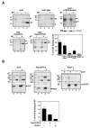
Fig. 6. Krit1 interacts directly with microtubules in vitro via two sites in its N- and C-termini and Rap1 and ICAP-1 inhibits this interaction
In each experiment, 40 pmoles of Krit1 were incubated in the absence or presence of taxol-stabilized MT polymerized in vitro from 150 pmoles of purified tubulin and centrifuged on sucrose gradient. Supernatant (S) and pellet (P) were analyzed by SDS-PAGE and the percentage of Krit1 bound to MT quantified by fluorometry. Krit1 precipitation in the absence of MT has been substracted. (A) Identification of MT binding sites on Krit1 using different Krit1 mutants. Arrowheads indicate Krit1 WT or mutants, T: Tubulin. Each experiment was repeated between two to four times. (B) Inhibition of Krit1 binding to MT by Rap1 and ICAP-1. 200 pmoles of Rap1 or ICAP-1 were added to 40 pmoles of Krit1 and to polymerized MT. Each experiment was repeated three times.
Fig 7
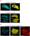
Fig 7
YFP-Krit1 co-localizes with CFP-labelled microtubules in transfected BHK cells and is delocalized by activated Rap1. BHK-cells were transfected with plasmids encoding YFP-Krit1 or YFP-ARNO and CFP-tubulin (A) together with pMT2HA-Rap1V12 (B) HA-Rap1V12 was detected with anti-HA 3F10 antibody. Scale bars: 15 μm.
Fig. 8
Fig. 8. Rap1 stimulates Krit1 binding to membranes
(A) GST-HypoKrit1 (0.5 μM) was incubated with asolectin vesicles (1 mg/ml) in absence or presence of Rap1GTPγS (3 μM). After centrifugation, the supernatant (S) and the pellet (P) were analyzed by SDS-PAGE and quantified by fluorometry. Protein precipitation in absence of liposomes has been substracted. (B) Dose-response of stimulation of Krit1 binding to asolectin vesicles by increasing concentrations of Rap1GTPγS. Each experiment was reproduced three times.
Fig 9
Fig 9. Model of regulation of Krit1 binding to microtubules and plasma membrane
In the cell, Krit1 would be transported in its closed conformation along the microtubules toward the plasma membrane. When reaching the membrane, Krit1 would detach from MT and be captured by activated Rap1 and ICAP-1 on the plasma membrane.

Similar articles

Cited by

References

    1. Bos JL. Linking Rap to cell adhesion. Curr Opin Cell Biol. 2005;17:123–8. - PubMed
    1. Caron E. Cellular functions of the Rap1 GTP-binding protein: a pattern emerges. J Cell Sci. 2003;116:435–440. - PubMed
    1. Kooistra MR. Rap1: a key regulator in cell-cell junction formation. J Cell Sci. 2007;120:17–22. - PubMed
    1. Katagiri K. RAPL, a Rap1-binding molecule that mediates Rap1-induced adhesion through spatial regulation of LFA-1. Nat Immunol. 2003;6:6. - PubMed
    1. Lafuente EM. RIAM, an Ena/VASP and Profilin ligand, interacts with Rap1-GTP and mediates Rap1-induced adhesion. Dev Cell. 2004;7:585–95. - PubMed

Publication types

MeSH terms

Substances